

Cryotherapy vs Surgical Removal for Skin Lesions: Why Pathology Sometimes Wins
Key Takeaways
- Cryotherapy destroys a skin lesion in place and leaves no tissue for a pathologist, which is the single biggest difference from surgical excision.
- Pigment-producing cells are more sensitive to cold than ordinary skin cells, so even a perfectly executed freeze commonly leaves a pale patch, especially on darker skin.
- Most clinicians will not freeze a mole, because a frozen melanoma loses its diagnosis, its measurable thickness and its margin assessment all at once.
- A Cochrane review found cryotherapy and salicylic acid preparations broadly similar for viral warts, and freezing often needs repeat sessions spaced weeks apart.
- After freezing, a blister and crust that clear within roughly one to three weeks is normal; a lesion that regrows afterwards is a reason to reassess rather than refreeze.
- Whole-body cold chambers, cold plunges and fat-freezing body contouring share the prefix but have nothing to do with liquid nitrogen treatment of skin lesions.
Cryotherapy freezes a skin lesion with liquid nitrogen and leaves nothing for a laboratory to examine, while surgical excision removes the tissue intact so a pathologist can confirm what it was and whether it is fully out. Freezing suits benign lesions a clinician can identify with confidence; excision is usually preferred when the diagnosis is uncertain or cancer is possible.
She had two spots on the same forearm. One, a rough scaly patch the size of a fingernail, was frozen in the clinic in under a minute and she went straight back to work. The other, a slightly darker, slightly odd mole, earned a numbing injection, a small ellipse of skin, three stitches and a two-week wait for a letter. Same arm, same morning, two completely different plans. She wanted to know why the quick option was not simply used for both.
That is the real heart of cryotherapy vs excision. It is less a contest between a cold spray and a scalpel than a question about certainty. Freezing destroys tissue; cutting preserves it. One of those gives the laboratory something to look at, and for some lesions that difference matters more than speed, comfort or the size of the eventual scar.
This explainer walks through how each method works, which lesions tend to go which way, what healing looks like, and why a seemingly slower route is sometimes the safer one.
Cryotherapy vs excision: why the real question is certainty, not speed
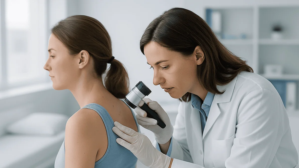
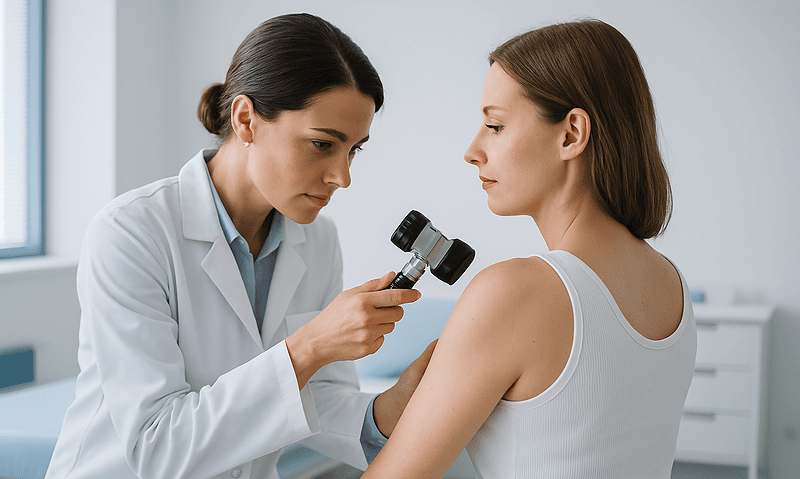
A skin lesion is simply any patch, bump or growth that looks different from the skin around it. Most are harmless. The difficulty is that a few serious ones are skilled at impersonating the harmless ones, and the eye, even a trained eye using a dermatoscope (a handheld magnifier with polarised light), cannot always tell them apart.
Cryotherapy, also called cryosurgery, destroys a lesion by freezing it, usually with liquid nitrogen. Excision removes it with a scalpel and the specimen goes to a laboratory. Those two facts drive almost every decision. Freezing is fast, needs no stitches and often no anaesthetic, which is exactly why it is popular for warts, skin tags and sun-damaged patches that a clinician can confidently name on sight (Cleveland Clinic, NHS).
But destruction has a cost: there is nothing left to test. If a frozen lesion was in fact an early skin cancer, nobody will ever know until it regrows, perhaps deeper, perhaps months later. That is the quiet risk behind an otherwise convenient treatment, and it is why guideline-level advice in dermatology consistently pairs any destructive method with a confident clinical diagnosis first (Mayo Clinic, NHS).
So when a clinician chooses excision for a lesion that could have been frozen, the choice is rarely about preferring surgery. It is about buying information. The slower route produces a histopathology report, a written description of what the tissue showed under the microscope, and that report is sometimes worth more than a week of healing time saved.
The sections below unpack both methods, the lesions each usually suits, what recovery looks like and the myths that tend to cluster around the word cryotherapy. The decision for any individual lesion always rests with the treating team, who can see the spot, its history and the person attached to it.
How cryotherapy actually works on a skin lesion
Liquid nitrogen boils at roughly minus 196°C (about minus 321°F). When it is sprayed or dabbed onto skin, water inside and between the cells freezes into ice crystals almost instantly. The crystals rupture cell membranes, and as the tissue thaws, the damaged cells leak, swell and die. Blood vessels in the frozen zone also clot, which starves any surviving cells of oxygen over the following hours (Cleveland Clinic).
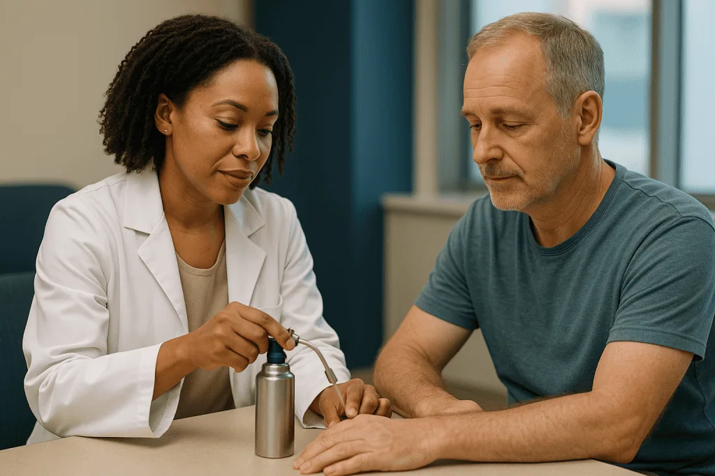
In practice the clinician either uses a handheld spray canister or a cotton-tipped applicator dipped in nitrogen. The lesion turns white as it freezes, including a small rim of surrounding skin. The freeze lasts seconds rather than minutes; the clinician judges the time by watching that white halo rather than by a stopwatch. Some lesions are frozen, allowed to thaw and frozen again in a single visit, because a second freeze-thaw cycle increases tissue destruction (Cleveland Clinic).
What it feels like: most people describe a sharp stinging or burning that peaks as the skin thaws, then settles within minutes. Children and people with many lesions sometimes find it harder to tolerate, which is one reason freezing is not automatically the easy option (NHS).
Over the next day the area reddens and often blisters. A blister is not a complication; it is the expected sign that the top layers of skin have separated from the dead tissue beneath. The blister dries, a crust forms and eventually falls away, taking the lesion with it. Thicker lesions such as viral warts frequently need more than one session, spaced weeks apart, because a single freeze does not reach deep enough to clear them (NHS).
Nothing is sent to the laboratory. That single sentence is the whole reason the rest of this article exists.
Shave excision vs cryotherapy vs full excision: what happens in each
Surgical removal is not one procedure but a small family of them, and the names matter because they decide what the pathologist receives.
A full excision, sometimes called an elliptical or fusiform excision, removes the lesion with a margin, meaning a rim of normal-looking skin around it, down to the fatty layer beneath. The wound is closed with stitches and the whole specimen goes to histopathology with its edges intact. This is the standard approach when a clinician wants to know both what a lesion is and whether it has been completely removed (Mayo Clinic, MedlinePlus).
A shave excision uses a blade held almost flat to the skin to slice off a raised lesion at or just below skin level. There are no stitches; the wound heals like a graze. The specimen still goes to the laboratory, so the diagnosis is usually confirmed, but the deep edge may not be assessable, which is why shaves are typically reserved for lesions thought to be benign or very superficial (Mayo Clinic).
Curettage and cautery scrapes the lesion away with a spoon-shaped instrument and seals the base with heat. The scraped fragments can be sent to pathology, though they arrive in pieces, so margins cannot be judged.
Mohs surgery is a specialised technique for certain skin cancers in cosmetically sensitive areas. Thin layers are removed and examined under the microscope during the same appointment until the edges are clear. It is not used for benign lesions (Mayo Clinic).
Set against this family, cryotherapy is the outlier: it is the only common option that produces no specimen at all. Shave excision vs cryotherapy is often the real choice for a raised benign lesion, and the deciding factor is usually whether anyone wants written confirmation of what it was.
Why pathology sometimes wins: what the laboratory sees that the eye cannot
Histopathology, the examination of thinly sliced, stained tissue under a microscope, answers three questions that no amount of staring at the skin can settle with certainty.
First, what is it? A pathologist can distinguish a seborrhoeic keratosis (a harmless, waxy, stuck-on growth of ageing skin) from a pigmented basal cell carcinoma, or a benign mole from an early melanoma, by the architecture and behaviour of the cells themselves. Clinical examination, even with a dermatoscope, produces a best estimate; the slide produces a diagnosis (Mayo Clinic).
Second, how deep did it go? For melanoma in particular, the measured thickness of the tumour on the slide is one of the most important factors in planning further care. A frozen melanoma has no measurable thickness, because it has been destroyed (Mayo Clinic).
Third, is it all out? When a full excision is examined, the pathologist inks the edges and checks whether abnormal cells reach them. Clear margins mean the lesion was removed intact. Involved margins mean a further procedure may be discussed. Cryotherapy offers no equivalent. The clinician sees white skin and hopes the freeze reached far enough.
There is a fourth, quieter benefit: documentation. A pathology report lives in the record. Years later, when a new lesion appears nearby, a clinician can read exactly what was removed and when. A frozen lesion leaves only a line in the notes and, often, a pale patch of skin.
None of this makes freezing a poor treatment. For a lesion that is confidently benign, pathology would simply confirm what everyone already knew, and the stitches and scar would be a price paid for no new information. The skill lies in recognising the lesion where the information is genuinely uncertain, and reaching for the scalpel there.
Who is usually offered freezing, and who is usually asked to wait or to have it cut out
Clinicians tend to sort lesions into three loose groups, and the groups map neatly onto the two treatments.
Freezing is commonly offered for lesions whose appearance is characteristic enough that a clinical diagnosis is secure: viral warts, skin tags, seborrhoeic keratoses, actinic keratoses (rough, sun-induced patches that are considered precancerous), and small benign vascular spots. The NHS and Cleveland Clinic both list these as typical cryotherapy targets, and in each case the lesion’s natural history is well understood (NHS, Cleveland Clinic).
Excision is commonly preferred when a lesion is pigmented and changing, when it bleeds or fails to heal, when it has features on dermoscopy that raise concern, when it is a recurrence after earlier treatment, or when the person has a history of skin cancer and the clinician wants a written answer rather than a visual impression (Mayo Clinic). A lesion that does not quite fit any familiar pattern often goes this way too; uncertainty itself is an indication.
Some people are asked to wait, or to choose a different route, for reasons unrelated to the lesion. Cryotherapy is used cautiously on people with very dark skin, where loss of pigment after freezing can be prominent and long-lasting; on areas with poor circulation such as the lower legs in people with diabetes or vascular disease, where any wound heals slowly; and over superficial nerves on the fingers, where a deep freeze can cause lasting numbness (Cleveland Clinic). People with cold-sensitivity conditions such as cryoglobulinaemia are typically steered away from freezing altogether.
Children can usually have warts frozen, but many clinicians prefer to wait, because most warts resolve on their own and the procedure stings (NHS). Pregnancy rarely changes the choice for skin lesions, though the team will decide case by case.
Cryotherapy vs excision at a glance
The table below pulls the key differences together. It is a summary of typical practice described by major health bodies, not a rule; individual lesions and individual skin break the pattern often enough that the treating team’s judgement always takes precedence (Cleveland Clinic, Mayo Clinic, MedlinePlus).
| Feature | Cryotherapy | Surgical excision |
|---|---|---|
| What happens to the tissue | Destroyed in place by freezing | Removed intact with a margin |
| Specimen for pathology | None | Yes, with margin assessment |
| Anaesthetic | Usually none | Local anaesthetic injection |
| Stitches | No | Usually yes (not for shave) |
| Typical appointment length | Minutes | Longer, including closure |
| Sessions needed | Often one; thick warts may need several | Usually one |
| Common early effects | Blister, crust, pale patch | Linear wound, bruising, line scar |
| Pigment change | Common, especially in darker skin | Possible along the scar |
| Confirms diagnosis | No | Yes |
| Confirms complete removal | No | Yes, if margins are clear |
| Typical lesions | Warts, skin tags, seborrhoeic and actinic keratoses | Suspicious moles, suspected skin cancers, uncertain lesions |
Two rows deserve emphasis. Pigment change is listed as common for cryotherapy because melanocytes, the pigment-producing cells, are more sensitive to cold than the surrounding skin cells, so a well-executed freeze frequently leaves a pale mark even when everything else goes perfectly (Cleveland Clinic). And the final two rows are the reason the title of this article reads the way it does.
Cryosurgery healing time: what the first days and weeks usually look like
Healing after freezing follows a predictable sequence, and knowing it in advance prevents a lot of unnecessary worry.
In the first few hours the area reddens and swells; the sting fades but the spot may throb. By the next day a blister often appears. It may be clear or, less commonly, filled with blood, which looks alarming but is a normal result of small vessels rupturing during the freeze. Guidance from Cleveland Clinic is not to pop it; the fluid cushions the new skin forming underneath (Cleveland Clinic).
Over the following days the blister flattens and dries into a crust. Beneath it, new skin grows across the base. The crust lifts and falls away on its own, usually within one to three weeks for a small lesion, with thicker or deeper freezes taking longer (Cleveland Clinic). The fresh skin underneath looks pink and shiny at first and gradually blends in.
Day-to-day care is simple: keep the area clean, wash gently, cover it if it rubs on clothing, and leave the crust alone. Picking it off early can lead to infection and a worse mark. Most people are back to normal activities the same day; it is work, exercise and showering as usual, with a plaster if needed.
Not every lesion clears first time. Viral warts in particular often need repeat sessions, spaced a few weeks apart so the skin can recover in between (NHS). If a lesion looks unchanged after the crust falls off, that is a reason to return, not a sign the treatment was done badly.
Cryosurgery healing time is also the window in which the one real weakness of freezing shows itself: a lesion that regrows quickly, or looks different from before, was perhaps never what it appeared to be, and that is the moment many clinicians switch to excision and a pathology request.
Cryotherapy side effects on the face: pigment, scars and nerves
The face is where people most want a lesion gone and least want a reminder of its removal, so it pays to be honest about what freezing can leave behind.
Hypopigmentation, a pale patch where pigment-producing cells have been destroyed, is the most common lasting effect. Because melanocytes are more cold-sensitive than keratinocytes (the ordinary skin cells), a freeze that cleanly removes a lesion can still leave a lighter mark. It is most noticeable in people with medium to dark skin and may be permanent, though many patches soften over months (Cleveland Clinic). Hyperpigmentation, a darker patch, can also occur as the skin heals, particularly with sun exposure during recovery.
Scarring is generally less than after excision, but it is not zero. Deep or repeated freezes can produce a slightly sunken or shiny area. Freezing near the eyelid margin or over the cartilage of the nose and ear carries extra risks, including notching of the ear rim, and many clinicians avoid or modify technique there.
Nerve injury is rare on the face but matters at sites where sensory nerves run close to the surface, such as the side of the finger or the angle of the jaw; a deep freeze can cause numbness that lasts weeks or occasionally longer (Cleveland Clinic).
Infection is uncommon because the freeze itself sterilises the surface, but it can follow a broken blister. Bleeding is minimal. Pain is usually short-lived.
The flip side is that excision on the face leaves a line scar, which with good technique along natural skin folds often fades well, and offers the certainty of a pathology report. For a confidently benign facial lesion, freezing’s smaller footprint is attractive; for a facial lesion with any doubt about it, most teams want the tissue examined, because a missed cancer on the face is costly to put right later (Mayo Clinic).
Can cryotherapy be used for skin tags, warts and seborrhoeic keratoses?
These three are the bread and butter of cryotherapy clinics, and each illustrates a slightly different reason freezing is reasonable.
Skin tags are soft, skin-coloured flaps on a narrow stalk, typically found where skin rubs against skin or clothing: the neck, armpits, groin and under the breasts. They are benign and have no cancer potential. The NHS notes they can be removed by freezing, by snipping with sterile scissors, or by cautery, and warns against home removal with thread or nail clippers because of bleeding and infection risk (NHS). Freezing works, though very small tags on thin stalks are sometimes simply snipped, which is quicker and avoids a blister. Nothing is sent for pathology because nothing needs confirming.
Viral warts are caused by human papillomavirus and are, in the end, a problem of persistence rather than danger. The NHS points out that most warts clear on their own over months to years, and that cryotherapy and over-the-counter salicylic acid preparations have broadly similar results, with freezing often needing repeat sessions (NHS). A Cochrane systematic review of topical wart treatments reached a similar conclusion, finding no clear advantage of cryotherapy over salicylic acid (NIH/PubMed). Freezing a wart is reasonable; expecting one freeze to finish the job is optimistic.
Seborrhoeic keratoses are the waxy, warty, stuck-on growths that multiply with age. They are harmless and are frozen or shaved off mainly when they catch on clothing or bother the person. Here is where judgement enters: a dark, irregular seborrhoeic keratosis can mimic melanoma, and the reverse is also true. When a clinician hesitates over a stuck-on lesion, a shave excision sent to the laboratory settles the question at little extra cost in healing, which is why cryotherapy for skin tags is routine while cryotherapy for an atypical keratosis is a judgement call (Mayo Clinic).
Actinic keratoses: where freezing and biopsy meet
An actinic keratosis is a rough, dry, sometimes scaly patch caused by years of ultraviolet exposure, most often on the face, scalp, ears, backs of the hands and forearms. It is classed as precancerous because a small proportion can progress to squamous cell carcinoma, a common form of skin cancer that arises from the flat cells of the outer skin layer (NHS).
Cryotherapy is one of the most widely used treatments for individual actinic keratoses, and for good reason. The patches are superficial, their appearance is usually characteristic, and freezing clears them in a brief appointment. The NHS lists freezing alongside prescription creams and gels, curettage, surgery and light-based therapy as standard options; the choice depends on how many patches there are, where they sit and how thick they have become (NHS).
The pathology question surfaces when a patch stops behaving like a typical keratosis. Features that prompt many clinicians to biopsy rather than freeze include a patch that is thickening or becoming tender, one that bleeds, one that persists after treatment, or one that has developed a firm base. These can be early signs that the lesion has already crossed into squamous cell carcinoma, and a frozen carcinoma is a carcinoma whose depth and extent nobody has measured (Mayo Clinic, NHS).
Many dermatology services therefore run a two-track approach: freeze the thin, typical patches; biopsy or excise the thick, stubborn or suspicious ones. Field treatments, meaning creams applied across a whole sun-damaged area, are used when patches are too numerous to treat one by one; the mechanism and course length are matters for the prescribing clinician.
Whatever the treatment, new keratoses tend to keep appearing in sun-damaged skin, so daily sun protection and periodic skin checks remain part of the plan rather than an optional extra (NHS).
Moles and anything pigmented: why most teams excise rather than freeze
If there is one rule most dermatologists hold firmly, it is this: do not freeze a mole. The reasoning deserves spelling out, because people often ask why a small, flat brown spot cannot simply be sprayed off like a wart.
A mole, or melanocytic naevus, is a cluster of pigment cells. Melanoma, the most dangerous common skin cancer, arises from the same cell type and frequently begins inside or beside an existing mole. Early melanoma can be flat, small, evenly coloured and entirely unremarkable to the untrained eye; even expert dermoscopy produces a probability rather than a certainty (Mayo Clinic).
Freezing a melanoma does three harmful things at once. It destroys the only tissue that could confirm the diagnosis. It removes the opportunity to measure tumour thickness, which guides every later decision about margins and further investigation. And it may leave cancer cells alive at the base or edge of the frozen zone, now hidden under scar tissue and harder to spot when they regrow. Mayo Clinic’s guidance on skin biopsy and on melanoma treats intact removal and microscopic examination as the foundation of care for any suspicious pigmented lesion (Mayo Clinic).
There is a cosmetic argument too. Freezing destroys pigment cells unevenly; a partially frozen mole can regrow with irregular colour and shape, which later mimics the very changes that worry clinicians, prompting an excision anyway.
The practical upshot: a mole that is new, changing, asymmetric, multicoloured, larger than its neighbours, itching or bleeding is a lesion for the surgical pathway, not the spray can. A clinician who declines to freeze a mole is not being cautious for its own sake; they are protecting the one piece of evidence that cannot be recovered once it is gone.
Recovery after excision: stitches, scars and waiting for the report
Excision asks more of the person in the short term and gives more back in information. Here is the usual shape of it.
The procedure itself takes longer than a freeze because the skin is numbed with a local anaesthetic injection, the lesion and a margin are removed, any small bleeding points are sealed, and the wound is closed, usually with stitches. Mayo Clinic describes a brief sting from the injection and then pressure rather than pain during the removal itself (Mayo Clinic).
Afterwards the wound is dressed. Most teams advise keeping it dry for a day or so, then washing gently and keeping it covered until stitches come out. Stitches on the face are typically removed sooner than those on the trunk or limbs, because facial skin heals quickly and early removal reduces track marks; the team will give a specific date. Dissolving stitches are sometimes used under the skin and need no removal (MedlinePlus).
Common early effects are bruising, mild swelling and a tight pulling sensation as the wound settles. Heavy lifting, stretching the area and swimming are usually paused until the wound has closed, because tension on a fresh scar widens it. Mayo Clinic notes that a biopsy or excision site may take several weeks to heal fully and that the scar continues to remodel and fade for months afterwards (Mayo Clinic).
Then comes the wait. Pathology results take days to a couple of weeks depending on the laboratory and whether special stains are needed. The report will state the diagnosis and, for a full excision, whether the margins are clear. A clear report usually ends the matter. An involved margin, or an unexpected diagnosis, leads to a conversation about next steps. That conversation is the whole point of having chosen excision in the first place.
What people often get wrong about cryotherapy and skin lesion removal
The word cryotherapy has been borrowed by several industries, and the confusion it causes is worth untangling.
Myth: cryotherapy for skin lesions is the same as whole-body cryotherapy chambers. It is not. Lesion cryotherapy applies liquid nitrogen directly to a spot of skin to destroy it. Whole-body chambers expose a person to very cold air for a few minutes and are marketed for recovery and wellbeing; they do not remove anything, and the evidence for their health claims is limited (Cleveland Clinic). Cold plunges and ice packs belong to that second world, not this one.
Myth: fat-freezing body contouring is a form of medical cryotherapy. Cryolipolysis, the technique behind fat-freezing devices, cools fat beneath intact skin to reduce fat cells. It does not treat skin lesions and is not what a dermatology clinic means by cryotherapy.
Myth: a frozen lesion cannot come back. It can. Warts commonly need several sessions, and any lesion whose base lies deeper than the freeze reached may regrow (NHS). Regrowth is a signal to reassess, not to freeze harder.
Myth: freezing is always the gentler, scar-free choice. Freezing often leaves a pale mark, sometimes permanent, and deep freezes can scar or numb. Excision leaves a line scar. Neither is invisible; they are different trade-offs (Cleveland Clinic).
Myth: if a doctor wants to cut rather than freeze, they must think it is cancer. Usually they think it is probably benign but cannot be sure, and want the laboratory to settle it. Most excised lesions turn out to be harmless. The report is reassurance, not a verdict.
Myth: home freezing kits for warts do the same job. Home kits use a different, far less cold refrigerant and are intended only for self-evident warts. Anything pigmented, changing or uncertain should never be treated at home (NHS).
Questions to ask your care team before choosing cryotherapy or excision
A good consultation about a skin lesion is a two-way exchange. These questions tend to surface the reasoning behind a recommendation and help you understand the trade-offs for your particular spot.
- What do you think this lesion is, and how confident are you from examination alone?
- If we freeze it, what would we lose by not sending tissue to the laboratory?
- Is there anything about this lesion, or my history, that makes you lean towards excision?
- If excision is recommended, will it be a shave or a full excision with stitches, and why?
- Where exactly would the scar sit, and how might it look after it has settled?
- Given my skin tone, how likely is a pale or dark patch after freezing, and would it fade?
- How many sessions might this lesion need if frozen, and how will we know if it has not worked?
- What does normal healing look like, and what would make you want to see me sooner?
- When and how will I receive the pathology result, and who will explain it?
- If the report shows the margins are not clear, what happens next?
- Are there alternatives I have not considered, such as a prescription cream, curettage or simply monitoring?
- Should I be having regular skin checks after this, and how often?
It helps to bring a photograph of the lesion from a few months earlier if one exists; change over time is often more informative than appearance on the day. Mention any previous skin cancers in you or close relatives, any blood-thinning medicines you take (do not stop them yourself; the team will advise), and any history of poor wound healing or raised scars.
The answers will differ from person to person and lesion to lesion. What matters is that the reasoning is clear to you, and that the final choice is made together with the clinician who has examined the skin.
When to call your doctor after cryotherapy or excision
Most people heal from either procedure without incident, but a few signs mean the site should be looked at rather than watched. Contact your care team, or seek urgent care if the symptoms are severe or you feel unwell, if you notice any of the following (Cleveland Clinic, MedlinePlus, Mayo Clinic):
- Spreading redness, increasing warmth or swelling around the wound after the first couple of days, especially with worsening pain.
- Thick yellow or green discharge, a foul smell, or a crust that keeps weeping.
- Fever, chills or feeling generally unwell.
- Bleeding from an excision wound that does not stop after firm pressure for several minutes.
- Stitches that have come apart or a wound edge that has opened.
- Numbness, tingling or weakness near the site that is new or not improving.
- A large or very painful blister after freezing, or a blister over the eyelid, ear or a joint.
- Red streaks tracking away from the wound along the limb.
Beyond the healing window, two situations deserve a prompt appointment. The first is a frozen lesion that regrows, changes colour or shape, bleeds or fails to clear after the expected sessions; this is the moment to reassess and often to request a biopsy rather than repeat the freeze. The second is a pathology result you have not received within the timeframe you were given; chase it, because unreturned results are a known gap in care.
For any skin lesion, treated or not, seek assessment if it is new and growing, if a mole changes in size, shape or colour, if a sore fails to heal within a few weeks, or if a spot itches, crusts or bleeds without an obvious cause (NHS, Mayo Clinic). These are not diagnoses; they are reasons for someone qualified to look.
Every decision about freezing, cutting, monitoring or doing nothing belongs with the treating team, who can see what no article can.
Frequently asked questions
How long does it take to heal after cryosurgery on the skin?
Small frozen lesions typically blister, crust and heal within about one to three weeks, according to Cleveland Clinic, with deeper or repeated freezes taking longer. The new skin looks pink at first and blends in over months, though a pale patch may persist. Keep the area clean, leave the crust alone, and contact your team if redness spreads or the site becomes very painful.
Is cryotherapy the same as fat-freezing body contouring?
No. Cryotherapy for skin lesions applies liquid nitrogen directly to a wart, keratosis or similar spot to destroy it. Fat-freezing body contouring, known medically as cryolipolysis, cools fat beneath intact skin with a device and does not treat skin lesions at all. Whole-body cold chambers and cold plunges are a third, separate category. Only the first is what a dermatology clinic means by cryotherapy.
Can cryotherapy be used to remove skin tags?
Yes. The NHS lists freezing as one accepted way to remove skin tags, alongside snipping with sterile scissors and cautery. Skin tags are benign with no cancer potential, so nothing needs to be sent for pathology. Very small tags on thin stalks are sometimes simply snipped because it avoids a blister. The NHS advises against home removal with thread or clippers because of bleeding and infection risk.
What are the side effects of cryotherapy on the face?
The most common lasting effect is a pale patch where pigment cells were destroyed, more noticeable in darker skin and sometimes permanent. Temporary darkening, mild scarring or a slightly sunken area can also occur, and freezing near the eyelid, ear cartilage or superficial nerves carries extra risk. Blistering and stinging in the first days are expected. Cleveland Clinic describes infection as uncommon but possible after a broken blister.
Why would a doctor choose excision over cryotherapy for a lesion that could be frozen?
Usually to obtain a pathology report. Excision preserves the tissue so a laboratory can confirm what the lesion was and whether it was fully removed; freezing destroys it. When a clinician is confident a lesion is benign, freezing is reasonable. When there is any doubt, most prefer to accept stitches and a line scar in exchange for a definite diagnosis, which freezing can never provide.
Does cryotherapy work on moles?
Freezing is generally avoided for moles. A mole is made of pigment cells, the same cell type that gives rise to melanoma, and early melanoma can look entirely innocent. Freezing destroys the only tissue that could confirm the diagnosis and measure tumour depth, and may leave cancer cells hidden under scar. Mayo Clinic guidance treats intact removal with microscopic examination as the foundation of care for suspicious pigmented lesions.
What is the difference between shave excision and cryotherapy?
A shave excision slices a raised lesion off at skin level with a blade and sends it to the laboratory; there are no stitches and the wound heals like a graze. Cryotherapy freezes the lesion so it blisters and falls away, with nothing sent for testing. For a raised lesion that is probably benign, the choice often comes down to whether written confirmation of the diagnosis is wanted.
How many cryotherapy sessions does a wart usually need?
Often more than one. The NHS notes that thick viral warts frequently need repeat freezing, with sessions spaced weeks apart so the skin recovers, and that most warts clear on their own over months to years anyway. A Cochrane review found freezing and salicylic acid preparations broadly similar in effect. The number of sessions for any individual wart is a decision for the treating clinician.
Can an actinic keratosis be frozen, or does it need a biopsy?
Thin, typical actinic keratoses are commonly frozen, and the NHS lists cryotherapy among standard treatments alongside prescription creams, curettage and light-based therapy. A biopsy or excision is usually preferred when a patch thickens, becomes tender, bleeds, develops a firm base or persists after treatment, because these can signal progression to squamous cell carcinoma, which needs to be examined rather than destroyed.
Is excision more painful than cryotherapy?
The experiences differ rather than one being clearly worse. Cryotherapy stings sharply for seconds to minutes as the skin freezes and thaws, usually without anaesthetic. Excision involves a brief sting from the local anaesthetic injection, after which the removal itself is felt as pressure, followed by a few days of tightness or bruising. Mayo Clinic describes both as generally well tolerated; your team can discuss comfort measures.
References
This article is for general information only and is not a substitute for professional medical advice. Please consult a qualified doctor about your individual situation.

More from the Blog
Dermabrasion Recovery: The Raw Days, Ointments and When Redness Finally Fades
Dermabrasion recovery time typically runs in stages: the treated skin is raw, red and weeping for the first days, new skin usually covers the…
Sun Protection After Dermabrasion: Why Strict Cover Matters for Months, Not Days
Sun protection after dermabrasion needs to continue for months, not days, because the newly grown skin is thin and its pigment cells are easily…
What Happens During an IPL Photorejuvenation Session: Cooling Gel, Flashes and Timing
During an IPL photorejuvenation session, a clinician cleanses the skin, applies a cool clear gel, gives you protective eyewear, and passes a handpiece across…

Why Your Cold Sore History Matters Before Laser Resurfacing and How Doctors Prepare for It
Your cold sore history matters before laser resurfacing because the herpes simplex virus that causes cold sores stays dormant in facial nerves and can…

What to Stop Before a Chemical Peel: Retinoids, Scrubs, Waxing and Sun Exposure
Before a chemical peel, people are usually asked to pause topical retinoids and exfoliating acids, stop facial scrubs, masks and exfoliating devices, avoid waxing,…

Laser Resurfacing Results: What Fine Lines and Acne Scars Realistically Look Like Afterward
Laser resurfacing results are usually a partial, gradual softening rather than a complete erasure. Fine lines around the eyes and mouth commonly look shallower,…