

EKG Explained: What the 12 Leads Show and What ‘Abnormal’ Usually Means

Key Takeaways
- ECG and EKG are two abbreviations for the same test; the K comes from the German spelling and was kept in US hospitals to avoid confusion with EEG.
- Ten electrodes generate twelve leads because a lead is a calculated viewpoint, not a wire, and each group of leads faces a specific wall of the heart fed by a specific coronary artery.
- A standard resting EKG records about ten seconds, so it can miss rhythm problems that come and go; Holter, patch monitors, and wearables exist to extend the window.
- Automated interpretations are deliberately tuned to over-call, which is why 'abnormal' or 'borderline' on a machine-generated report is often a normal variant or an electrode artifact once a physician reviews it.
- The US Preventive Services Task Force recommended in 2018 against routine screening EKGs in adults at low cardiovascular risk with no symptoms, because false alarms lead to further testing without proven benefit.
- In a 2019 study of about 420,000 smartwatch users, an irregular-pulse alert corresponded to confirmed atrial fibrillation roughly 84 percent of the time, yet only about 0.5 percent of participants were ever notified.
An EKG (electrocardiogram, also written ECG) records the heart's electrical activity for about ten seconds using ten electrodes that produce twelve views, or leads, of the heart. It can show rhythm problems, signs of a current or past heart attack, and thickened or strained heart muscle. An 'abnormal' result is common and often benign; a clinician must interpret it alongside symptoms, history, and other tests.
A colleague slid her phone across the lunch table last week. On the screen: a jagged green line from her smartwatch and, underneath it, one word in gray, “Inconclusive.” Beside it she had saved a screenshot going around social media, a clinic printout with “ABNORMAL ECG” stamped across the top and a caption that read, “Doctors said it was nothing. Was it?”
That pairing explains why the ekg is having a moment. As of spring 2025, searches for the test have climbed alongside a wave of shared watch strips and clinic readouts, many posted with more alarm than context. Wearables now hand millions of people a single-lead tracing they were never taught to read, and machine-printed interpretations use words like “borderline” and “nonspecific” that sound far more ominous than they usually are.
So here is the calm version. What the squiggles actually measure, why there are twelve of them, what “abnormal” tends to mean in practice, and where the evidence is solid, thin, or still being written.
Are ECG and EKG the same? The ECG vs EKG difference explained
Yes. The two abbreviations describe one test. “Electrocardiogram” is the English word; “EKG” comes from the German spelling, Elektrokardiogramm, which stuck in American hospitals partly because it sounds distinct from “EEG,” the brain-wave test, when shouted across a busy ward. British and international sources lean toward ECG. Your report may carry either label and the tracing is identical.
The test itself is quieter than its reputation. A technician places ten sticky electrodes on your skin, four on the limbs and six across the chest, then asks you to lie still and breathe normally for around ten seconds while a machine records. Cleveland Clinic puts the whole visit at roughly ten minutes, most of it setup. Nothing is injected, nothing is switched on inside you, and no electricity flows into your body. The electrodes are microphones, not speakers; they listen for the tiny voltages your heart already produces.
A few practical points matter for accuracy. Chest hair may be trimmed so the electrodes stick. Lotion, cold skin, and shivering blur the signal, which is why you may be asked to remove a sweater and wait a minute under a blanket. Talking or shifting position during the recording can produce wobbles that a computer misreads as rhythm problems, a theme that returns later in this article.
The result appears as a page of twelve short strips plus a longer rhythm strip along the bottom, usually with a computer-generated interpretation at the top. That interpretation is a draft. A physician’s over-read, ideally by someone who also knows why the test was ordered, is the version that counts.
What does an EKG actually measure?
Every heartbeat begins as electricity. A cluster of cells in the right upper chamber called the sinus node, the heart’s natural pacemaker, fires on its own about 60 to 100 times a minute at rest. That impulse spreads across the two upper chambers (atria), pauses briefly at a relay station called the AV node, then races down two wiring bundles into the lower chambers (ventricles), which do the heavy pumping.
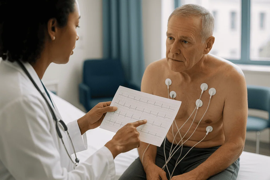
As each region of muscle activates, its cells flip electrical charge, a process called depolarization; as they recover, they flip back, called repolarization. The sum of millions of cells switching creates voltage differences small enough to be measured in thousandths of a volt at the skin. The EKG machine amplifies those differences and draws them over time, much as a seismograph turns ground tremors into a line on paper.
The paper has rules. It moves at 25 millimeters per second, so each small square represents 0.04 seconds and each large square 0.2 seconds. Height is calibrated so that 10 millimeters equals one millivolt. Those conventions let a clinician count boxes and calculate, for example, that a heart beating once every five large squares is running at 60 beats per minute.
Two facts follow from this. First, the EKG reports electrical events, not blood flow or valve function directly; it infers structural problems from the way electricity behaves in damaged or thickened tissue. Second, it is a snapshot. Ten seconds of a heart that beats roughly 100,000 times a day is a very small sample, excellent for what is happening right now and blind to anything that happened an hour ago or will happen tonight.
What the 12 leads show: ten electrodes, twelve camera angles
The arithmetic confuses almost everyone. Ten electrodes, twelve leads? A lead is not a wire; it is a view, calculated by comparing the voltage between two points. Ten electrodes, combined in standard pairs and averages, yield twelve viewpoints. Think of twelve cameras ringed around a stadium, each filming the same play from a different seat.
Six of those views come from the limb electrodes and look at the heart in the frontal plane, as if you were facing the patient. They are labeled I, II, III, aVR, aVL, and aVF. Leads II, III, and aVF look up from the feet at the underside, or inferior wall, of the heart. Leads I and aVL look in from the left side. Lead aVR looks from the right shoulder and mostly serves as a check on electrode placement.
The other six, V1 through V6, come from the chest electrodes and slice the heart horizontally, from the right of the breastbone (V1) around to the left armpit (V6). V1 and V2 sit over the septum, the wall dividing the ventricles. V3 and V4 look at the front, or anterior, wall. V5 and V6 join I and aVL in viewing the lateral wall.
Why the fuss about geography? Each wall of the heart is fed mainly by one coronary artery. When a heart attack shows on an EKG, the leads that change point toward the blocked vessel: inferior leads toward the right coronary artery in most people, anterior leads toward the left anterior descending artery, lateral leads toward the circumflex. Emergency teams use that map to plan treatment before a single imaging scan is done.
The standard twelve leave one blind spot. The back, or posterior, wall has no camera aimed directly at it, so clinicians sometimes add extra chest electrodes, V7 to V9, or right-sided leads when the story suggests trouble there.
Reading the waves: P, QRS, T, and the intervals between them
Once you know the alphabet, a normal EKG beat reads like a short sentence. Each letter marks one electrical event.
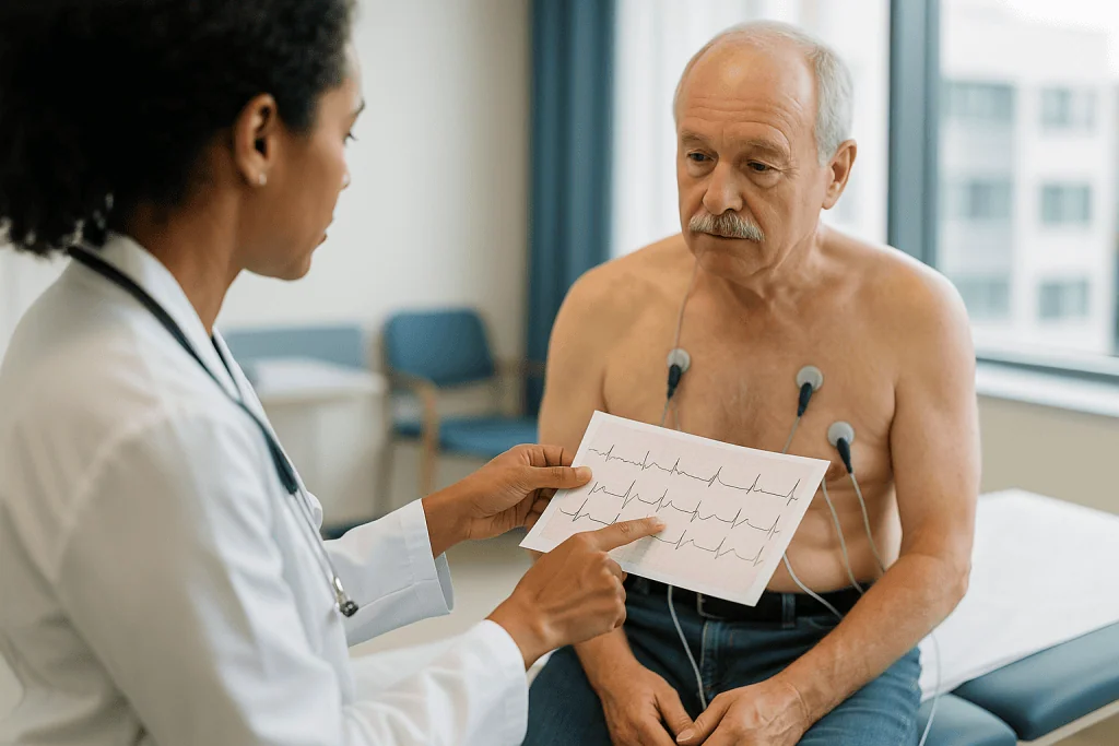
- P wave. A small rounded bump. The atria depolarizing, which triggers them to contract and top off the ventricles.
- PR interval. The flat stretch after the P wave. The impulse is waiting at the AV node. Normally 0.12 to 0.20 seconds, three to five small squares.
- QRS complex. The tall spike. Both ventricles depolarizing in a coordinated burst, normally within 0.12 seconds. A wide QRS suggests the impulse took a detour, as in a bundle branch block.
- ST segment. The flat line after the spike. The ventricles are fully activated and about to recover. Elevation or depression here is what clinicians scan for first when chest pain is the complaint.
- T wave. A broad hump. Ventricular repolarization, the muscle resetting for the next beat.
- QT interval. From the start of the QRS to the end of the T wave, the ventricles’ full electrical cycle. Because it shortens as heart rate rises, it is reported as a rate-corrected value, QTc.
Sometimes a small dip called a U wave trails the T; it is often normal. A physician’s read moves through rate, rhythm, axis (the average direction of electrical travel), intervals, and then wave shapes lead by lead. Rate is estimated quickly by dividing 300 by the number of large squares between two beats.
The point of learning this is not self-diagnosis. It is so that when a report says “prolonged PR” or “nonspecific T-wave abnormality,” you know which part of the sentence is being described and can ask a sharper question.
Why would a doctor order an EKG?
Mostly because you told them something. Chest pain, pressure, or tightness is the classic trigger. So are palpitations (an awareness of your heartbeat as fast, pounding, or irregular), unexplained shortness of breath, fainting or near-fainting, and new dizziness. MedlinePlus and Mayo Clinic list these as the leading reasons, and in an emergency department the American Heart Association describes the EKG as the standard first test for a suspected heart attack, with guidelines calling for it within ten minutes of arrival.
Symptoms are not the only door. Clinicians also order an EKG to establish a baseline before starting certain medicines that can lengthen the QT interval, before some surgeries, or when high blood pressure has been present long enough to strain the heart muscle. People with known heart disease, a pacemaker, or an inherited rhythm condition get periodic tracings to track change over time. A family history of sudden cardiac death at a young age can prompt one. Some athletic programs include an EKG in pre-participation screening, a practice whose value remains debated in the evidence.
An EKG is also a comparison tool. A single tracing tells you what the heart looks like today; two tracings a year apart tell you whether anything moved. That is why physicians ask for old copies, and why it is worth keeping a photo of yours.
What the order does not mean is that the clinician suspects disaster. The test is cheap in time, harmless, and reversible, so the threshold for ordering it is deliberately low. Many EKGs are ordered precisely to document that the heart’s electrical system is fine and move on to a more likely explanation, such as reflux, anxiety, a muscle strain, or thyroid trouble.
What will an EKG test show, and what can it miss?
The test is strongest on rhythm and conduction. It can identify whether the heart is beating too slowly, too fast, or irregularly, and it can usually say which pattern is responsible: atrial fibrillation, in which the upper chambers quiver chaotically; atrial flutter; supraventricular tachycardia; various degrees of heart block, where the AV node relay slows or drops beats; and extra beats arising from the atria or ventricles.
It is also the front-line detector of an acute heart attack. When an artery is fully blocked, the starving muscle changes how it conducts electricity and the ST segment lifts in the leads facing that wall, the pattern called ST-elevation myocardial infarction, or STEMI. Old, healed heart attacks leave scars that show as deep Q waves. Thickened muscle from long-standing high blood pressure produces taller voltages and shifted axes. Very high or low potassium reshapes T waves. Inflammation of the heart’s lining, pericarditis, has a recognizable signature. Pacemakers announce themselves with sharp spikes.
Now the limits, which the top-ranking patient pages tend to soften. A normal EKG does not rule out coronary artery disease; arteries can be substantially narrowed while resting electricity looks pristine. Even during a heart attack, the first tracing is normal or nondiagnostic in a meaningful share of people, which is why emergency teams repeat the EKG and add troponin blood tests, markers of heart-muscle injury, before deciding. Intermittent rhythm problems that appear for minutes a day will slip through a ten-second window. Valve disease, heart-failure pumping strength, and the size of the chambers are assessed by echocardiogram, an ultrasound, not by electricity.
The honest summary: the EKG is superb at what is happening electrically right now and only suggestive about structure or about arteries that have not yet closed.
What do abnormal EKG results usually mean?
Start with what the word does not mean. On an EKG report, “abnormal” means the tracing differs from a statistical template built from thousands of reference recordings. It does not mean disease, and it does not mean danger. In practice a large share of tracings flagged abnormal by the machine turn out to be normal variants, technical artifacts, or findings that are real but unimportant for that person.
Common benign sources of the label include a slow resting rate in fit adults (sinus bradycardia); a rate that speeds and slows with breathing, especially in young people (sinus arrhythmia); a pattern called early repolarization, frequent in young men and athletes; an incomplete right bundle branch block, a minor wiring quirk; and a slightly long PR interval, called first-degree AV block, which often needs nothing more than a note in the chart.
Then there is the machine itself. Automated interpretation is tuned to over-call rather than under-call, a sensible bias for a safety net but a recipe for alarming printouts. Swapped limb electrodes can fabricate an apparent old heart attack. Muscle tremor, a cold room, or a chatty patient can mimic atrial fibrillation. Poor skin contact produces wandering baselines that get read as ST changes.
This is why reports are labeled “unconfirmed” until a physician signs them. The over-read asks the question the computer cannot: does this pattern fit this person, at this age, with these symptoms and this history? A widened QRS in a healthy 25-year-old and the same finding in a 70-year-old with breathlessness are different conversations.
If your report says abnormal, the useful next step is a single question to the ordering clinician: which finding, and does it change anything? Often the answer is a specific name and a reassuring no.
Common EKG findings decoded in plain words
Reports reuse a small vocabulary. Here is what the frequent phrases describe, with the caveat that meaning depends on the person, not the phrase.
- Sinus rhythm. The normal pacemaker is in charge. Good news, and the most common line on any report.
- Sinus bradycardia / tachycardia. Normal rhythm, but slower than 60 or faster than 100 beats per minute. Sleep, fitness, fever, pain, anxiety, caffeine, dehydration, and thyroid activity all move this number.
- First-degree AV block. The relay pause is a little long. Frequently harmless; higher-degree blocks, where beats are dropped, need evaluation.
- Right or left bundle branch block. One of the two cables to the ventricles conducts slowly, widening the QRS. Right-sided blocks are often incidental; a new left-sided block prompts a closer look at the heart muscle.
- Left ventricular hypertrophy (LVH). Electrical signs suggesting thickened pumping muscle, usually from years of high blood pressure. EKG criteria are imprecise; an echocardiogram confirms or refutes.
- Nonspecific ST-T changes. The most maddening phrase in cardiology. Subtle shifts in the recovery phase that can reflect medicines, electrolytes, breathing, or nothing at all. Context decides.
- Q waves. Possible scar from a past heart attack, sometimes silent. Also produced by electrode misplacement, so repeat tracings matter.
- Prolonged QTc. The ventricles take longer to reset, which can raise the risk of a dangerous rhythm, particularly with certain medicines. Reviewed carefully by the prescribing clinician; never a reason to stop a medicine on your own.
- Premature atrial or ventricular contractions. Extra beats. Nearly everyone has some; frequency and symptoms guide whether more testing is needed.
- Atrial fibrillation. An irregular, often fast rhythm from chaotic atrial signals. Clinically important because it raises stroke risk, and always deserves a clinician’s confirmation and plan.
Why don't doctors do EKGs anymore? The screening question
The premise is half wrong. The EKG remains one of the most frequently performed tests in medicine; anyone with chest pain, palpitations, or fainting will meet one quickly. What has faded is the ritual EKG for healthy adults with no symptoms, the strip that used to be part of every annual physical.
The shift followed evidence, not fashion. In 2018 the US Preventive Services Task Force reviewed screening EKGs, resting or exercise, in adults with no symptoms and recommended against them for people at low risk of cardiovascular disease, concluding that harms outweigh benefits. For adults at intermediate or high risk it judged the evidence insufficient to recommend for or against. The reasoning ran like this: in a healthy person, an abnormal-looking tracing is more likely to be a false alarm than a true warning, and false alarms lead to stress tests, imaging, and sometimes invasive angiography, each carrying its own small risks, without evidence that anyone lives longer or avoids a heart attack as a result.
Risk in these guidelines is estimated from the ordinary factors, such as age, blood pressure, cholesterol, smoking, and diabetes, which predict events better than a resting tracing does. Similar logic trimmed routine pre-operative EKGs before low-risk surgery in otherwise healthy patients.
None of this applies once symptoms appear, once a medicine that affects heart rhythm is being considered, or once heart disease is known. The distinction is between screening, testing people who feel well, and diagnosis, testing people with a complaint or a specific reason. The EKG lost ground in the first role and kept every inch of the second.
Whether wearables, which effectively screen continuously, change this calculus is the live question, and the next two sections take it up.
What changed recently
Three dated developments explain the current surge of interest.
2018: single-lead EKGs reached wrists. The US Food and Drug Administration cleared the first consumer smartwatch electrocardiogram feature that year, and several manufacturers followed. These devices record one lead, roughly equivalent to lead I, for 30 seconds and classify the rhythm as sinus, atrial fibrillation, or inconclusive. Cleveland Clinic and Mayo Clinic patient pages, updated since, now describe wearables as a legitimate way to capture a rhythm during symptoms while stressing that a clinician must confirm the result.
2018 to 2023: screening guidance tightened, then met wearables. The 2018 US Preventive Services Task Force statement discouraged routine screening EKGs in low-risk adults. In late 2023 the joint American College of Cardiology and American Heart Association atrial fibrillation guideline addressed the wearable era directly, treating device-detected irregular rhythm as a prompt for clinician-reviewed EKG confirmation rather than a diagnosis in itself, and emphasizing that management decisions rest on confirmed rhythm plus individual stroke risk.
2019 onward: artificial intelligence began reading the strip. Researchers trained neural networks on hundreds of thousands of twelve-lead tracings to spot patterns humans cannot see, including a weak pumping heart, a condition the EKG was never thought capable of detecting. A 2019 retrospective analysis showed the approach could flag low ejection fraction with useful accuracy, and a 2021 pragmatic cluster-randomized trial in primary care found that showing clinicians the AI-EKG result increased new diagnoses of low ejection fraction compared with usual care. As of spring 2025 these tools remain largely in research and early clinical use, and no guideline yet calls for AI-EKG screening of the general population.
Put together: more people are seeing their own tracings, and the tracing itself is being asked to do more. The vocabulary on the printout has not changed, which is why explanation is overdue.
What the evidence actually says about the EKG
Not every claim about this test rests on the same footing. Grading them separately is the only honest way to read the headlines.
Diagnosing a heart attack in someone with chest pain: strong. The EKG’s role is embedded in decades of observational data and in randomized trials of treatment pathways that used EKG findings to select patients, notably the trials establishing emergency artery-opening procedures for ST-elevation heart attacks. Guidelines from the American Heart Association and international bodies agree. The caveat, also well documented, is that a single normal tracing does not exclude a heart attack, hence serial EKGs plus troponin.
Detecting rhythm disorders when symptoms are present: strong, with a timing problem. If the arrhythmia is happening during the recording, the twelve-lead EKG is the reference standard. If it is intermittent, the ten-second snapshot has poor sensitivity, which is why longer monitors exist.
Screening adults with no symptoms: weak for benefit. Observational studies consistently show that certain EKG abnormalities predict future cardiac events, but prediction is not the same as improvement. No randomized trial has shown that screening EKGs reduce heart attacks or deaths in low-risk adults, and downstream testing carries measurable harms. This is expert consensus built on the absence of trial benefit.
Wearable single-lead detection of atrial fibrillation: moderate and mixed. A 2019 study of roughly 420,000 smartwatch users found that when the watch flagged an irregular pulse and a confirmatory patch was worn, atrial fibrillation was present about 84 percent of the time; only about 0.5 percent of participants received any notification. Randomized screening trials in older adults using intermittent EKG devices have shown a small reduction in strokes in one large Swedish study and no significant reduction in another using implanted monitors, so whether detecting more atrial fibrillation prevents strokes remains unsettled.
AI interpretation: promising, early. Mostly retrospective validation, one pragmatic randomized trial showing more diagnoses, and no outcome trials yet.
12-lead EKG vs Holter monitor vs smartwatch: which test for which question
Each tool trades breadth for time. The twelve-lead gives the fullest picture of the shortest moment; wearables give the thinnest picture of the longest stretch. The table sets them side by side, with a Holter monitor defined as a portable recorder worn for one to two days and an event or patch monitor as a small adhesive device worn for one to four weeks.
| Tool | Leads | Typical duration | Best for | Main limitation |
|---|---|---|---|---|
| Resting 12-lead EKG | 12 | About 10 seconds | Chest pain evaluation, baseline tracing, conduction blocks, signs of strain or prior damage | Misses anything intermittent; says little about arteries that are narrowed but open |
| Holter monitor | 3 to 12 | 24 to 48 hours | Daily palpitations, dizziness, or extra-beat counting | Symptoms that occur weekly or less may not appear in the window |
| Event or patch monitor | 1 to 2 | 1 to 4 weeks | Infrequent palpitations or unexplained fainting | Fewer leads, so location of a problem is harder to pin down |
| Smartwatch single-lead EKG | 1 | 30 seconds on demand; passive pulse checks in background | Capturing a rhythm during symptoms; flagging possible atrial fibrillation | Cannot diagnose heart attack, blocks, or strain; “inconclusive” readings are common; confirmation required |
Two reading tips. Choose the monitor by how often the symptom happens, not by which device sounds most advanced; a monthly faint calls for weeks of recording, not a better ten seconds. And treat a watch strip as evidence to bring to an appointment, not as a report. Clinicians increasingly welcome those exports because a rhythm caught in the wild during a real episode can be more informative than a pristine tracing recorded on a calm afternoon in the clinic.
Common myths about the EKG, corrected
The viral posts driving this trend repeat a handful of misunderstandings. Each one has a short, evidence-based answer.
“My EKG was normal, so my heart is fine.” A normal resting tracing means the electrical system looked healthy for ten seconds. Coronary arteries can be significantly narrowed with a normal EKG, and intermittent rhythms hide easily. The test is one instrument in the band, not the whole orchestra.
“Abnormal EKG means I’m having, or about to have, a heart attack.” Most flagged findings are normal variants, artifacts, or stable quirks of wiring. A heart attack produces specific patterns that clinicians recognize immediately and act on within minutes; a report you receive days later that says “nonspecific” is not one of them.
“The machine shocks you.” It only records. The device that delivers a shock is a defibrillator, a different machine used in a different situation.
“A watch EKG is the same as the hospital one.” A watch records one lead for 30 seconds and answers one question, mainly whether the rhythm looks like atrial fibrillation. The twelve-lead answers dozens. Neither replaces the other.
“Everyone should get a yearly EKG.” Guidelines advise against routine screening in low-risk adults without symptoms because false alarms lead to unnecessary testing and no trial has shown benefit. Risk is better estimated from blood pressure, cholesterol, smoking, diabetes, and age.
“The EKG shows blocked arteries.” It shows the electrical consequences of a blockage once muscle is injured or starved, which is a different and later thing. Stress testing and imaging assess the arteries themselves.
“Abnormal results mean I should stop my medicine.” Some medicines legitimately change the tracing, and stopping them abruptly can be far riskier than the finding. Any change belongs to the prescribing clinician.
When to see a doctor about an EKG result or heart symptoms
Some situations do not wait for an appointment. Call emergency services, 911 in the United States, for chest pain, pressure, or squeezing that lasts more than a few minutes or comes and goes, especially if it spreads to the arm, jaw, neck, or back, or arrives with sweating, nausea, lightheadedness, or shortness of breath. Do the same for fainting, a racing or irregular heartbeat accompanied by chest discomfort or near-collapse, sudden severe breathlessness, or symptoms of stroke such as facial droop, arm weakness, or slurred speech. The American Heart Association stresses that women, older adults, and people with diabetes more often have less typical symptoms, so unusual fatigue or breathlessness with a sense that something is wrong deserves the same urgency.
Book a prompt visit, within days rather than weeks, if you have new palpitations that recur, exertional breathlessness that is worsening, episodes of dizziness on standing, or a smartwatch notification of an irregular rhythm, even without symptoms. Bring the exported strips.
If you have already had an EKG and the report says abnormal, borderline, or unconfirmed, contact the clinician who ordered it and ask two questions: which specific finding, and does it change the plan. Most will be explained in a sentence. Do not stop, skip, or adjust any prescribed medicine on the basis of a printout or a watch reading; that decision, including whether an EKG finding relates to a medicine at all, belongs to the prescribing clinician who can weigh the whole picture.
Anyone with a family history of sudden cardiac death before age 50, unexplained fainting during exercise, or a known inherited heart condition should have an ongoing relationship with a clinician who tracks their tracings over time. And if a result is worrying you and the explanation you received did not land, asking for the over-read to be walked through, lead by lead, is a reasonable request. Good clinicians expect it.
Frequently asked questions
What does an ECG show?
An ECG shows the heart’s electrical activity: its rate, whether the rhythm is regular, how quickly the signal travels through the heart’s wiring, and whether patterns suggest a current or past heart attack, thickened muscle, or electrolyte problems. It does not directly show blood flow, valve function, or artery narrowing, which need other tests such as an echocardiogram or stress imaging.
Are ECG and EKG the same test?
Yes. ECG is the English abbreviation for electrocardiogram and EKG comes from the German Elektrokardiogramm. American hospitals often say EKG so it is not mistaken for EEG, the brain-wave test, in noisy settings. The procedure, the electrodes, the twelve leads, and the printed report are identical regardless of which label appears on your paperwork.
Why would a doctor order an EKG?
Most often because of symptoms such as chest pain, palpitations, shortness of breath, dizziness, or fainting. Other reasons include establishing a baseline before certain medicines or surgery, evaluating long-standing high blood pressure, checking a pacemaker, tracking known heart disease, or investigating a family history of early sudden cardiac death. A low threshold for ordering reflects that the test is quick and harmless.
What do abnormal EKG results usually mean?
Usually less than the word suggests. ‘Abnormal’ means the tracing differs from a reference template, and many flagged findings are normal variants such as a slow rate in fit people, early repolarization, or minor conduction delays, or artifacts from electrode placement and movement. A physician’s review in the context of your symptoms and history determines whether any finding matters.
Why don't doctors do EKGs anymore at checkups?
Routine screening EKGs for healthy adults with no symptoms fell out of favor after evidence reviews, including the 2018 US Preventive Services Task Force statement, found no proven benefit and real harms from false alarms leading to further tests. EKGs remain a standard, frequently used test whenever symptoms, medicines, surgery, or known heart disease provide a specific reason.
Can a normal EKG miss a heart attack?
Yes. The first EKG is normal or nondiagnostic in a meaningful share of people who turn out to be having a heart attack, particularly when an artery is partly rather than fully blocked. Emergency teams therefore repeat the tracing over time and measure troponin, a blood marker of heart-muscle injury, before making a decision. A single normal strip never ends the evaluation of ongoing chest pain.
Is a smartwatch EKG as accurate as a 12-lead EKG?
No, but it answers a different question. A watch records a single lead for about 30 seconds and classifies rhythm as sinus, atrial fibrillation, or inconclusive; in a large 2019 study, irregular-pulse alerts matched confirmed atrial fibrillation about 84 percent of the time. It cannot detect heart attacks, conduction blocks, or muscle strain, and any flag needs confirmation by a clinician-reviewed EKG.
What is the difference between an EKG and an echocardiogram?
An EKG records electricity; an echocardiogram is an ultrasound that films structure and motion. The EKG tells you about rhythm, conduction, and electrical signs of injury or strain. The echocardiogram shows chamber size, wall thickness, valve function, and how strongly the heart pumps. Clinicians often use the first to decide whether the second is needed, and an EKG finding such as suspected hypertrophy is typically confirmed by echo.
How long does an EKG take and does it hurt?
The recording itself lasts about ten seconds and the whole visit roughly ten minutes, most of it attaching ten sticky electrodes to the chest and limbs. It is painless and no electricity enters the body; the machine only listens to voltages the heart produces. Chest hair may be trimmed for contact, and the electrodes can leave brief red marks that fade within hours.
Should I stop a medicine if my EKG shows a prolonged QT or other change?
No. Some medicines legitimately alter the tracing, and stopping a prescribed medicine abruptly can be more dangerous than the finding itself. Bring the report to the clinician who prescribed the medicine and ask whether the change is expected, whether it needs monitoring, and whether any adjustment is warranted. That judgment belongs to the prescribing clinician, who can weigh the benefit of the medicine against the electrical finding.
References
- Electrocardiogram (EKG or ECG): MedlinePlus Medical Test
- Electrocardiogram (ECG): NHS
- Electrocardiogram (EKG): Cleveland Clinic
This article is for general information only and is not a substitute for professional medical advice. Please consult a qualified doctor about your individual situation.

More from the Blog

Celiac Testing: The Blood Tests, the Biopsy, and Why You Must Not Go Gluten-Free First
Celiac disease is diagnosed in two steps: a blood test that looks for antibodies, most often tissue transglutaminase IgA (tTG-IgA), followed, for most adults,…

Calprotectin Stool Test: How It Separates IBS From Inflammatory Bowel Disease
Calprotectin is a protein released by white blood cells into the gut when the bowel lining is inflamed. A stool test measures it in…
FibroScan: The Liver Stiffness Test Explained
FibroScan is a painless, non-invasive scan that uses a brief vibration and ultrasound to measure how stiff the liver is, reported in kilopascals (kPa),…

The PSA Test: What Levels Mean by Age, and Why Context Is Everything
There is no single normal PSA. Commonly used age-referenced upper limits are roughly 2.5 ng/mL for men in their 40s, 3.5 in their 50s,…
The FIT Test: Home Stool Screening That Actually Works
The fecal immunochemical test (FIT) is a yearly at-home colorectal cancer screening that uses antibodies to detect hidden blood in a small stool sample.…

Gut Microbiome Tests: What the Science Supports and What It Does Not, Yet
At-home gut microbiome tests can describe which bacteria appear in a stool sample, but current evidence does not show they can diagnose disease, predict…