

Fiducial Markers Before SBRT: Why Some Tumors Need Them and How They Are Placed

Key Takeaways
- Fiducial markers are inert gold or polymer objects smaller than a grain of rice, defined by the National Cancer Institute as implants that guide radiation; they contain no radioactivity and do not treat the cancer themselves.
- They are used mainly for tumors that move with breathing or are invisible on treatment-room images, such as liver, pancreas, peripheral lung and prostate targets, while spine and brain tumors are aligned to bone instead.
- Placement routes include a needle through the skin, an endoscope through the stomach, a bronchoscope through the airway, or an ultrasound-guided approach for the prostate, with site-specific risks such as pneumothorax, bleeding, infection and pancreatitis.
- Standard gold markers are not magnetic, so MRI is usually possible with only a small artifact, but every implant should be declared on the scanner safety questionnaire.
- SBRT is delivered in one to five sessions according to Mayo Clinic, yet treatment does not begin immediately after placement because the markers must settle before the planning scan.
- Radiotherapy tiredness can continue for several weeks after the final session according to the NHS, and response is judged on follow-up imaging arranged by the oncology team rather than in the first days.
Fiducial markers before SBRT are tiny implanted objects, usually gold, placed in or near a tumor so the radiation team can see exactly where it sits on each treatment day. They are most often used for tumors that move with breathing or are hard to see on X-ray images, such as liver, pancreas, lung and prostate tumors. Placement is a short image-guided procedure done before planning begins.
The radiation oncologist has just finished explaining stereotactic body radiotherapy, and the patient across the desk is nodding along until one sentence lands strangely: “First, we will need to place some markers.” Markers? Nobody said anything about a second procedure. The plan was radiation, not needles.
That small moment of confusion plays out in clinics every week. People arrive ready to talk about radiation and leave with an appointment in interventional radiology, endoscopy or urology instead. The reason is a problem of geography. SBRT aims very high doses at a small target in a handful of sessions, and a tumor in the liver, pancreas or lung does not hold still. It rides up and down with every breath.
Fiducial markers before SBRT solve that problem in a plain, mechanical way: give the machine something it can see. This explainer walks through what they are, who typically needs them, how the placement procedure actually feels, and what the following weeks usually look like.
What are fiducial markers, and why does a few millimeters matter so much?
A fiducial marker is a small object, deliberately placed inside the body, that shows up clearly on imaging and serves as a fixed reference point. The National Cancer Institute defines it as a marker implanted in or near a tumor to help guide radiation treatment. Most are made of gold because gold is dense enough to appear bright white on X-ray images and does not react with tissue. Each one is smaller than a grain of rice; some look like short cylinders, others like tiny coiled wires or seeds.
Their purpose is to answer one question, over and over: where is the tumor right now? Conventional radiation planning tolerates a bit of uncertainty because doses per session are modest and treatment runs over many weeks. SBRT flips that logic. It concentrates a very large dose into one to five sessions, according to Mayo Clinic, which means the beam must be shaped tightly around the target. Any extra margin added to cover uncertainty is healthy tissue receiving a dose it does not need.
Soft-tissue tumors are the difficulty. On the low-quality X-ray or cone-beam CT images taken at the treatment machine, a liver tumor and the liver around it look almost identical. Bones are obvious; a pancreatic mass is not. A gold marker turns an invisible target into a visible one.
This is why I would argue the markers matter more than most patients expect. They are not a decorative extra. They are the thing that lets the team shrink the margin around the tumor with confidence, and shrinking that margin is what protects the bowel, the stomach, the healthy lung or the rectum sitting a centimeter away.
How fiducial markers before SBRT actually work on treatment day
Picture the treatment room. The patient lies on the couch in the same position used for the planning CT, often held by a custom mold or cushion. Before any radiation is delivered, the machine takes images. Image-guided radiotherapy, described by the National Cancer Institute as the use of imaging during treatment to improve accuracy, is the general term for this step. The software then compares where the markers appear today with where they sat on the planning scan.
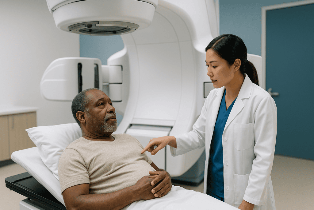
Three things can happen next. If the markers line up, treatment proceeds. If they are shifted, the couch moves by the needed few millimeters so that the tumor, not the skin or the bones, sits at the center of the beams. If the markers are in a completely unexpected arrangement, the team stops and investigates rather than treating.
For tumors that move with breathing, the markers do a second job. Some systems track them continuously, either from repeated X-ray snapshots or from their motion on planning scans, and either switch the beam on only during a particular part of the breathing cycle (a technique called gating) or steer the beam to follow the target. Other programs use the markers to measure how far the tumor travels with each breath and then build that range into the plan.
Typically three or more markers are placed, arranged in a spread rather than a straight line, so the software can detect rotation as well as simple shifts. If one migrates or is too close to another, the remaining ones can still define the target. The exact number is a planning decision that varies by site and by the tracking system a department uses.
Which tumors usually need fiducial markers before SBRT, and who is asked to wait?
The short version: markers are for targets that move and that the machine cannot see on its own. Four situations come up most often.
- Liver tumors, whether primary liver cancer or metastases. The liver slides several centimeters with breathing and has no internal landmarks visible on treatment images.
- Pancreatic tumors, which sit next to the stomach and small bowel, organs that tolerate high doses poorly. Precision here is a safety issue, not a refinement.
- Lung tumors, especially small ones near the chest wall or low in the lungs where breathing motion is largest. Central tumors near the airways are sometimes tracked using the spine or airway instead.
- Prostate, which shifts from day to day depending on how full the bladder and rectum are, even though it does not move with breathing.
Tumors anchored to bone are the classic exception. Spine metastases and brain targets are aligned to the skeleton or the skull, so markers add nothing. Some lung tumors that stand out sharply against dark, air-filled lung can be tracked directly without markers.
Who is usually asked to wait? Anyone whose bleeding risk is temporarily high, most commonly because of blood-thinning medicines that the prescribing team may decide to pause or continue; that decision belongs to them, weighed against the reason the medicine was started. Large amounts of fluid in the abdomen, an active infection, a very low platelet count, or a tumor that cannot be reached safely by any route are other reasons to delay or choose a different approach. A patient whose overall plan is still being decided, for example between surgery and radiation, will often be asked to hold off until the path is clear.
Fiducial marker placement procedure: what happens, step by step
The route depends on where the tumor is. What all routes share is imaging guidance, a thin needle, and a marker released at the tip.

Through the skin (percutaneous). This is the usual route for liver and many lung tumors, performed by an interventional radiologist. After local anesthetic, a needle is guided with CT or ultrasound into or beside the tumor, and a preloaded marker is pushed out. The needle is repositioned for each additional marker. A follow-up scan confirms the arrangement.
Through the digestive tract (endoscopic ultrasound). For pancreatic tumors and some liver or lymph node targets, a gastroenterologist passes a flexible scope with an ultrasound tip down the throat into the stomach or duodenum. The tumor is imaged through the gut wall and the needle passes a short distance to reach it. No skin puncture is involved.
Through the airway (bronchoscopy). Some lung tumors are reached with a navigational bronchoscope, avoiding the chest wall entirely.
Through the perineum or rectum. Prostate markers are placed by a urologist or radiation oncologist using ultrasound guidance, in a setting similar to a prostate biopsy.
How long does fiducial marker placement take? Programs describe the marker step itself as brief, usually a matter of minutes per marker, with the total visit stretched by preparation, sedation and observation afterward. Expect to set aside a good part of a day, and to be told to arrange a ride home if sedation is planned. The precise schedule is set by the performing department and varies enough between routes that any single number would mislead.
Is fiducial placement painful? What sedation usually looks like
People ask this more than any other question, and the honest answer is that it depends mostly on the route, and that the experience is usually far milder than the word “implant” suggests.
Percutaneous placement in the liver or lung is done under local anesthetic injected into the skin and the tissue along the needle path. The anesthetic itself stings briefly. After that, most people describe pressure or a dull ache rather than sharp pain, sometimes with a moment of discomfort when the needle passes the capsule of the liver or the lining of the lung. Light sedation, given through a vein and decided by the anesthesia or radiology team, is common and leaves many people with little memory of the middle part.
Endoscopic placement for the pancreas is typically performed under sedation because a scope in the stomach is uncomfortable without it. Patients wake with a scratchy throat and mild bloating from the air used to see inside.
Prostate placement mirrors a biopsy: local anesthetic to the area, a sensation of pressure, and occasionally a brief sting as each marker is placed.
Afterward, tenderness at the site for a day or two is expected. Over-the-counter pain relief is often enough, but which medicine, and whether it is compatible with other prescriptions, is a question for the team, since some common pain relievers thin the blood slightly and may be discouraged right after a needle procedure. Anyone with a history of difficult sedation, sleep apnea, or a strong needle fear should raise it beforehand so the plan can be adjusted, not endured.
Fiducial marker side effects: the realistic list of risks
Every needle carries risk, and the point of listing these is not to alarm but to make the consent conversation concrete. Serious complications are uncommon; minor ones are ordinary.
Bleeding. The liver is a blood-rich organ, and the lung’s edge is lined with small vessels. A small bruise inside the organ is common and usually silent. Significant bleeding requiring treatment is rare but is the main reason blood thinners are reviewed beforehand.
Pneumothorax. When a needle crosses the lung lining, air can leak into the space around the lung, causing it to partially collapse. Many small leaks seal on their own and are simply watched; larger ones may need a chest drain. This is the specific reason lung placements are followed by a chest X-ray and a period of observation.
Infection. Rare with skin routes; a recognized consideration with endoscopic and transrectal routes, where the needle passes through a bacteria-rich lining. Teams may give antibiotics around the procedure; whether and which is their call.
Pancreatitis. Inflammation of the pancreas is an uncommon complication of endoscopic placement into or near pancreatic tissue.
Marker migration. A marker can shift or, rarely, travel away from the tumor, most often in the first days. This is why planning scans wait for the markers to settle and why more than the minimum number is usually placed. A migrated marker is a nuisance rather than a danger; it is excluded from tracking.
Tumor seeding along the needle track is theoretical and reported very rarely. It sits in the same category of risk as any biopsy.
Marker types and placement routes compared by tumor site
Departments choose the route and the marker design based on anatomy, the tracking system they use, and who has the safest access. The table below summarizes the common patterns; a particular program may differ, and none of these choices is a matter of one being “better” in general.
| Tumor site | Usual route | Who typically performs it | Main site-specific risk | Common alternative to markers |
|---|---|---|---|---|
| Liver | Percutaneous, CT or ultrasound guided | Interventional radiologist | Bleeding | Breath-hold or compression with liver outline as surrogate |
| Pancreas | Endoscopic ultrasound through stomach or duodenum | Gastroenterologist | Pancreatitis, infection | Rarely omitted; stent or bone surrogates in select cases |
| Lung, peripheral | Percutaneous or bronchoscopic | Interventional radiologist or pulmonologist | Pneumothorax | Direct tumor tracking if visible; motion-encompassing plan |
| Lung, central | Often none needed | N/A | N/A | Spine or airway alignment |
| Prostate | Transperineal or transrectal, ultrasound guided | Urologist or radiation oncologist | Infection, urinary symptoms | Daily soft-tissue imaging on newer machines |
| Spine, brain | Not used | N/A | N/A | Bone landmarks |
Marker materials also vary. Solid gold cylinders and coiled gold wires are the workhorses. Some centers use carbon or polymer markers when they want less streaking on CT images, or a liquid marker that is injected and sets into a soft, visible blob. Coils tend to anchor better in soft tissue than smooth cylinders, which is one reason they appear in liver and lung work. None of this changes the patient’s experience much; it changes what the physicist sees on the planning screen.
What the following days and weeks usually look like, from placement to first SBRT session
The marker procedure is the start of a sequence, and knowing the sequence removes much of the anxiety.
The first day. Observation for a few hours, longer after lung placements so a delayed air leak can be caught. A chest X-ray or a quick scan confirms marker position. Discharge instructions usually cover wound care for a tiny puncture, activity limits, and what warrants a call. Soreness peaks that evening and eases over a day or two.
The settling interval. Teams deliberately leave a gap before the planning scan so that any small shift in marker position has finished and swelling from the needle has settled. This interval is measured in days rather than weeks in most programs, but its exact length is a departmental decision; ask what yours will be.
Simulation. The planning appointment, often called a CT simulation, is where the immobilization device is made and the reference scan is taken, sometimes with breathing recorded across the cycle. Contrast may be used. Skin marks or small tattoo dots may be placed. This visit is typically longer than any single treatment.
Planning. Behind the scenes, physicists and dosimetrists build the plan around the markers and the organs nearby. Patients see nothing of this stage but should expect it to take time.
Treatment. SBRT itself is delivered in one to five sessions, according to Mayo Clinic, often on alternating days. Each session is dominated by imaging and alignment; the beam-on time is a fraction of the appointment.
Follow-up imaging afterward is scheduled by the oncology team and is where the response to treatment is judged, usually not in the first weeks, when inflammation can blur the picture.
Fiducial markers and MRI: can you still have scans, and will airport security notice?
Yes, an MRI is usually possible with fiducial markers, and this is worth stating plainly because the worry stops people from mentioning the markers to scanning staff at all, which is the one thing they should not do.
Gold is not magnetic. Platinum, carbon and the polymers used in some markers are not magnetic either. The danger with MRI comes from ferromagnetic metal, the kind that can heat up or be pulled by the scanner’s field. Standard fiducials do not behave that way. What they do produce is a small dark spot or artifact on the images, a few millimeters across, which radiologists are used to reading around.
Two practical cautions apply. First, every implanted device should be declared on the MRI safety questionnaire, and the scanning department will check the specific marker type against its records; some newer markers contain tiny metal components beyond the main body, and the safety label for the exact product governs. Second, if a marker was placed alongside another device, such as a rectal spacer, a stent or a port, each item is assessed separately.
CT and PET scans are unaffected beyond a bright streak near the marker. Ultrasound sees markers as small bright dots.
Airport scanners are not an issue. The markers are far too small to trigger a metal detector, and no card is needed. The one document worth keeping is a note from the placing team stating the marker material and number, useful if imaging is done at an unfamiliar site years later. Markers are permanent; they are not removed after treatment and cause no known long-term harm sitting in place.
Do you always need fiducial markers before SBRT? Alternatives and where the evidence is shifting
No. Markers are one solution to the visibility problem, and radiation departments have others. Which is used depends on the tumor, the machine, and how well a patient can tolerate a given technique.
Motion-encompassing plans record the tumor’s full breathing excursion on a four-dimensional CT and treat the whole envelope. Simple and robust, at the price of a larger volume of healthy tissue.
Breath-hold asks the patient to hold a deep breath for short periods while the beam is on, freezing the target. It works well for people with good lung function and steady breathing, and poorly for those who cannot hold their breath reliably.
Abdominal compression uses a plate pressed gently on the upper abdomen to shrink breathing motion. Uncomfortable for some, effective for many liver and lower-lung tumors.
Anatomic surrogates such as the diaphragm dome, a biliary stent already in place, or a clearly visible tumor edge can stand in for markers when they move in lockstep with the target.
MRI-guided radiotherapy machines image soft tissue directly during treatment. Where these are available, markers are sometimes unnecessary because the tumor itself is visible.
The prostate is where practice is changing fastest. For years, three gold seeds were routine. Improved cone-beam CT that shows the prostate outline directly, and MRI-guided systems, have led some programs to stop placing them. The evidence is still developing, and many departments continue to use markers because they are reliable and inexpensive in terms of treatment-room time. Neither approach is wrong. What matters is that a department uses the method it has validated on its own equipment, and that is exactly the question a patient is entitled to ask.
What people often get wrong about fiducial markers
“The markers are radioactive.” They are not. Confusion comes from brachytherapy seeds, which look similar and are placed with similar tools but contain a radioactive source. Fiducials are inert metal or polymer. Nothing about them emits anything.
“The markers treat the cancer.” They are a navigational aid, nothing more. If the SBRT plan were canceled tomorrow, the markers would sit harmlessly and do nothing. Their entire value lies in what the treatment machine can do because of them.
“Placing them will spread the tumor.” Needle-track seeding is a real but very rare phenomenon, reported mainly with biopsies of certain liver and abdominal tumors. Teams weigh it against the precision the markers provide, and for most patients the balance favors placement. It is a fair question to ask about your particular tumor type.
“Once they are in, treatment can start right away.” The opposite. Placement is followed by a settling gap, then simulation, then planning. Anyone expecting to start radiation the following week may be disappointed, and knowing this in advance avoids the sense that something has gone wrong.
“If a marker moves, the whole thing has failed.” A migrated marker is checked and excluded. The remaining markers, or a repeat placement in rare cases, carry on.
“I will feel them.” After the first days, virtually nobody is aware of the markers. They are far smaller than most people picture and sit in organs with no fine touch sensation.
“They must come out afterward.” They stay permanently, and this is intended. Removal would require another procedure with its own risks for no benefit.
How long does it take to recover from SBRT radiation once the markers have done their job?
Recovery from the marker procedure and recovery from SBRT are two different things, and patients often blend them together.
The marker procedure leaves a puncture site and a day or two of soreness. Most people are back to ordinary activity quickly, with lifting and strenuous exercise limited briefly after liver or lung placement according to the discharging team’s instructions.
SBRT is different. Because the sessions are few, the treatment course itself is short, yet the body’s reaction unfolds over weeks. Tiredness is the most common effect of any radiotherapy, and the NHS notes it can build during treatment and continue for several weeks after the last session. Site-specific effects depend entirely on what lies near the target: nausea or a queasy feeling after upper-abdominal treatment, a cough or mild chest discomfort after lung treatment, more frequent or urgent urination after prostate treatment. These usually appear in the first days to a couple of weeks and then ease, though the exact course varies and the treating team will describe what to expect for a specific plan.
Late effects, meaning changes that appear months later, are the reason SBRT plans are so exacting. Scarring in a patch of lung, a change in liver blood tests, or a stretch of narrowed bowel are possible outcomes the planning process is designed to minimize. This is where the markers earn their place: tighter margins mean smaller volumes of normal tissue exposed.
Response is judged on follow-up scans arranged by the oncology team, typically not in the first weeks. Early images can show inflammation that looks worse before it looks better, which is why clinicians ask for patience before reading anything into them.
Questions to ask your care team before fiducial marker placement
A good consent conversation is a two-way exchange. These questions tend to surface the information that actually changes how a patient feels about the procedure.
- Why do I need markers rather than one of the marker-free techniques, and is that a decision about my tumor or about your equipment?
- Which route will you use, who will perform it, and how often does your team do this particular procedure?
- How many markers will be placed, and what happens if one of them shifts?
- Should I stop, continue or adjust any of my medicines, especially anything that affects bleeding or blood sugar, and who is responsible for that instruction?
- What kind of anesthesia or sedation is planned, and can it be changed if I have had trouble before?
- How long will I be observed afterward, and do I need someone to drive me home?
- What are the specific risks for my site, and what signs would mean I should call rather than wait?
- How many days will pass between placement and the planning scan, and when do you expect treatment to begin?
- Will the markers affect any future MRI, and is there documentation I should keep?
- Are there other procedures, such as a spacer or a stent, being done at the same time?
- If I decide against markers, what would the plan look like instead, and what would change about the margins?
Write the answers down, or ask whether a companion can join the appointment. The information density of these visits is high, and the marker step is easy to lose amid the larger conversation about the cancer itself. Nobody on the team will mind being asked to repeat something.
When to call your doctor after fiducial marker placement
Most people go home with nothing more than a small dressing and a tender spot. A short list of symptoms should prompt a call to the team that performed the procedure, or emergency care if severe, rather than waiting for the next appointment.
Seek urgent care for:
- Sudden or worsening shortness of breath, sharp chest pain, or a rapid heartbeat after lung placement, which can indicate an air leak around the lung.
- Severe or steadily increasing abdominal pain, a rigid or swollen abdomen, dizziness, fainting, or a racing pulse after liver or pancreatic placement, which can signal internal bleeding or pancreatitis.
- Fever, chills or shaking after any route, especially endoscopic or transrectal placement, since these can indicate infection.
- Coughing or vomiting blood, or black or bloody stools.
- Inability to pass urine, or heavy blood in the urine that does not clear, after prostate placement.
- Redness, warmth, swelling or discharge spreading from the puncture site.
Milder concerns, such as bruising, a low-grade ache lasting beyond a few days, or uncertainty about wound care or medicines, deserve a routine call rather than silence. Departments would rather hear about a symptom that turns out to be nothing than miss one that mattered.
During and after SBRT itself, the radiation team will provide its own list of side effects to report. Nothing in this article replaces that guidance. The markers, the plan, the timing and every decision about medicines sit with the treating clinicians, who know the specifics of the tumor, the anatomy and the equipment involved.
Frequently asked questions
How long does a fiducial marker placement take?
The marker step itself is brief, typically minutes per marker, but the full visit is much longer because of preparation, sedation and observation afterward, and lung placements add a chest X-ray and a longer watch for air leaks. Plan for most of a day and arrange a ride home if sedation is expected. The performing department sets the schedule, and it varies by route.
Is fiducial placement painful?
Most people describe pressure or a dull ache rather than sharp pain. Local anesthetic numbs the skin and needle path, and light sedation is common for liver, lung and endoscopic routes. Prostate placement feels similar to a biopsy. Soreness at the site for a day or two is normal; ask the team which pain relievers are appropriate, since some can affect bleeding.
Can you have an MRI if you have fiducial markers?
Usually yes. Gold and the other common marker materials are not magnetic, so they do not heat or move in the scanner and produce only a small dark artifact on the images. Always declare the markers on the MRI safety form so staff can confirm the specific product, especially if other devices such as a stent, port or spacer were placed as well.
What are fiducial markers made of, and are they radioactive?
Fiducial markers are made of dense, inert materials, most often gold, sometimes platinum, carbon or a polymer, and some are injected as a liquid that sets into a visible gel. None are radioactive. The confusion arises from brachytherapy seeds, which look alike but contain a radiation source. Fiducials emit nothing and simply reflect X-rays so the treatment machine can see the target.
How long does it take to recover from SBRT radiation?
The treatment course is short, but effects unfold over weeks. The NHS notes radiotherapy tiredness can continue for several weeks after the last session. Site-specific effects such as nausea, cough or urinary changes typically appear within the first days to weeks and then ease. Late changes months later are possible, and follow-up imaging arranged by the oncology team is how response is judged.
What are the main fiducial marker side effects?
Minor bruising and soreness at the site are common and settle within days. Less common risks depend on the route: air leak around the lung (pneumothorax) after lung placement, bleeding after liver placement, infection or pancreatitis after endoscopic placement, and infection or urinary symptoms after prostate placement. Marker migration can occur, which is why more than the minimum number is usually placed.
Do fiducial markers need to be removed after radiation?
No. Fiducial markers are permanent and are left in place for life. They cause no known long-term harm, are not felt once the first days of soreness pass, and removal would require another needle or scope procedure with its own risks for no benefit. Keep a note of the marker type and number for any future imaging at an unfamiliar site.
Why do liver and pancreas tumors so often need fiducial markers before SBRT?
Both organs move several centimeters with breathing and look almost identical to their surrounding tissue on the X-ray and cone-beam CT images taken at the treatment machine. Without a marker there is no reliable landmark. The pancreas also sits beside the stomach and small bowel, which tolerate high doses poorly, so precise alignment is a safety requirement rather than a refinement.
Are fiducial markers still needed for prostate SBRT?
Practice is shifting. Improved cone-beam CT that shows the prostate outline and MRI-guided treatment machines allow some programs to treat without markers, while many departments continue to use them because they are reliable on their equipment. The evidence is still developing, and the right answer is whichever method a department has validated on its own systems. Ask which approach yours uses and why.
What is the fiducial marker placement procedure like for lung tumors?
Lung markers are placed either with a needle through the chest wall under CT guidance or through the airway with a navigational bronchoscope. Local anesthetic and light sedation are common. Because the needle route crosses the lung lining, a chest X-ray and a period of observation follow to check for an air leak. Central tumors near the airways are sometimes aligned to the spine instead and skip markers.
References
This article is for general information only and is not a substitute for professional medical advice. Please consult a qualified doctor about your individual situation.

More from the Blog
How CT, MRI and PET-CT Stage Kidney Cancer Before a Treatment Plan Is Written
Kidney cancer is staged mainly with a contrast-enhanced CT scan of the abdomen and chest, which measures the tumor, checks whether it has grown…

Monitoring Response in Lymphocytic Leukemia: What Blood Counts Show Between Visits
CLL treatment response monitoring uses regular complete blood counts, a physical examination of lymph nodes, spleen and liver, and sometimes imaging or a bone…
Why Chemotherapy and Radiation Often Come Before Esophageal Cancer Surgery
Chemotherapy and radiation are often given before esophageal cancer surgery because the combination can shrink the tumor, treat cancer cells that may have spread…

Transfusions and Infection Precautions During Acute Myeloid Leukemia Treatment
During acute myeloid leukemia treatment, chemotherapy drives red cells, platelets and infection-fighting white cells to very low levels for weeks. Red cell and platelet…

Radiation Therapy Side Effects by Treated Area: What Is Expected and When to Call the Team
Radiation side effects are expected and follow the treated area: skin changes, fatigue, and local effects such as a sore mouth or loose stools.…

What Happens During Wilms Tumor Surgery? A Parent’s Guide to the Operation and Anesthesia
Wilms tumor surgery usually means a nephrectomy: the surgeon removes the affected kidney, the tumor inside it, and nearby lymph node samples through an…