

How CT, MRI and PET-CT Stage Kidney Cancer Before a Treatment Plan Is Written
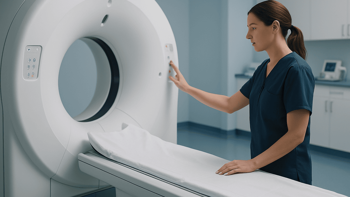
Key Takeaways
- A contrast-enhanced CT of the abdomen and chest is the standard staging test for kidney cancer, and measurable enhancement of a mass is the key sign that it is a solid tumor rather than a cyst.
- Stage I tumors measure 7 cm or less and stay inside the kidney; the 4 cm line within Stage I often decides whether a kidney-sparing partial nephrectomy is possible.
- MRI replaces or supplements CT when iodinated contrast is unsafe, in pregnancy, and when the extent of tumor growing inside the renal vein or vena cava needs precise mapping.
- PET-CT is not routine for kidney cancer because the tracer is excreted by the kidneys and clear cell tumors take it up inconsistently; it is reserved for equivocal lesions and suspected recurrence.
- Imaging assigns a clinical stage that pathology can change after surgery, most often at the Stage II to III boundary where fat and vein invasion are hard to judge.
- No diet or supplement has been shown to treat kidney cancer; after nephrectomy the priorities are hydration, balanced protein, moderate salt and avoiding medicines that reduce kidney blood flow.
Kidney cancer is staged mainly with a contrast-enhanced CT scan of the abdomen and chest, which measures the tumor, checks whether it has grown into surrounding fat, veins or lymph nodes, and looks for spread to the lungs and bones. MRI is used when CT contrast is unsafe or vein involvement is unclear. PET-CT is reserved for selected cases, and the final stage is confirmed after surgery.
The radiology waiting room has a particular hush. A man in his sixties sits with a paper cup of water he was told to finish, a cannula taped to his forearm, reading the same magazine page for the fourth time. Two weeks ago an ultrasound for back pain found a shadow on his left kidney. Today he is here for the scan that will decide what happens next.
That scan is the heart of kidney cancer CT scan staging. Before any surgeon, oncologist or nurse can write a treatment plan, the team needs to know three things: how big the tumor is, whether it has pushed beyond the kidney, and whether it has traveled anywhere else. Imaging answers those questions faster and more safely than any other tool.
What follows explains, without jargon or drama, how CT, MRI and PET-CT each contribute, why one is chosen over another, where the pictures can mislead, and what the weeks between the first scan and the first treatment decision usually look like.
What kidney cancer CT scan staging actually measures
Staging is a shorthand language. It compresses everything a scan shows into a code that any cancer team in the world can read. For kidney cancer, that code is the TNM system: T for the primary tumor, N for nearby lymph nodes, and M for metastasis, which means cancer that has spread to a distant organ.
A CT scan is a rotating X-ray that builds cross-sectional pictures of the body, slice by slice, like looking at a loaf of bread one piece at a time. Add an iodine-based contrast dye through a vein and blood vessels light up, which matters enormously here. Kidney tumors are often richly supplied with blood, so they brighten in a way that cysts and scar tissue do not. Radiologists call that brightening “enhancement,” and measurable enhancement is one of the strongest clues that a kidney mass is a solid tumor rather than a harmless fluid-filled cyst.
From the same set of images the radiologist records the tumor’s largest diameter, whether its edge stays inside the kidney capsule, whether it reaches into the fatty tissue around the kidney, whether it extends into the renal vein or the inferior vena cava (the large vein carrying blood back to the heart), and whether lymph nodes near the kidney look enlarged. Each observation maps directly onto a T or N category.
The scan does not deliver a diagnosis on its own. It produces a clinical stage, which is the team’s best estimate before treatment. The pathological stage, assigned after a surgeon removes the tumor and a pathologist examines it under a microscope, can shift that estimate up or down. Mayo Clinic describes imaging as the first step in confirming a kidney mass and determining its extent, with surgery and pathology completing the picture.
How does a CT scan stage kidney cancer, step by step?
A staging CT is not one picture but a sequence timed to the flow of contrast through the kidney. The radiology team usually acquires images before contrast, then again at several points after it is injected.

The pre-contrast pass gives a baseline. Without it, the radiologist cannot tell whether a mass that looks bright after dye was already bright beforehand, which happens with blood or calcium inside a lesion. Then come the contrast phases. In the early arterial phase, the outer cortex of the kidney brightens first and many tumors glow against it. A later nephrographic phase, when the whole kidney is evenly enhanced, is the one most radiologists rely on for measuring the tumor and judging whether it has breached the kidney’s edge. A delayed phase shows the collecting system and can reveal whether the tumor has grown into the urine-draining structures.
At each step the reader asks a short list of questions: Is this a cyst or a solid mass? How large is it, in centimeters? Does it stay inside the kidney? Does it touch or invade the perinephric fat, the cushion of fat wrapped around the organ? Is there a tongue of tumor inside the renal vein? Are the lymph nodes along the aorta enlarged? Does the adrenal gland sitting on top of the kidney look normal?
Cysts get their own grading, the Bosniak classification, which ranks fluid-filled kidney lesions from clearly benign to likely malignant based on their walls, septations and enhancement. Many people who hear “kidney mass” are relieved to learn their lesion sits in the simple-cyst category and needs no further action.
Cleveland Clinic lists CT, often with contrast, as the standard test for characterizing a kidney mass before any treatment is discussed. The whole acquisition typically takes only minutes once the person is on the table; the preparation and the reading take longer.
Kidney cancer stages explained: what T1 to T4 mean on a scan
The T category is largely about size and boundaries, and the thresholds are specific. According to the National Cancer Institute’s patient summary, a Stage I kidney cancer is 7 centimeters or smaller and confined to the kidney. Stage II is larger than 7 centimeters but still confined. Stage III has grown into a major vein or the tissues around the kidney, or has reached nearby lymph nodes. Stage IV means the cancer has spread beyond the fascia that wraps the kidney, into the adrenal gland on the same side, or to distant organs.
| Stage | What the scan usually shows | Typical imaging clues |
|---|---|---|
| Stage I | Tumor 7 cm or smaller, inside the kidney | Sharp edge, intact capsule, clean fat |
| Stage II | Tumor larger than 7 cm, still inside the kidney | Large but contained, no vein or fat invasion |
| Stage III | Into renal vein or vena cava, perinephric fat, or regional nodes | Filling defect in the vein, strands in the fat, enlarged nodes |
| Stage IV | Beyond the fascia, into the adrenal, or distant spread | Lung nodules, bone lesions, liver deposits |
Within Stage I, radiologists subdivide at 4 centimeters because tumors of 4 centimeters or less are often candidates for kidney-sparing approaches such as partial nephrectomy, which means removing the tumor while leaving the rest of the kidney in place. That single measurement can change the entire conversation with a surgeon.
Two reminders keep these stages honest. First, a number on a scan is a measurement with some uncertainty, especially for tumors near a threshold. Second, stage describes extent, not behavior; a pathologist’s grade, which judges how abnormal the cells look, adds the second half of the story and is only available after biopsy or surgery.
MRI vs CT for a kidney tumor: when is MRI the better choice?
MRI uses a strong magnet and radio waves rather than X-rays to build images, and it shows soft tissue with a different kind of clarity. For most people with a kidney mass, CT remains the workhorse, and the NHS lists CT scanning as the standard way to assess kidney cancer and its spread. MRI steps in for specific reasons.

The first is contrast safety. Iodinated CT contrast can be a problem for people with reduced kidney function, which is common in exactly the population that develops kidney tumors. Blood tests before the scan check filtration, and if the numbers are low the team may switch to MRI. MRI contrast, gadolinium, is also used with care in severe kidney disease, and the radiology team weighs that risk case by case; in some situations MRI without contrast still answers the question.
The second is a known allergy to iodinated contrast. The third is pregnancy, where avoiding radiation matters. The fourth is anatomical: MRI is especially good at showing how far a tumor thrombus, meaning a plug of tumor growing inside a vein, extends up the inferior vena cava toward the heart. That detail shapes surgical planning because a thrombus reaching the chest requires a different operation than one confined to the renal vein.
MRI also helps with indeterminate cysts, where CT cannot decide whether a thickened wall is enhancing, and with small masses where CT measurements sit on the fence. It has drawbacks: longer scan times, more breath-holds, and difficulty for people who are claustrophobic or who have certain implanted devices. MedlinePlus notes that an MRI typically takes longer than a CT scan and that patients must lie still inside a tube-shaped machine for the duration.
In practice the two are complementary rather than rivals. The question a team asks is not which scan is best in general, but which one answers the specific unknown in front of them.
What does a PET scan for kidney cancer add, and when is it skipped?
A PET-CT combines a CT scanner with a detector for a radioactive tracer, most often a form of glucose called FDG. Cancer cells that burn sugar quickly take up the tracer and glow on the images. For many cancers, PET-CT is a routine staging tool. For kidney cancer it is not, and the reason is biology, not budget.
Clear cell renal cell carcinoma, the most common type, often takes up FDG inconsistently, so a tumor can be present without lighting up. Worse, the kidneys themselves excrete the tracer into urine, which means the organ being studied is bathed in a bright signal that can hide a lesion. Major urology and oncology guidelines therefore do not recommend PET-CT for routine staging of a newly found kidney mass.
Where it earns its place is in selected problems: an equivocal lung nodule or bone lesion that CT cannot classify, suspected recurrence after surgery when conventional scans are inconclusive, or planning for a person with advanced disease where the pattern of spread will change treatment. The treating team decides on a case-by-case basis.
The rest of the metastatic search relies on simpler tools. A chest CT checks the lungs, the most common site of distant spread. A bone scan, which uses a different tracer that collects in areas of active bone turnover, is added when there is bone pain or a blood test suggests bone involvement. Brain imaging, usually MRI, is reserved for people with headaches, weakness or other neurological symptoms. MedlinePlus explains that a PET scan involves a waiting period after the tracer injection before imaging begins, which is why the appointment is longer than the scan itself.
Who is sent for staging scans, and who is usually asked to wait?
Anyone with a solid, enhancing kidney mass on ultrasound or an incidental CT is typically referred for full staging imaging. Many kidney cancers today are discovered this way, found on a scan ordered for something unrelated, which is one reason the person in the waiting room often feels perfectly well.
People with blood in the urine, a persistent flank pain, or an unexplained lump are also imaged promptly. So are people with a known hereditary syndrome, such as von Hippel-Lindau disease, where surveillance of the kidneys is part of lifelong care.
Waiting is also a legitimate plan, chosen rather than defaulted to. For very small masses, generally those under 4 centimeters, especially in older people or those with other serious illnesses, the team may propose active surveillance: repeat imaging at intervals to watch for growth, with treatment only if the mass changes. Small kidney tumors frequently grow slowly, and the risks of surgery can outweigh the risks of observation in someone whose heart or lungs are fragile. Mayo Clinic lists active surveillance as an option for small kidney cancers.
Timing can be adjusted for other reasons. Someone with acute kidney injury may have contrast imaging deferred until function recovers. Someone who is pregnant will have the sequence reordered to favor ultrasound and MRI. Someone with a recent contrast reaction may need premedication or a different modality, a decision that rests with the radiology and clinical teams, not with a blog.
A short word on urgency. Being asked to wait for a scan slot of a few weeks is usually not a sign of neglect. Kidney tumors found on routine imaging rarely change meaningfully over that interval, and the team sequences blood tests, imaging and specialist review so the first treatment conversation has complete information.
What actually happens on the day of a staging CT or MRI
The morning starts with questions, not machines. A radiographer confirms identity, asks about allergies, diabetes medicines, pregnancy, prior contrast reactions and kidney problems. A recent blood test for kidney function is checked, and if none exists, one may be drawn on the spot.
Most centers ask people to drink water beforehand and to avoid solid food for a few hours, mainly so that any nausea from contrast is less unpleasant. A cannula is placed in a forearm or hand vein. On the table, the person lies on their back with arms raised above the head, which keeps bone out of the field of view.
When the contrast runs in, a warm flush spreads through the chest and pelvis, and many people feel a brief, odd sensation of having passed urine. They have not. It fades within a minute. The radiographer gives breath-hold instructions through a speaker, and the table slides through the ring several times to capture each contrast phase. MedlinePlus notes that the scanning itself is usually complete within a short span of minutes, though the whole appointment runs longer.
An MRI appointment is quieter in preparation and louder in execution. Metal screening is thorough. Earplugs or headphones are provided because the machine knocks and buzzes loudly. The person lies inside the bore for a longer session with repeated breath-holds, and a coil, essentially a padded antenna, is strapped over the abdomen. Gadolinium contrast, if used, produces no warm flush.
Afterward, people are usually asked to drink extra fluids to help clear the contrast and are free to drive and eat normally unless sedation was used. Results go to the referring clinician, generally within days, and are discussed at an appointment rather than handed over in the scanner room.
Why kidney cancer CT scan staging can be wrong, and how teams correct for it
No imaging test is perfect, and knowing where CT stumbles is part of reading it well. The weakest link is the boundary between Stage I or II and Stage III. Deciding whether a tumor has invaded the perinephric fat is notoriously hard, because inflammation, previous infection and simple tumor bulk can all produce streaky changes in that fat without actual invasion. Pathology after surgery sometimes upgrades a tumor the scan called contained, and occasionally downgrades one the scan called invasive.
Lymph nodes are the second soft spot. CT judges nodes by size, yet reactive nodes can be enlarged and cancerous nodes can be normal in size. A node that looks suspicious on imaging may be removed at surgery for a definitive answer.
Small lung nodules are a third. Many adults carry tiny benign nodules from old infections, and a staging chest CT may find one or two that cannot be labeled. The usual response is a repeat scan after an interval to see whether anything grows, not an immediate change of stage.
Teams correct for these limits in three ways. They review images together at a multidisciplinary meeting, where a radiologist, urologist, oncologist and pathologist look at the same pictures and argue the details. They add a second modality, typically MRI for venous or fat questions and bone scan or PET-CT for equivocal distant lesions. And they use biopsy selectively: a needle sample of a kidney mass can settle whether it is cancer and what subtype, which matters most when the result would change the plan, for example before active surveillance or thermal ablation. Cleveland Clinic notes that biopsy is not needed for every kidney mass and is chosen when the information will alter management.
The practical lesson is that a clinical stage is a well-informed starting point. It is written in pencil until pathology inks it in.
What the days and weeks after staging scans usually look like
The scan is done. Now comes a period that many people find harder than the imaging itself, because it is mostly waiting while others work.
First, the radiologist writes a structured report, typically within a few working days, recording tumor size, enhancement, fat and vein status, nodes, adrenal appearance and any findings elsewhere. Second, the report is matched with blood tests: kidney function, blood count, calcium, liver enzymes, and a marker of bone turnover that may trigger a bone scan if raised.
Third, the case goes to a multidisciplinary team meeting. These usually run weekly in cancer centers. The group assigns a clinical stage, decides whether further imaging or biopsy is needed, and sketches options: active surveillance, ablation, partial nephrectomy, radical nephrectomy, or, for advanced disease, systemic therapy. Fourth, the person meets a specialist, often a urologist, to hear the stage and the options in plain language.
If surgery is planned, the weeks that follow involve pre-operative assessment of heart and lung fitness, and sometimes a repeat CT to map the blood vessels in detail for the operation. If systemic treatment is planned, an oncologist discusses the drug classes and arranges baseline tests. If surveillance is chosen, the next scan date is set, often several months ahead.
The NHS notes that referral for suspected cancer is designed to deliver a specialist appointment quickly, and the staging process then unfolds over a span of weeks rather than days. Within that span, the most useful thing a person can do is write questions down as they arise, bring a companion to appointments, and ask the team to explain the stage in their own words before leaving the room.
What is metastatic kidney cancer, and how is it staged differently?
Metastatic kidney cancer is cancer that has traveled from the kidney to a distant site, most commonly the lungs, bones, liver, brain or distant lymph nodes. In TNM language it is M1 disease, and any M1 finding places the cancer in Stage IV regardless of how small the kidney tumor is.
Staging shifts emphasis at this point. The exact size of the primary tumor matters less; the number, location and burden of distant deposits matter more, because they determine whether surgery on the kidney still makes sense and which treatments are likely to help. Chest CT is reviewed for the count of lung nodules. Bone scan or MRI defines bone lesions that might need protection from fracture. Brain MRI is added when symptoms suggest it. PET-CT may be considered to clarify a single ambiguous site.
Treatment for metastatic disease is mainly systemic, meaning medicines that travel through the bloodstream. Two drug classes dominate current guidelines. Immune checkpoint inhibitors release a brake on the immune system so that T cells can recognize and attack cancer cells. Tyrosine kinase inhibitors block signals, especially those driven by VEGF, that tumors use to grow new blood vessels. The National Cancer Institute’s patient summary describes both immunotherapy and targeted therapy as standard options for Stage IV kidney cancer, often in combination, with the choice depending on risk group, kidney function and other illnesses.
Responses vary widely from person to person, and no article can predict an individual’s course. What imaging does in this setting is track change over time: repeat CT scans at intervals show whether deposits are shrinking, stable or growing, and the oncologist adjusts the plan accordingly. All such decisions sit with the treating team.
What people often get wrong about kidney cancer staging scans
“A PET scan is the most advanced test, so it must be the best.” For kidney cancer it is usually the least useful first-line scan, for the biological reasons already described. Contrast CT finds and characterizes kidney tumors far more reliably.
“If the scan says Stage I, that is final.” Clinical stage from imaging is an estimate. Pathology after surgery can move it, most often across the Stage II to III boundary where fat and vein invasion are judged.
“Contrast dye will destroy my kidneys.” Iodinated contrast carries a small risk of temporary kidney injury, mainly in people whose function is already reduced, and that is precisely why blood tests are checked beforehand and hydration is encouraged. For most people the benefit of an accurate scan outweighs the risk, and the radiology team can switch to MRI when it does not.
“A biopsy will spread the cancer.” Needle-track seeding is rare with modern technique, and biopsy is used when it changes management. Fear of it should not drive the decision; the team’s judgment should.
“A small tumor means nothing needs to happen.” A small tumor often means more options, including surveillance, but it still needs characterization and a plan.
“Scans can tell me the type of kidney cancer.” Imaging suggests, pathology confirms. Some features point toward clear cell versus other subtypes, but only tissue under a microscope settles it.
“A lung nodule on my staging scan means Stage IV.” Tiny nodules are common and frequently benign. Interval follow-up, not alarm, is the standard response.
Johns Hopkins emphasizes that kidney cancer is diagnosed by combining imaging with laboratory tests and, when needed, tissue sampling, rather than by any single test alone.
What to eat after kidney surgery, and are there foods to avoid with kidney cancer?
Nutrition questions arrive the moment a plan is written, and the evidence-based answer is calmer than many online lists suggest.
There is no diet shown to shrink a kidney tumor or to prevent it from spreading. Claims about specific “anti-cancer” foods, juices or supplements are not supported by controlled evidence, and some supplements interact with the medicines used for advanced disease. The NIH Office of Dietary Supplements advises telling the care team about every supplement taken, because herbal products can affect drug metabolism and kidney function.
After a nephrectomy, whether partial or complete, the practical priorities are healing and protecting the remaining kidney tissue. Adequate protein supports wound repair, and most surgical teams encourage a normal balanced diet as soon as the bowel is working again. Fluids matter, since dehydration stresses a kidney already adapting to extra workload. If a whole kidney was removed, the remaining one takes over the job, and clinicians generally advise moderate salt intake, avoiding excessive protein loading, and caution with non-steroidal anti-inflammatory painkillers, which can reduce kidney blood flow. Those are conversations for the surgical team and, where available, a renal dietitian, rather than rules to apply alone.
People living with metastatic disease on systemic therapy may face appetite loss, taste changes or diarrhea. A dietitian can tailor small, frequent meals and manage weight, and the oncology team should hear about any significant change in eating.
What to avoid, then, comes down to common sense grounded in kidney physiology: heavy alcohol, very high-salt processed foods, unregulated supplements, and any product marketed with promises it cannot keep. Mayo Clinic’s guidance on kidney cancer notes that no alternative medicine has been shown to treat the disease, while gentle supportive practices may help with coping.
Questions to ask your care team about your staging results
A good consultation is a two-way exchange. These questions help people leave the room understanding their own scan rather than just hearing a stage number.
- What size is the tumor in centimeters, and does that put it above or below the 4 cm and 7 cm thresholds?
- Does the scan show the tumor contained within the kidney, or is there any sign it has reached the fat, the veins or the adrenal gland?
- Did the chest CT show anything, and if there are small nodules, what is the plan for following them?
- Is my kidney function good enough for contrast imaging, and if not, what alternative was used and how confident is the team in the result?
- Would an MRI, bone scan or PET-CT change the plan, or has the team decided they are not needed and why?
- Is a biopsy being recommended, and what decision would the result change?
- Is active surveillance a reasonable option for me, and what would prompt a move to treatment?
- If surgery is proposed, can the kidney be partially preserved, and what does that depend on?
- How will the stage be confirmed after surgery, and how might it differ from today’s estimate?
- Who is my point of contact if a new symptom appears before the next appointment?
- How often will scans be repeated after treatment, and which type?
- Does my family history suggest a hereditary syndrome, and should relatives be offered screening?
Writing the answers down, or asking permission to record the conversation, turns an overwhelming appointment into a document that can be reread calmly at home. Many teams also offer a specialist nurse contact for questions between visits, and the NHS describes this role as a standard part of cancer care pathways.
When to call your doctor during staging or treatment
Most of the staging journey is uneventful, but some situations should not wait for the next scheduled appointment. Contact the care team the same day, or seek emergency care, if any of the following occur.
- Heavy or persistent blood in the urine, clots, or difficulty passing urine, especially if accompanied by faintness.
- Sudden, severe flank or abdominal pain, which can signal bleeding into or around a tumor.
- Fever, chills or rigors, particularly after a biopsy or contrast injection, which may indicate infection.
- Any reaction after contrast: hives, swelling of the face or throat, wheeze, chest tightness or dizziness. Severe reactions are rare but need immediate treatment.
- New leg swelling on one side, or sudden breathlessness and chest pain, which can be signs of a blood clot and are more likely when a tumor involves the large veins.
- New headache that is severe or different from usual, confusion, weakness on one side, or seizures, which require urgent assessment for possible brain involvement.
- New bone pain that wakes you at night, or pain after a minor knock, which should prompt imaging for bone lesions.
- A marked drop in urine output, or rapidly rising weight and swelling, which can indicate kidney injury after contrast or surgery.
Mayo Clinic advises making an appointment for any persistent symptom that causes concern, and the NHS urges urgent review for visible blood in the urine at any age. None of these signs confirm cancer progression; many have simple explanations. The point of calling is to let the treating team decide, with the full picture in front of them, rather than to carry uncertainty alone.
Frequently asked questions
What does a kidney cancer CT scan staging report actually say?
It records the tumor’s largest diameter, whether it enhances with contrast, whether it stays inside the kidney or reaches the surrounding fat, veins or adrenal gland, whether nearby lymph nodes are enlarged, and whether the chest or other organs show suspicious lesions. Those findings are translated into T, N and M categories to give a clinical stage. The report is a starting point that the team reviews alongside blood tests and, where needed, biopsy or further imaging.
Can kidney cancer stages be explained simply?
Yes. Stage I is a tumor 7 cm or smaller contained within the kidney; Stage II is larger than 7 cm but still contained; Stage III has grown into a major vein, the fat around the kidney, or nearby lymph nodes; Stage IV has spread beyond the kidney’s outer fascia, into the adrenal gland, or to distant organs such as the lungs or bones. These definitions come from the TNM system used worldwide and summarized by the National Cancer Institute.
MRI vs CT for a kidney tumor: which is more accurate?
Neither is universally more accurate; they answer different questions. Contrast CT is the standard first test and reliably measures the tumor and checks the chest. MRI is better at showing how far a tumor thrombus extends inside a vein and at characterizing complex cysts, and it avoids iodinated contrast and radiation. The team picks the modality that resolves the specific uncertainty, and sometimes uses both.
Is a PET scan for kidney cancer ever recommended?
Only selectively. PET-CT is not part of routine staging for a new kidney mass because the FDG tracer is excreted into urine, which obscures the kidney, and clear cell tumors do not consistently take it up. It may be considered to clarify an ambiguous lung or bone lesion, to investigate suspected recurrence when standard scans are inconclusive, or in some cases of advanced disease. The decision rests with the treating team.
What is metastatic kidney cancer?
Metastatic kidney cancer is cancer that has spread from the kidney to distant sites, most often the lungs, bones, liver, brain or distant lymph nodes. In staging terms it is M1 disease, which is always Stage IV regardless of the size of the kidney tumor. Treatment is mainly systemic, using immune checkpoint inhibitors and tyrosine kinase inhibitors, with repeat CT scans used to track response over time.
Can kidney cancer be treated successfully and controlled long term?
Many kidney cancers, particularly those found at an early stage and confined to the kidney, are treated with surgery or ablation and need only surveillance afterward. Advanced disease is managed with systemic therapies that can control it for extended periods in some people. Outcomes depend on stage, subtype, grade and overall health, and only the treating team can discuss what is realistic for an individual. No reliable source promises a specific result.
How long do staging scans take?
The CT scan itself takes only minutes on the table, though the appointment, with preparation, cannula placement and post-contrast observation, runs longer. MRI sessions are longer and involve repeated breath-holds inside the machine. A PET-CT appointment includes a waiting period after the tracer injection before imaging starts. MedlinePlus provides typical durations for each, and the radiology department will give a specific estimate when booking.
What should I eat after kidney surgery?
Most surgical teams encourage a return to a normal balanced diet once the bowel is working, with adequate protein to support healing and plenty of fluids to protect the remaining kidney tissue. If a whole kidney was removed, moderate salt intake and avoiding excessive protein loading are commonly advised. Ask the surgical team about pain relief choices, since some over-the-counter anti-inflammatory drugs can reduce kidney blood flow, and request a renal dietitian referral if available.
What foods should I avoid if I have kidney cancer?
No specific food has been shown to worsen kidney cancer, and no food or supplement has been shown to treat it. Sensible limits apply to heavy alcohol and very high-salt processed foods, which burden the kidneys. Unregulated supplements deserve particular caution because some interact with targeted therapies and immunotherapy or affect kidney function. Tell the care team about everything you take, including herbal products.
Can a CT scan be wrong about the stage of kidney cancer?
Yes, within limits. CT is very good at finding and measuring kidney tumors but less reliable at judging invasion into the fat around the kidney and at distinguishing reactive from cancerous lymph nodes. Pathology after surgery can revise the stage upward or downward. Teams reduce the chance of error by reviewing images together, adding MRI or other scans when needed, and using biopsy when the result would change management.
References
This article is for general information only and is not a substitute for professional medical advice. Please consult a qualified doctor about your individual situation.

More from the Blog

Monitoring Response in Lymphocytic Leukemia: What Blood Counts Show Between Visits
CLL treatment response monitoring uses regular complete blood counts, a physical examination of lymph nodes, spleen and liver, and sometimes imaging or a bone…
Why Chemotherapy and Radiation Often Come Before Esophageal Cancer Surgery
Chemotherapy and radiation are often given before esophageal cancer surgery because the combination can shrink the tumor, treat cancer cells that may have spread…

Transfusions and Infection Precautions During Acute Myeloid Leukemia Treatment
During acute myeloid leukemia treatment, chemotherapy drives red cells, platelets and infection-fighting white cells to very low levels for weeks. Red cell and platelet…

Radiation Therapy Side Effects by Treated Area: What Is Expected and When to Call the Team
Radiation side effects are expected and follow the treated area: skin changes, fatigue, and local effects such as a sore mouth or loose stools.…

What Happens During Wilms Tumor Surgery? A Parent’s Guide to the Operation and Anesthesia
Wilms tumor surgery usually means a nephrectomy: the surgeon removes the affected kidney, the tumor inside it, and nearby lymph node samples through an…

What a Bone Marrow Test Shows Before Leukemia Treatment and How It Shapes the Plan
A bone marrow test before leukemia treatment samples the spongy tissue inside the hip bone to confirm the diagnosis, count immature cells called blasts,…