

How a PEG Tube Is Placed: The Endoscope, the Sedation and What Happens in the Room

Key Takeaways
- PEG stands for percutaneous endoscopic gastrostomy: an opening into the stomach created through the skin, guided by a camera passed through the mouth, without an abdominal operation.
- Safe placement depends on two checks done together: the scope's light glowing through the skin and a finger press on that spot producing a visible dent inside the inflated stomach.
- Most placements use moderate sedation plus local anesthetic in the abdominal wall, not general anesthesia, and take about 30 to 45 minutes according to MedlinePlus.
- The first tube is almost always a bumper type because it cannot be dislodged by accident; balloon tubes and low-profile buttons are usually swapped in only after the tract has healed over several weeks.
- A gastrostomy is generally considered when tube feeding is expected to last beyond roughly four to six weeks; shorter needs are usually met with a nasogastric tube.
- For advanced dementia, major guideline bodies have not found that tube feeding prolongs life or prevents aspiration pneumonia, so careful hand feeding is often recommended instead.
A PEG tube is placed during a short endoscopic procedure, usually under moderate sedation with a local anesthetic in the abdominal wall. A thin camera is passed through the mouth into the stomach, the stomach is inflated with air, a light marks the exit point on the skin, a needle and guidewire create the path, and a soft tube is drawn or pushed into position and secured with an internal bumper. Most placements take about 30 to 45 minutes.
The question usually arrives in a hallway, not an office. A speech therapist has just explained that swallowing is no longer safe, and a family member hears the word “gastrostomy” for the first time. Within an hour someone is typing how is a PEG tube placed into a phone, scrolling past diagrams that look more alarming than the procedure actually is.
It helps to know what the room will look like. There is no operating theater, no breathing tube, no incision longer than a fingertip. There is an endoscopist, a nurse watching monitors, a dimmed light so a glow can be seen through the skin, and a person who is drowsy rather than unconscious.
This explainer walks through that room step by step: the sedation, the scope, the moment the tube comes through, and the days that follow. It also covers who is usually a good candidate, who is often asked to wait, and the myths that make the decision harder than it needs to be.
What is a gastrostomy, and what does PEG actually stand for?
A gastrostomy is a small, deliberately created opening between the stomach and the skin of the abdomen. A tube sits in that opening so that liquid nutrition, fluids and medicines can reach the stomach without passing through the mouth and throat.
PEG stands for percutaneous endoscopic gastrostomy. Each word describes part of the method. “Percutaneous” means through the skin. “Endoscopic” means guided by an endoscope, a thin flexible tube with a camera and a light at its tip. “Gastrostomy” is the opening itself. Put together, the phrase simply describes a gastrostomy created through the skin with the help of a camera inside the stomach, rather than through a surgical incision.
That distinction matters because the first gastrostomies, performed in the nineteenth century, required open abdominal surgery. The endoscopic technique, first described in 1980 in a pediatric surgical journal, turned a major operation into a procedure that is routinely done in an endoscopy suite with sedation rather than general anesthesia (see the PubMed reference below). It is now the most common way a long-term feeding tube is placed in adults.
The tube that results is often called a G-tube. People searching for an “AG tube” are almost always looking for information about a G-tube, or occasionally an NG tube, which is a nasogastric tube passed through the nose rather than the abdominal wall. The two are not interchangeable: an NG tube is a short-term bridge, typically for days to a few weeks, while a gastrostomy is intended for people who are expected to need tube feeding for longer than roughly four to six weeks, a threshold cited by MedlinePlus and Cleveland Clinic.
How is a PEG tube placed? The procedure in plain language
Here is the whole thing in one paragraph, before the details. You lie on your back or slightly to one side. Medicine through an intravenous line makes you drowsy. The endoscopist passes a camera through your mouth into your stomach and inflates the stomach with air so its front wall presses against the inside of your abdominal wall. The room lights go down, and the scope’s light glows through your skin, marking the spot. The skin there is numbed and a tiny cut, about a centimeter, is made. A needle passes through the skin into the stomach, a guidewire follows, and the tube is drawn into place along that wire. A soft internal disc holds it against the stomach wall; an external disc holds it against the skin. The camera confirms the position, the air is let out, and you go to recovery.
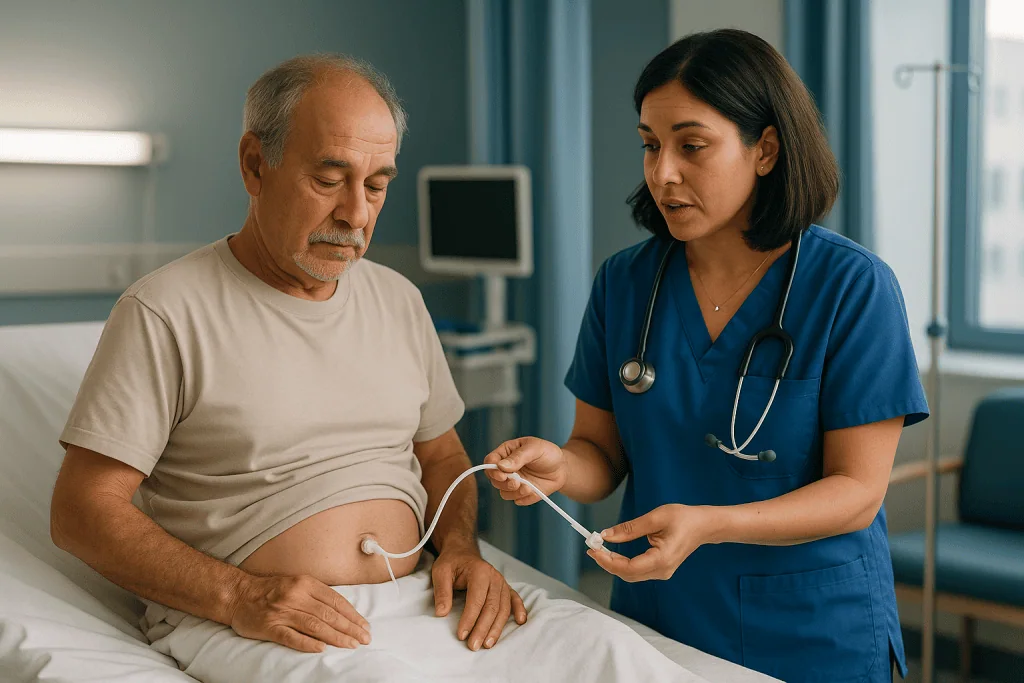
Why inflate the stomach? Because the stomach normally sits loosely behind the abdominal wall, and other organs, including a loop of colon, can drift between them. Filling it with air pushes it forward and flat, so the needle has a clear, short path. The endoscopist also presses a finger on the skin and watches the camera image for a matching indentation inside the stomach. That double check, glow from inside plus dent from outside, is the heart of safe placement.
The entire sequence typically takes 30 to 45 minutes according to MedlinePlus, and much of that time is preparation and sedation rather than the placement itself. The tube is usually in position within a few minutes of the skin being numbed.
What happens before you enter the endoscopy room
Preparation is quieter than the procedure. You will be asked not to eat or drink for a set period beforehand, usually overnight, so the stomach is empty and the camera view is clear (MedlinePlus). The care team reviews your medicines, with particular attention to blood thinners and diabetes medicines; any changes are decided by the prescribing clinician, never by the patient alone, and this article does not suggest stopping or adjusting anything.
Blood tests are commonly checked to confirm that clotting is adequate. Many units give a single dose of an antibiotic through the intravenous line shortly before placement, because the tube passes through the mouth and throat, which carry bacteria, before it comes to rest in a fresh wound. This is a well-established step for reducing infection at the tube site, and it is given by the team as part of the procedure rather than as something you arrange.
Consent is a conversation, not a signature. A good one covers why tube feeding is being proposed, what the alternatives are, what the realistic risks look like for you specifically, and what happens if the tube later turns out not to be wanted. If the person receiving the tube cannot take part in that conversation, for example because of a stroke or advanced illness, the discussion happens with a legally recognized decision-maker, guided by any advance directive.
On the day, a small intravenous cannula is placed in the hand or arm, monitoring stickers go on the chest, a clip measures oxygen on a finger, and a nurse walks through the plan once more. Then the sedation begins.
PEG tube placement sedation: how awake will I be?
Most PEG placements use what clinicians call moderate or conscious sedation. The medicines, typically a short-acting benzodiazepine for relaxation and a short-acting opioid for comfort, are given through the intravenous line in small amounts, adjusted to your response. The aim is a state where you are drowsy, comfortable and able to respond to a touch or a simple instruction, but unlikely to remember much afterward. Decisions about which agents and how much are made by the sedating clinician in the room, based on your age, weight, breathing and other medicines.
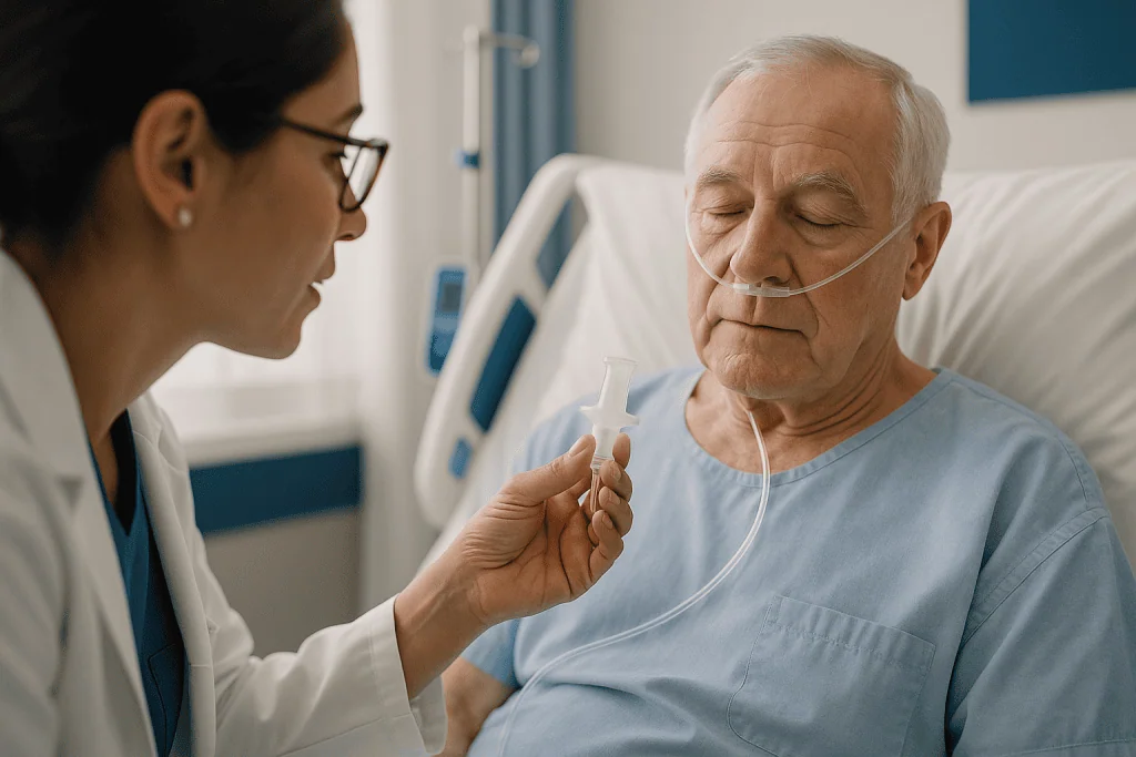
Some units use deeper sedation with an anesthetic drug given by an anesthesia professional, particularly for people with significant breathing or heart conditions, severe anxiety, or difficulty cooperating. General anesthesia with a breathing tube is uncommon for PEG placement and is reserved for specific circumstances.
Throughout the procedure a nurse watches heart rhythm, blood pressure and oxygen saturation continuously, and supplemental oxygen is often given through a small nasal tube. A plastic mouth guard protects your teeth and keeps the airway open for the scope.
The local anesthetic in the abdominal wall is separate from the sedation and is what makes the needle pass and the small skin cut comfortable. People who remember the procedure usually describe pressure and a feeling of fullness from the air in the stomach rather than sharp pain.
Because sedative medicines linger for hours, you will not be allowed to drive, operate machinery or make important decisions until the following day, and an adult must accompany you home if you are not staying overnight. The NIH’s description of upper GI endoscopy covers these same sedation principles.
PEG tube procedure steps: the pull technique, moment by moment
The pull technique is the most widely used method, so it is worth following in sequence.
- Scope in. The endoscope passes over the tongue and down the esophagus into the stomach. The endoscopist inspects the lining and checks that the stomach can be reached and inflated.
- Insufflation. Air or carbon dioxide inflates the stomach until its front wall meets the abdominal wall.
- Transillumination. With room lights dimmed, the scope light shows as a glow on the skin, usually in the left upper abdomen below the rib margin.
- Finger indentation. An assistant presses the glowing spot; the endoscopist sees the dent inside and confirms the match.
- Local anesthetic and incision. The skin is cleaned, numbed and opened by about a centimeter.
- Needle and wire. A needle enters the stomach under direct camera view. A guidewire is threaded through it and grasped by a snare passed down the scope.
- Pull through. Scope, snare and wire are drawn back out through the mouth. The feeding tube is attached to the wire outside the mouth and pulled back down the esophagus, into the stomach and out through the abdominal wall, tapered end first.
- Seat and secure. The soft internal bumper rests against the stomach wall. The external fixation disc slides down to the skin, snug but not tight, so blood flow to the tissue between them is preserved.
- Final check. The scope is often reinserted briefly to confirm the bumper position, then the air is suctioned out.
The tube’s length at skin level is recorded in centimeters so that later checks can detect whether it has migrated inward or outward. That single number becomes one of the most useful items in your discharge paperwork.
Push and introducer techniques: when the tube goes in from the outside
Not every PEG is pulled through the mouth. Two alternatives exist, and the choice usually depends on anatomy rather than preference.
The push technique mirrors the pull method up to the guidewire stage. Instead of attaching the tube to the wire and pulling, the endoscopist threads a stiffer tube over the wire and pushes it down through the mouth, esophagus and stomach until the tapered end emerges through the abdominal wall. The final arrangement is the same, with an internal bumper and external disc.
The introducer technique, sometimes called the Russell technique, avoids the mouth and throat entirely for the tube itself. After the stomach is inflated and the site confirmed, the endoscopist places small sutures or T-fasteners that temporarily anchor the stomach wall to the abdominal wall. A dilator widens the needle track from the outside, and a balloon-type tube is inserted directly through the skin into the stomach. The balloon is filled with water to hold it in place.
Why would a team choose the introducer route? The most common reason is a cancer of the mouth, throat or esophagus. Dragging a tube past a tumor risks carrying tumor cells to the fresh abdominal wound, a rare but documented complication of the pull method, and may be physically difficult if the passage is narrowed. The introducer method is also chosen when the throat is heavily colonized with resistant bacteria, since the tube never touches it.
The trade-off is that the introducer technique relies on a balloon, which can deflate or rupture, and the initial tract is less mature when the anchoring sutures are removed. Your endoscopist will explain which route is planned and why, and that reasoning should be part of your consent discussion.
Types of PEG tubes and related feeding tubes compared
People searching for the different types of PEG tubes usually mean one of three things: how the tube is held in the stomach, how it is placed, or where its tip ends up. The table below separates those ideas.
| Tube or route | How it is held in place | How it is placed | Typical reason it is chosen |
|---|---|---|---|
| Bumper-type PEG | Soft internal disc or dome | Endoscopic, pull or push | Standard first tube; durable, less likely to dislodge |
| Balloon-type gastrostomy | Water-filled balloon | Endoscopic introducer, or as a replacement through a mature tract | Easier to exchange at the bedside; used after the tract has healed |
| Low-profile (button) device | Balloon or bumper, flush with skin | Replacement once tract is mature | Comfort and discretion, common in children and active adults |
| PEG-J (jejunal extension) | PEG bumper plus a thinner inner tube reaching the small intestine | Endoscopic | Severe reflux, delayed stomach emptying, aspiration risk |
| RIG (radiologically inserted gastrostomy) | Usually balloon | X-ray or fluoroscopy guidance, no endoscope | When the scope cannot pass, or endoscopy is unavailable |
| Surgical gastrostomy | Various | Laparoscopic or open surgery | When abdominal anatomy or prior surgery makes other routes unsafe |
A few practical notes. The first tube placed endoscopically is almost always bumper-type because the bumper cannot be removed by accident while the tract is still healing. Balloon tubes and buttons come later, when the opening has become a stable channel that can be re-entered easily. A PEG-J is a PEG with an extra, narrower tube threaded through it into the jejunum, the middle part of the small intestine, for people whose stomachs do not empty well.
Cleveland Clinic and MedlinePlus both describe these options in patient-facing language, and your team’s choice depends on your anatomy, your expected duration of feeding and your ability to manage the device at home.
Where is the tube located, and what does the site look like?
The exit point is usually in the left upper quadrant of the abdomen, a hand’s width or so below the ribs and to the left of the midline. That location is not arbitrary. It sits over the body of the stomach, away from the liver on the right, the ribs above, and the transverse colon, which tends to hang lower. The glow test and finger-indentation test described earlier are what confirm, for each individual, that this expected position is actually clear of other organs.
What you see on the skin after placement is modest: a soft tube about the width of a drinking straw emerging through a small opening, held by a flat plastic disc that rests against the skin, with a clamp and a feeding port on the end. A light dressing may cover it for the first day or two. Some redness and a little clear or slightly blood-tinged drainage are expected in the first week as the wound settles.
Inside, the internal bumper sits against the stomach wall. Between the bumper and the external disc is a sandwich of stomach wall, a thin layer of fat, muscle and skin. Over the following weeks, the body forms scar tissue around the tube, sealing stomach to abdominal wall. This is the “tract” or “stoma” that clinicians refer to, and its maturation is what eventually allows the tube to be swapped without another endoscopy.
The question “where is the AG tube located” that appears in search data almost certainly reflects a misheard or mistyped “G-tube.” If someone describes a tube in the nose, that is a nasogastric tube and a different device altogether. If it is in the abdomen, it is a gastrostomy tube, and this section describes where it should be.
Who is a PEG tube usually for, and who is often asked to wait?
A gastrostomy is proposed when someone has a working digestive system but cannot safely or adequately take food by mouth, and this is expected to last more than a few weeks. MedlinePlus and Cleveland Clinic list the common situations: swallowing impairment after a stroke, neurological conditions such as motor neuron disease or Parkinson’s disease, cancers of the head and neck or esophagus (sometimes placed before treatment begins, when swallowing may become difficult during radiotherapy), severe injuries, and children with conditions that prevent adequate oral intake.
The stomach and intestine must be able to absorb what is delivered. If the gut itself is not working, for example in a complete bowel obstruction, tube feeding is not the answer and nutrition is given intravenously instead.
Some people are asked to wait, or offered a different route, because the endoscopic method would be unsafe at that moment. Reasons include a clotting problem that has not been corrected, infection in the abdominal wall at the planned site, peritonitis (inflammation of the abdominal lining), a large amount of fluid in the abdomen (ascites), or a blockage in the throat or esophagus that the scope cannot pass. A stomach that cannot be brought against the abdominal wall, because of prior surgery, an enlarged liver or an unusual anatomy, may need a radiological or surgical approach instead.
There is also a harder category. For people with advanced dementia, major guideline bodies have concluded that tube feeding has not been shown to prolong life, prevent aspiration pneumonia or improve comfort, and careful hand feeding is generally recommended instead. Raising this is not a judgment about anyone’s worth; it is an honest statement about what the evidence does and does not support, and the decision rests with the person, their representatives and the treating team.
What are the risks of PEG tube placement?
PEG placement is a low-risk procedure in the hands of an experienced team, but “low risk” is not “no risk,” and a candid list is more useful than reassurance.
The most frequent problems are minor and local. Infection around the tube site, showing as spreading redness, warmth, pain or pus, is the commonest complication and the reason pre-procedure antibiotics are standard. Leakage of stomach contents around the tube can irritate the skin. A small amount of bleeding at the site is expected; significant bleeding is uncommon.
Less common but more serious events include injury to the colon or another organ that has slipped between stomach and abdominal wall, which is exactly what the glow and indentation tests are designed to prevent; peritonitis if stomach contents leak into the abdominal cavity, usually because the stomach pulls away from the abdominal wall before the tract has sealed; and aspiration during the procedure, meaning stomach contents entering the lungs. Sedation itself carries risks of slowed breathing and low blood pressure, which is why monitoring is continuous.
Two complications appear later. “Buried bumper syndrome” occurs when the internal bumper is pulled too tightly against the stomach wall and the lining grows over it, blocking the tube; keeping the external disc snug but not tight, and rotating the tube as instructed, reduces this. Accidental removal, particularly of a balloon tube or by a confused patient, can close a fresh tract within hours.
MedlinePlus and Cleveland Clinic both describe these risks in their patient materials. What matters most is that your team explains which of them apply particularly to you, given your medicines, your anatomy and your general health, before you consent.
What the first days after PEG placement usually look like
Recovery begins in the endoscopy unit. You wake gradually over 30 to 60 minutes while monitoring continues. The tube site is checked, and you may notice a dull ache around it, often described as similar to a bruise, which is managed with pain relief chosen by the team.
Feeding does not usually start immediately. Many units begin with water through the tube after a few hours to confirm it passes freely, then start liquid nutrition the same day or the next morning; MedlinePlus notes that the tube is often first used within 24 hours. Feeds are increased in steps toward the goal volume set by a dietitian over a day or two. Some people go home the same day; others stay one or two nights, particularly if the PEG is part of a longer hospital admission for the condition that made it necessary.
In the first week, expect a little drainage and some redness that should fade rather than spread. The dressing is typically removed after a day or two and the site left open to air, cleaned once daily with mild soap and water and dried thoroughly. Showering is generally permitted after the first couple of days; soaking in a bath or pool is usually delayed until the tract has healed.
Before discharge, you or your caregiver should be taught, and should practice, three skills: giving a feed or flush, checking the centimeter marking at skin level, and gently rotating the tube a full turn once a day (or as your team instructs) to prevent the bumper from embedding. Written instructions, a 24-hour contact number and a follow-up appointment are standard. If any of those three is missing when you leave, ask for it.
Weeks two to six: healing, feeding routines and tube changes
Over the following weeks the tract matures. Scar tissue forms a stable channel between stomach and skin, and the tube begins to feel like part of the day rather than an intrusion. Discomfort fades. The skin around the site should look calm and dry; a small ring of pink, moist tissue called granulation tissue sometimes forms and is managed by the care team if it becomes bulky or bleeds easily.
Feeding settles into one of two patterns. Bolus feeding delivers a measured amount of formula over 15 to 30 minutes several times a day using a syringe, mimicking meals (MedlinePlus patient instructions describe this method). Continuous or overnight feeding uses a small pump to deliver formula slowly over many hours, often chosen when boluses cause bloating or when a PEG-J is in place. The dietitian chooses the formula and schedule; if medicines are given through the tube, the pharmacist advises which can be crushed or dissolved and which cannot, and the tube is flushed with water before and after each one to prevent blockage.
The original bumper tube is left in place for several weeks at minimum, and often for many months, until the tract is fully established. After that, if a change is wanted, for comfort, for a low-profile button, or because the tube has worn, it can usually be exchanged. A balloon tube is deflated and withdrawn and a new one inserted through the tract, often at the bedside or in clinic without sedation. A bumper tube is removed either by firm traction or endoscopically, depending on its design.
If the underlying condition improves and swallowing becomes safe again, the tube can simply be removed. The opening typically closes on its own within a day or two with a dressing over it. Nothing about a PEG commits you to keeping it.
What people often get wrong about PEG tubes
“It means I can never eat again.” Not necessarily. Many people with a gastrostomy continue to eat and drink for pleasure in amounts their speech therapist judges safe, using the tube to top up calories, fluids and medicines. The tube removes the pressure to meet every nutritional need by mouth; it does not forbid the mouth.
“It is major surgery.” The endoscopic technique replaced the open operation specifically to avoid that. There is a centimeter-sized skin opening, sedation rather than general anesthesia in most cases, and the procedure typically runs 30 to 45 minutes.
“It is permanent.” A PEG is removable. If swallowing recovers, the tube comes out and the opening closes on its own.
“A feeding tube will stop aspiration pneumonia.” It does not. Pneumonia from aspiration often comes from saliva and reflux rather than food, and a tube does not change either. People with tubes still need oral care and positioning advice.
“It always prolongs life.” For many conditions, adequate nutrition clearly supports treatment and recovery. In advanced dementia, however, the evidence reviewed by major guideline bodies has not shown that tube feeding extends life or improves comfort. The honest answer depends on why the tube is being proposed.
“I will be unconscious.” Most people are sedated, not anesthetized. They are drowsy and comfortable and usually remember little, but they are breathing on their own.
“A little redness means infection.” Mild redness and slight drainage in the first week are part of healing. Spreading redness, pus, fever or increasing pain are the signs that warrant a call.
Correcting these ideas does not make the decision easy. It does make it a decision about the real procedure rather than an imagined one.
Alternatives to PEG: when another route makes more sense
A PEG is one of several ways to provide nutrition when eating by mouth is not enough, and a good team will have considered the others before proposing it.
A nasogastric tube, passed through the nose into the stomach, is the usual choice when feeding is expected to be needed for a short time, typically days to a few weeks. It requires no procedure beyond bedside placement and an X-ray or pH check of position, but it is uncomfortable for many people, visible, prone to dislodging and associated with nasal and throat irritation if left for long. A nasojejunal tube is a longer version ending in the small intestine.
A radiologically inserted gastrostomy, or RIG, achieves the same end result as a PEG using X-ray guidance rather than an endoscope. It is the route of choice when the scope cannot pass a narrowing in the throat or esophagus, and in some units it is simply the local standard. The tube is usually balloon-type and anchored with temporary fasteners.
A surgical gastrostomy, placed laparoscopically or through a small open incision, is reserved for situations where the stomach cannot be safely reached from inside or outside without direct vision, for example after extensive abdominal surgery. It carries the risks of an operation and general anesthesia.
Parenteral nutrition delivers nutrients directly into a vein, bypassing the gut. It is used when the intestine itself cannot absorb food, and it carries its own infection and metabolic risks, so it is not a substitute for a working gut.
Finally, careful hand feeding with texture-modified food and thickened fluids, supported by a speech therapist, is itself an alternative, and in some situations the one most consistent with a person’s goals. Which route fits is a clinical and personal judgment that belongs with you and your treating team.
Questions to ask your care team before PEG placement
Consent conversations go better with a list in hand. These are the questions that tend to matter most, drawn from what patients and families report wishing they had asked.
- Why is tube feeding being recommended for me specifically, and what would happen if we did not place a tube?
- Is this expected to be short-term, long-term or uncertain? What would trigger a review of whether the tube is still wanted?
- Which technique is planned, pull, push, introducer or radiological, and why that one for my anatomy?
- What kind of sedation will I have, and who will be giving and monitoring it?
- Which of the risks apply particularly to me, given my medicines and health conditions?
- Will any of my current medicines need to be changed before the procedure, and who will make and communicate that decision?
- Will I be able to eat or drink anything by mouth afterward, and who will reassess my swallowing?
- When will feeding start, how will the formula and schedule be chosen, and who adjusts them?
- What will I or my caregiver be taught before discharge, and will there be written instructions and a 24-hour contact number?
- How do I recognize a problem at the tube site, and what should I do if the tube comes out?
- When and how will the tube be replaced, and could I eventually have a low-profile device?
- If my condition improves, how is the tube removed and what does the site look like afterward?
Write down the answers, or ask whether someone can join you to take notes. The person giving consent should leave the conversation able to describe, in their own words, what will happen in the room and why.
When to call your doctor after PEG tube placement
Most people recover without incident, but a gastrostomy is a direct connection between the outside world and the stomach, so certain signs should never wait for a routine appointment.
Seek urgent medical attention, by emergency services if severe, for: sudden or worsening abdominal pain, especially with a rigid or very tender abdomen, fever and vomiting, which can indicate leakage into the abdominal cavity (peritonitis); vomiting blood or passing black, tarry stools; bleeding from the tube site that soaks a dressing or does not stop with gentle pressure; difficulty breathing, choking or a new cough with fever after a feed, which may suggest aspiration; or the tube coming completely out within the first few weeks, when the tract can close within hours. In that last case, do not try to reinsert it yourself; cover the site and contact the team immediately.
Call your care team the same day for: redness spreading beyond a thumb’s width from the site, pus, a bad smell or increasing pain at the site; persistent leakage of stomach contents around the tube; a tube that will not flush despite the technique you were taught; a change in the centimeter marking at skin level, suggesting the tube has moved; diarrhea, bloating or vomiting after feeds that does not settle; or signs of dehydration such as very dark urine, dizziness or confusion.
Routine contact is appropriate for questions about skin care, a small amount of granulation tissue, or adjusting the feeding schedule. MedlinePlus and Cleveland Clinic both list these warning signs in their patient guidance. When in doubt, call: a brief conversation is far cheaper, in every sense, than a complication that was noticed too late. Every decision about treatment, including whether and when to remove the tube, remains with you and your treating team.
Frequently asked questions
What is the PEG procedure in simple terms?
It is a way of placing a feeding tube into the stomach through the abdominal wall using a camera for guidance rather than surgery. Under sedation, an endoscope is passed through the mouth, the stomach is inflated, a light marks the exit point on the skin, a needle and wire create a path, and the tube is drawn into place and held by an internal bumper. MedlinePlus puts the typical duration at 30 to 45 minutes.
What is a gastrostomy and how is it different from a PEG?
A gastrostomy is the opening itself, a channel between the stomach and the skin through which a feeding tube sits. PEG describes one method of creating that opening, using an endoscope. Other methods include radiological placement with X-ray guidance and surgical placement. All three produce a gastrostomy; they differ in how the stomach is reached and what anesthesia and equipment are needed.
What are the different types of PEG tubes?
Tubes differ mainly in how they are held inside the stomach. Bumper-type tubes have a soft internal disc and are the usual first tube. Balloon-type tubes are held by a water-filled balloon and are easier to exchange once the tract has healed. Low-profile buttons sit flush with the skin. A PEG-J adds a thinner inner tube reaching the small intestine for people with severe reflux or slow stomach emptying.
Where is the G tube located on the body?
Usually in the left upper abdomen, a hand’s width or so below the rib margin and to the left of the midline, directly over the body of the stomach. That position keeps it clear of the liver, ribs and colon. The exact spot is confirmed during the procedure by the glow of the scope light through the skin and a matching finger indentation seen from inside the stomach.
What kind of sedation is used for PEG tube placement?
Most placements use moderate sedation given through an intravenous line, typically a short-acting sedative and a short-acting pain reliever, adjusted by the clinician to keep you drowsy and comfortable while still breathing on your own. Local anesthetic numbs the abdominal wall separately. Deeper sedation with an anesthesia professional is used for some people, and general anesthesia is uncommon. You cannot drive for the rest of the day afterward.
What are the PEG tube procedure steps in order?
Scope into the stomach; inflate the stomach with air; dim the lights and find the glow on the skin; press the spot and see the dent inside; numb the skin and make a centimeter cut; pass a needle, then a guidewire, which is snared and pulled out through the mouth; attach the tube and pull it down and out through the abdominal wall; seat the internal bumper and external disc; confirm position and remove the air.
Does a PEG tube hurt?
During placement, sedation and local anesthetic mean most people feel pressure and fullness from the air in the stomach rather than sharp pain. Afterward, soreness around the site, often compared to a bruise, is common for a few days and is managed with pain relief chosen by the care team. Pain that worsens rather than eases, or is accompanied by fever or a rigid abdomen, is a warning sign and needs urgent assessment.
How soon can feeding start after a PEG is placed?
Typically within 24 hours, and often the same day, according to MedlinePlus. Many units first flush the tube with water after a few hours to confirm it is clear, then begin liquid nutrition in gradually increasing amounts over a day or two. The dietitian sets the formula and schedule, and medicines given through the tube are chosen with pharmacist advice about which can safely be crushed or dissolved.
Can I still eat by mouth with a PEG tube?
Often yes, depending on why the tube was placed. Many people continue to take small amounts of food or drink for pleasure in textures their speech therapist has judged safe, using the tube to meet the rest of their needs. For others, swallowing is unsafe at any amount. This is reassessed over time, and if swallowing recovers fully the tube can be removed and the opening closes on its own.
What is the difference between a PEG and a RIG?
Both create a gastrostomy, but a PEG is guided by an endoscope passed through the mouth while a RIG, or radiologically inserted gastrostomy, is guided by X-ray imaging without a scope. A RIG is often chosen when a narrowing in the throat or esophagus prevents the endoscope from passing, or when the mouth route should be avoided. RIG tubes are usually balloon-type and anchored with temporary fasteners.
References
- MedlinePlus: Feeding tube insertion – gastrostomy
- MedlinePlus: Gastrostomy feeding tube – bolus
- Cleveland Clinic: Percutaneous Endoscopic Gastrostomy (PEG)
- NIH NIDDK: Upper GI Endoscopy
This article is for general information only and is not a substitute for professional medical advice. Please consult a qualified doctor about your individual situation.

More from the Blog

Autoimmune Hepatitis Diagnosis: Antibody Tests, Imaging and When a Liver Biopsy Is Needed
Autoimmune hepatitis is diagnosed by combining blood tests (raised liver enzymes and immunoglobulin G), autoantibody tests such as ANA, anti-smooth muscle and anti-LKM1, and…

Living Gluten-Free With Celiac Disease: Label Reading, Eating Out and Travel Without Slips
A gluten free diet with celiac disease means removing all wheat, barley and rye, every day, for life, because even small amounts can damage…
Preparing for Gallstone Surgery: Blood Tests, Fasting and Which Medicines Are Paused
Preparing for gallstone surgery usually means a pre-assessment visit with blood tests (blood count, kidney and liver function, sometimes clotting), a fasting window of…

Why Variceal Banding Is Often Repeated: How Many Sessions and What Follow-Up Endoscopy Checks
Variceal banding is usually repeated every 2 to 4 weeks until the varices are flattened or gone, a point endoscopists call eradication. Liver guidance…

IBS Myths: Is It All in Your Head and Does Cutting Out Gluten Always Help?
IBS is not all in your head. It is a recognized disorder of gut-brain interaction in which the bowel and nervous system miscommunicate, producing…

Living With Chronic Liver Disease: Weight, Alcohol, Sleep and the Check-Ups That Matter
Living with chronic liver disease usually means protecting the working tissue you still have: avoiding alcohol if your team advises it, aiming for gradual…