

How Cryoablation (CRYO) Works: Image-Guided Probes, the Freeze Cycles and What You Feel
Key Takeaways
- Cells reliably die when tissue reaches roughly minus 40 degrees Celsius, so operators aim for that temperature at the tumor edge, not just at the probe tip.
- The visible edge of an ice ball is near zero degrees while the lethal zone lies several millimeters inside it, which is why doctors freeze beyond the tumor margin.
- Two freeze-thaw cycles destroy more tissue than one because refreezing partly thawed cells forms larger, more damaging intracellular ice crystals.
- Unlike heat ablation, freezing does not seal blood vessels, so bleeding is the complication cryoablation teams watch for most closely.
- Cold numbs nerves as it chills them, which is why the freeze itself is usually felt as pressure rather than pain, with soreness arriving afterward as tissue thaws.
- Because nothing is removed, success is judged by follow-up contrast imaging over months, and a remnant found at the edge can often be re-treated.
Cryoablation destroys a targeted patch of tissue by freezing it. A doctor guides one or more thin probes into the target using ultrasound, CT or MRI, then pumps a pressurized gas through the probe tip to form an ice ball. Two or more freeze and thaw cycles rupture cell membranes and cut off local blood supply. Most people feel pressure or cold, not sharp pain, because the tissue is numbed or sedation is used.
The radiologist turns the screen so you can see it: a gray smudge the size of a grape on your kidney, and beside it a thin white line where a needle might go. Then a phrase you did not expect to hear in a hospital: “We would freeze it.” You nod, because nodding is easier than asking the question forming in your chest, which is, roughly, how on earth does freezing something inside me work, and what will I feel while it happens?
That question deserves a real answer, not a brochure. If you want to understand the cryoablation procedure and how it works, you need three things: a sense of why extreme cold kills cells, a picture of how a probe is steered to the right spot without a scalpel, and an honest account of the freeze cycles, the recovery, and the parts medicine still cannot promise.
This explainer covers cryoablation for tumors, the balloon version used in the heart, and its skin-surface cousin, all through the same lens: what the evidence shows, what it does not, and which decisions stay with your treating team.
Cryoablation procedure: how it works in plain language
Ablation is the medical word for deliberately destroying a small area of tissue while leaving the surrounding structures intact. Cryoablation (the prefix “cryo” comes from the Greek for frost) does that job with cold rather than heat, radiation or a surgeon’s knife.
The basic sequence is simple to describe. A doctor, usually an interventional radiologist (a specialist who treats conditions through tiny skin punctures using imaging as a map), numbs the skin and slides a slender, hollow needle called a cryoprobe toward the target. Live imaging confirms the tip is sitting where it should. Pressurized gas then flows through the probe, and the metal tip drops to temperatures far below anything in a household freezer. An ice ball forms around the tip and grows outward, swallowing the target lesion with a small safety margin. After a set number of minutes the tissue is allowed to thaw, and the freeze is repeated. The probe is withdrawn and a small dressing goes over a puncture that is often no wider than a drinking straw.
What makes cryoablation distinctive among image-guided treatments is that the ice ball itself shows up on the scan. Mayo Clinic and the National Cancer Institute both describe doctors watching the ice form in real time on ultrasound, CT or MRI, which lets them judge whether the frozen zone has reached the edges of the target and whether it is creeping toward anything it should not touch.
Nothing is cut out. The frozen tissue stays in place and is gradually cleared by the body’s own repair cells over weeks to months, which is why follow-up scans, not the day of treatment, tell the story of whether the ablation did its job.
Why does extreme cold kill cells? The biology of the ice ball
Cold damages tissue through two separate routes, and understanding both explains why the procedure uses repeated cycles rather than one long freeze.

The first route is direct. According to an NIH-indexed review of cryoablation mechanisms, as temperature falls past roughly minus 20 degrees Celsius, ice crystals form in the fluid between cells. That outside ice draws water out of the cells, shrinking them and concentrating their internal salts to toxic levels. Push the temperature lower, toward minus 40 degrees, and ice begins forming inside the cells themselves. Intracellular ice crystals physically shear the membranes and the organelles. The same review describes this as the reliably lethal threshold for most tumor types, which is why operators aim for the lethal temperature at the tumor edge, not merely at the probe tip.
The second route is indirect and takes longer. Freezing injures the lining of the tiny blood vessels inside the ice ball. When the tissue thaws, those vessels leak, clot and collapse. The target is starved of oxygen over the following hours and days, a process called ischemic necrosis. In plain terms, the plumbing fails and the neighborhood dies.
There is a nuance that matters clinically. The visible edge of the ice ball sits at about zero degrees. The reliably lethal zone lies several millimeters inside that edge. The NIH review notes that operators therefore extend the ice ball beyond the tumor margin so the lethal zone, not just the frosty edge, covers the disease.
Researchers have also observed that freezing can leave tumor proteins intact for the immune system to inspect, raising interest in immune effects. The evidence here remains early and is not a reason to choose or expect any particular outcome.
Image-guided probes: how the needle finds the target without surgery
The accuracy of cryoablation lives or dies on imaging. Three technologies do the steering, and each has a personality.
Ultrasound is fast, cheap and radiation-free, and it shows the moving needle in real time. Its weakness is that ice reflects sound strongly, so the near edge of the ice ball appears as a bright curve with a dark shadow behind it. The doctor can see where the ice begins but not what lies beyond it, which is one reason ultrasound is often paired with another modality for deeper targets.
CT (computed tomography, a rotating X-ray that builds cross-sectional images) is the workhorse for kidney, lung and bone ablations. Frozen tissue appears darker than its surroundings, so the whole ice ball can be measured in three dimensions. The trade-off is X-ray exposure and a slight lag, because the doctor advances the needle, steps back, scans, and adjusts.
MRI shows the ice ball as a crisp dark void and gives superb soft-tissue detail, which suits targets in the prostate, liver and breast. It demands MRI-compatible equipment and longer scanning time.
Whatever the modality, the steps are the same. A planning scan maps the target, the safest path and any at-risk neighbors such as bowel, nerves or the ureter. Where a vital structure sits too close, doctors can inject fluid or carbon dioxide to push it away, a technique called hydrodissection. One or several probes are then placed; larger or irregular targets often need multiple probes arranged so their individual ice balls merge into one shape. Cleveland Clinic describes doctors confirming probe position with imaging before the first freeze begins, then re-imaging during the freeze to track the ice.
The entire approach is percutaneous, meaning through the skin. For a few locations the same probes are placed during laparoscopic or open surgery, but the freezing physics does not change.
What actually happens during the freeze cycles
The engine behind the cold is a piece of physics called the Joule-Thomson effect: when a compressed gas is allowed to expand suddenly through a tiny opening, it cools. Modern cryoprobes send high-pressure argon down the shaft to a chamber in the tip, where it expands and chills the metal to the lethal range within seconds. The gas never leaves the probe; it circulates back up and vents away from the patient. To thaw, many systems switch to helium, which warms on expansion, or simply stop the argon and let body heat do the work.
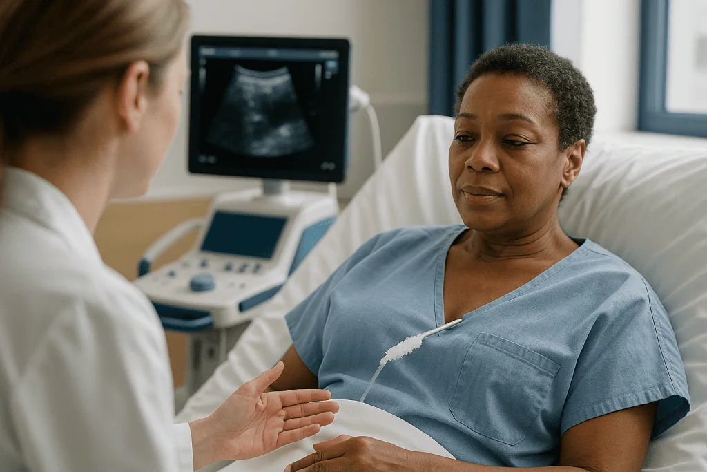
A typical cycle, as described in the NIH-indexed review, runs roughly like this:
- First freeze, often around ten minutes, until the ice ball covers the target with its margin.
- Passive or active thaw, long enough for the ice to soften and refreeze later with larger crystals.
- Second freeze of similar length, which the review notes causes more complete cell destruction than a single freeze.
- Final thaw and probe removal, sometimes with the tract warmed or sealed to reduce bleeding.
Why repeat? The first freeze pulls water out of cells and disrupts circulation. When the tissue thaws and then refreezes, water re-enters the already damaged cells and forms larger intracellular crystals, and the compromised blood vessels fail more completely. Two cycles are the common standard; some protocols use three for stubborn or large targets. The number is a clinical judgment, not a fixed rule.
During the freeze the doctor is not idle. Each imaging check confirms the ice is growing where expected and stopping before it reaches anything precious. If a nerve or a loop of bowel sits close, the team may pause, add protective fluid, or accept a slightly smaller margin. Those trade-offs are decided in the room, by the people who can see the scan.
Cryoablation vs radiofrequency ablation and surgery: a side-by-side view
Patients often meet cryoablation as one option among several, so a neutral comparison helps. Radiofrequency ablation (RFA) uses an alternating electrical current to heat tissue; microwave ablation uses electromagnetic waves to do the same, faster. Surgery removes the target outright. None of these is universally better; each has situations where guidelines favor it.
| Feature | Cryoablation | Heat ablation (RFA or microwave) | Surgical removal |
|---|---|---|---|
| How tissue is destroyed | Ice crystals and vessel collapse | Protein coagulation from heat | Physical excision |
| Visibility during treatment | Ice ball visible on CT, MRI, ultrasound | Treated zone harder to see live | Direct view |
| Anesthesia commonly used | Local plus sedation, or general | Sedation or general; heat is more painful | General |
| Tissue available for pathology | Biopsy only | Biopsy only | Whole specimen |
| Typical in-hospital time | Same day or one night (Mayo Clinic) | Same day or one night | Several days |
| Notable limits | Bleeding risk, large targets, cold injury to neighbors | Heat loss near large vessels, more procedural pain | Longer recovery, higher upfront risk |
The NIH review highlights the visibility point as cryoablation’s signature advantage: a doctor can see exactly how far the destruction extends while it is happening. The National Cancer Institute, in turn, lists the chief disadvantage: because nothing is removed, whether every cell was treated is uncertain until follow-up imaging. Heat-based methods carry their own reverse problem; large blood vessels nearby carry heat away like a radiator and can leave an undertreated rim, a phenomenon called heat sink. Cold suffers a smaller version of the same effect.
Which column applies to you depends on the organ, the size and location of the target, your overall health, and what the team is trying to achieve. That weighing is theirs to do with you.
Who is cryoablation usually for, and who is asked to wait
Cryoablation is typically considered when a target is small, well defined and reachable by a needle, and when the person either cannot have or prefers not to have a larger operation. Mayo Clinic and the National Cancer Institute list the common uses: small kidney tumors, certain liver tumors, bone and soft-tissue tumors causing pain, selected lung lesions, prostate cancer in specific circumstances, and some precancerous or benign growths. It is also used to numb or destroy nerves that transmit chronic pain, a smaller niche with growing study.
People who tend to be good candidates share a few features. The lesion is modest in size; the NIH review describes ice balls being planned to exceed the tumor by a margin, which becomes impractical for very large masses. There is a safe needle path. The person has reasons that make surgery less attractive: age, heart or lung disease, a single remaining kidney, prior operations, or a wish to preserve as much organ as possible.
Being asked to wait, or to choose a different route, is also common and should not be read as rejection. Typical reasons include:
- Blood that does not clot normally, or anticoagulant medicines that cannot safely be paused for the procedure. Decisions about pausing any blood thinner belong solely to the prescribing team.
- A target pressed against bowel, major nerves or the central collecting system of the kidney, where cold would damage neighbors.
- Disease that has spread beyond what a local treatment can address, where systemic therapy or surgery is more appropriate.
- Active infection or recent illness that makes sedation unwise for now.
A frank multidisciplinary discussion, often involving a radiologist, a surgeon and an oncologist or urologist, is the usual route to the decision. Ask to understand their reasoning; a good team will explain why cold was chosen, or why it was not.
Cryoablation for the heart: a different kind of freeze
If your doctor mentioned cryoablation for an irregular heartbeat, you are hearing about a cousin of the tumor procedure that shares the physics but little else about the experience.
Atrial fibrillation, a chaotic rhythm in the heart’s upper chambers, is often triggered by rogue electrical signals from the four pulmonary veins where they join the left atrium. Cardiac cryoablation aims to electrically isolate those veins. A cardiac electrophysiologist (a heart specialist in rhythm disorders) threads a catheter from a vein in the groin up to the heart, then inflates a small balloon at the mouth of each pulmonary vein. Coolant inside the balloon freezes a ring of tissue, creating scar that blocks the stray signals. MedlinePlus describes cardiac ablation procedures lasting roughly three to six hours, most of it spent on mapping and positioning rather than freezing.
What you feel also differs. The procedure is usually done under general anesthesia or deep sedation. Afterward, MedlinePlus notes that people lie flat for several hours so the groin puncture can seal, and that fatigue and a sore groin are common for a day or two. Palpitations in the first weeks are expected while the heart tissue heals and do not automatically mean the procedure failed.
Cryoballoon ablation is listed alongside radiofrequency ablation in American Heart Association patient materials as an established rhythm-control option. Specific risks include injury to the phrenic nerve, which runs near the right pulmonary veins and controls the diaphragm; temporary weakness of this nerve is a recognized complication, and operators monitor it during the freeze. Rare but serious risks include damage to the esophagus, stroke and fluid around the heart.
Whether rhythm control, rate control, medicines or ablation is right for a given person is an individual decision guided by symptoms, heart structure and other conditions, and it rests with the cardiology team.
Cryotherapy on the skin and face: the liquid nitrogen cousin
Readers who arrived here from a dermatology appointment may know “cryo” as the quick spray of liquid nitrogen used on a facial spot. The National Cancer Institute groups this under cryosurgery, and while the biology is identical, the method is different enough to deserve its own paragraph.
On the skin, no probe enters the body. Liquid nitrogen, which boils at about minus 196 degrees Celsius, is sprayed or dabbed onto the lesion for seconds rather than minutes. It is commonly used for actinic keratoses (rough, sun-induced patches that can progress to skin cancer), seborrheic keratoses, warts and some small, superficial skin cancers. MedlinePlus describes it as a routine office procedure usually done without injected anesthetic.
What you feel is a sharp sting followed by a burning or throbbing sensation as the tissue thaws, typically settling within minutes. Over the next day a blister often forms, sometimes blood-tinged, and a crust follows that falls away within roughly one to three weeks depending on the site, according to MedlinePlus. Facial skin tends to heal fastest.
Because this is the Facial Aesthetics section, two cosmetic realities deserve plain statement. First, cryotherapy can leave the treated spot lighter than the surrounding skin, a change called hypopigmentation, because pigment cells are more cold-sensitive than other skin cells. The risk is higher in people with deeper skin tones, and the change can be permanent. Second, a faint scar or textural change is possible, especially with longer freezes. For lesions in cosmetically sensitive areas, a dermatologist may discuss alternatives such as topical treatments, curettage or excision, each with its own balance of effectiveness and appearance. The choice is individual, and the right person to weigh it is the clinician who can see the lesion.
Is cryoablation painful? What you actually feel, step by step
The honest answer is that most people describe pressure, cold and dull aching rather than sharp pain, and that the sensations differ by phase.
Before the freeze, you will feel the sting of local anesthetic in the skin, a sensation most people compare to a dental injection. If sedation is used, which Mayo Clinic notes is typical for percutaneous ablation, you may remember little after this point. General anesthesia is chosen for some locations, for children, or when probes must pass near sensitive structures.
During the freeze, cold has a built-in anesthetic property: nerves stop conducting as they chill, which is part of why cryoablation is generally considered more comfortable than heat-based ablation. People who are awake often report deep pressure or an odd internal coldness. Bone targets and areas near the diaphragm or chest wall are the exceptions, where a dull ache or referred shoulder pain can occur as the ice approaches the lining of the organ.
Afterward, the picture shifts. As the tissue thaws and inflammation sets in, aching at the site is common for a few days. Cleveland Clinic describes soreness, bruising and tiredness as the usual aftermath. Kidney ablations can cause flank discomfort and a little blood in the urine; liver ablations can cause right-sided or shoulder ache; lung ablations may cause a cough or chest-wall soreness.
Pain control afterward is individualized. Teams generally use over-the-counter analgesics or short courses of prescription pain relief, and the type, amount and duration are decisions for the prescribing clinician. Tell your team candidly how you are feeling; unexpected or escalating pain is information, not a complaint.
Cryoablation recovery time: what the following days and weeks usually look like
Recovery is often shorter than people fear, but it is not instant, and the first follow-up scan matters more than the first week.
On the day itself, Mayo Clinic describes a few hours of observation after the procedure. Nurses watch the puncture site, your blood pressure and, for kidney or liver work, signs of bleeding. Many people go home the same day; others stay one night, particularly after lung or liver ablation or when sedation ran deep. Someone else should drive you home.
The first week brings the inflammatory phase. Soreness peaks around day one to three and then eases. Low-grade fever, fatigue and general achiness can occur as the body begins clearing the dead tissue, a pattern sometimes called post-ablation syndrome. Bruising around the puncture is normal. Cleveland Clinic suggests most people resume light daily activities within a few days, with heavier lifting or strenuous exercise held back for roughly one to two weeks, though your team will set limits for your situation.
Weeks two through eight are quiet on the outside and busy on the inside. Frozen tissue softens, shrinks and is replaced by scar. You will likely feel normal long before the ablation zone finishes changing.
Follow-up imaging is the real checkpoint. Teams commonly schedule a contrast CT or MRI within the first several months to confirm the treated zone shows no blood flow, then periodic scans thereafter. The National Cancer Institute notes that because treated tissue is not removed for pathology, imaging is how success is judged. A spot that still takes up contrast may be inflammation or may be residual disease; the radiologist’s reading, sometimes with a repeat scan, decides which.
Timelines above are typical ranges from the cited sources, not guarantees. Your own course will be shaped by the organ treated, the size of the ice ball and your overall health.
What are the disadvantages and side effects of cryoablation?
Every effective treatment carries risk, and the cryoablation side effects worth knowing fall into three groups: general, organ-specific and the one that has nothing to do with harm.
General risks come with any needle procedure. Bleeding is the most important for cryoablation specifically, because ice damages blood vessels inside the ablation zone and, unlike heat, does not cauterize the needle track as it leaves. Infection at the site or in the dead tissue, reactions to sedation and the post-ablation flu-like syndrome round out the list, all described by Mayo Clinic and Cleveland Clinic as recognized complications.
Organ-specific risks follow the anatomy:
- Kidney: blood in the urine, urine leak if the collecting system is frozen, injury to the ureter.
- Liver: bile leak, injury to the gallbladder or bowel, bleeding into the abdomen.
- Lung: pneumothorax (air leaking into the space around the lung, sometimes needing a chest tube), coughing up blood, pneumonia.
- Bone: fracture of a weakened bone, injury to adjacent nerves with numbness or weakness.
- Prostate: urinary symptoms, erectile dysfunction, rarely a connection forming between the urethra and rectum.
- Skin: blistering, pigment loss, scarring.
The third disadvantage is not a complication but an uncertainty. Because the target is destroyed in place, there is no specimen to confirm the edges are clear. The National Cancer Institute frames this plainly: imaging follow-up is required, and some people will need a repeat ablation if a remnant is found. For some tumors, high-quality long-term comparative data against surgery are still limited, which is one reason guidelines reserve cryoablation for defined situations rather than offering it universally.
Risks must be weighed against the risks of doing nothing or of alternatives, and that balance differs for every person. It is a conversation, not a formula.
How many years does cryoablation last, and what counts as success?
Two of the most searched questions about this procedure rest on a misunderstanding worth untangling gently: cryoablation is not a device with a service life, and “success rate” is not a single number.
Start with durability. When an ice ball covers the entire target with an adequate margin, the cells inside are dead and do not come back. There is no wearing off. What can happen is one of three things: a sliver at the edge survived the freeze and regrows (residual disease); a new lesion appears elsewhere in the same organ (new disease); or, for a cancer, cells that had already left before treatment show up in another site. Only the first is really about the ablation itself. Follow-up imaging is designed to catch all three, and a small residual area can often be re-treated with a second session. So “how many years does it last” is better rephrased as “how will we know it worked, and what is the plan if it did not fully?”
Now success. Published outcomes vary enormously by organ, tumor size, how success is defined (complete ablation on the first scan, absence of regrowth over years, survival) and how long patients were followed. A number quoted for a small kidney tumor tells you nothing about a liver metastasis or a lung nodule. The National Cancer Institute and Mayo Clinic both describe cryoablation as an established local treatment option for selected small tumors while noting that long-term comparisons with surgery remain an area of ongoing research for some uses.
Rather than ask your team for a percentage, ask what the evidence shows for your tumor type and size, how they define success, and how they will monitor. Those answers are more useful than any single figure, and they are the ones a good team can actually stand behind.
What people often get wrong about cryoablation
Myths gather around any treatment with a dramatic name. Here are the ones that cause the most confusion, corrected against the evidence.
“They freeze the tumor and it falls out.” Nothing is removed. The frozen tissue stays in place and is absorbed and scarred over by the body across weeks to months, as described by the National Cancer Institute. The lump may still be visible on scans for a long time while shrinking.
“Cold is gentler than heat, so it is risk-free.” Cold is often less painful during treatment because it numbs nerves, but the NIH review and clinical sources are clear that bleeding risk is higher with cryoablation than with heat ablation, precisely because ice does not seal vessels. Gentler is not the same as harmless.
“If the scan at three months shows something, it failed.” Inflammation around an ablation zone can mimic residual tumor on early imaging. Radiologists read patterns over time, and a stable or shrinking area without blood flow is reassuring even if it has not vanished.
“Cryoablation is only for people who cannot have surgery.” That is one common pathway, but guidelines also list it for small kidney tumors and certain other targets as a reasonable option in its own right, chosen for organ preservation or recovery speed.
“It is experimental.” Percutaneous cryoablation has been in clinical use for decades and is listed by Mayo Clinic, Cleveland Clinic and the National Cancer Institute as an established treatment for selected indications. Some newer uses, such as certain breast or pain applications, are still being studied, and a good team will tell you which category yours falls into.
“Freezing a wart is the same procedure.” Same physics, different world. Skin cryotherapy is seconds of surface spray; tumor cryoablation is minutes of imaging-guided internal freezing under sedation.
Questions to ask your care team before cryoablation
Consultations move quickly, and the best questions are the ones that surface the reasoning behind the plan. Bring a notebook or a companion, and consider asking some of these.
- Why is cryoablation being recommended for me rather than surgery, heat ablation, radiation, active surveillance or medicines, and what does the evidence show for my specific target size and location?
- Which imaging will you use to guide and monitor the ice ball, and how will you protect the structures next to the target?
- How many probes and how many freeze cycles do you expect, and what would make you change that plan during the procedure?
- Will I have local anesthetic with sedation or general anesthesia, and why?
- Which of my current medicines, including any blood thinners or supplements, need to be reviewed before the procedure, and who will make those decisions with me?
- What are the specific risks for this organ, and what is your plan if bleeding or another complication occurs?
- Should I expect to go home the same day, and what should I arrange at home for the first few days?
- What symptoms afterward are expected, which ones mean I should call, and whom exactly do I call, including after hours?
- When is the first follow-up scan, how will you decide whether the ablation was complete, and what happens if a remnant is seen?
- Will a biopsy be taken at the same time so we know the pathology?
- If I have a cancer, how does this fit with any other treatment I am receiving?
You are not obliged to ask everything in one sitting. A second conversation, or a written summary from the team, is a reasonable request. Clear answers to these questions are a good sign; vague ones are a prompt to ask again, not a reason to proceed on faith.
When to call your doctor after cryoablation: red-flag signs
Mild soreness, bruising, fatigue and a low-grade temperature in the first few days are expected. The following are not, and each deserves a same-day call to your care team or, where marked, emergency care.
Seek emergency care immediately for:
- Sudden or worsening shortness of breath, chest pain, or coughing up more than streaks of blood, which after lung ablation may signal a collapsed lung and after any procedure could indicate a clot.
- Dizziness, fainting, a racing heartbeat, or cold, clammy skin, which can indicate internal bleeding.
- Heavy bleeding or rapidly expanding swelling at the puncture site that does not stop with firm pressure.
- New weakness, numbness, trouble speaking, or facial drooping, especially after cardiac cryoablation.
- Severe abdominal pain with a rigid belly, or vomiting that will not stop.
Call your team the same day for:
- Fever above the threshold your team gave you, or shaking chills, particularly after the first two days.
- Pain that is increasing rather than easing, or not controlled by the plan you were given.
- Redness, warmth, pus or a foul smell at the puncture site.
- Blood in the urine that is heavy, contains clots, or persists beyond what you were told to expect after a kidney ablation.
- Yellowing of the skin or eyes, dark urine, or pale stools after a liver ablation.
- Inability to pass urine, or new burning and urgency after prostate ablation.
- After skin cryotherapy, a blister that becomes increasingly painful, spreads, or drains cloudy fluid.
Keep the phone number for your procedure team somewhere visible, and do not wait for a scheduled appointment if something feels wrong. Cleveland Clinic and Mayo Clinic both emphasize that most complications are manageable when reported early. Every decision about further tests or treatment belongs to the clinicians who know your case.
Frequently asked questions
How painful is cryoablation?
Most people describe pressure, cold or a dull ache rather than sharp pain. Local anesthetic stings briefly, sedation or general anesthesia is typical for the procedure itself, and the cold numbs nerves as it works. Soreness, bruising and tiredness usually follow for a few days as tissue thaws and inflames. Your team will individualize pain relief; report any pain that escalates instead of easing.
How many years does a cryoablation last?
Cryoablation does not wear off. Tissue fully inside the lethal zone is destroyed permanently. What can occur later is regrowth of a surviving edge, new disease elsewhere in the organ, or spread that predated treatment. Follow-up imaging over months and years is designed to detect each of these, and a small remnant can often be treated with a second session.
What is the success rate of cryoablation for cancer?
There is no single figure. Outcomes depend on the organ, tumor size, how success is defined and how long people were followed, so a number for a small kidney tumor says nothing about a liver or lung lesion. Ask your team what the evidence shows for your exact situation, how they define success and how they will monitor; those answers are more reliable than any headline percentage.
What are the disadvantages of cryoablation?
The main drawbacks are bleeding risk, because ice does not seal blood vessels; organ-specific complications such as collapsed lung, urine or bile leak, or nerve injury; and uncertainty, because the treated tissue is not removed for pathology, so imaging follow-up is essential. For some tumor types, long-term comparisons with surgery remain limited, which is why guidelines reserve it for defined situations.
How does cryoablation compare with radiofrequency ablation?
Both destroy tissue through a needle under imaging guidance, but cryoablation freezes while radiofrequency heats. Cold produces an ice ball that is visible on CT, MRI or ultrasound during treatment and tends to be less painful; heat seals vessels and lowers bleeding risk but is harder to monitor live. Organ, tumor location, nearby vessels and team expertise decide which is used.
What is the cryoablation recovery time?
Many people go home the same day or after one night, feel sore for a few days and return to light activity within about a week, with strenuous exertion held back for roughly one to two weeks according to Cleveland Clinic. Internally, the frozen zone takes weeks to months to shrink and scar. The first follow-up scan, not the first week, confirms whether the treatment was complete.
What are the common cryoablation side effects in the first week?
Expect aching at the site, bruising, fatigue and sometimes a low-grade fever as the body starts clearing dead tissue, a pattern called post-ablation syndrome. Kidney ablations may cause mild blood in the urine; lung ablations a cough; liver ablations shoulder-tip ache. Symptoms that worsen rather than improve, high fever, heavy bleeding or breathlessness are not routine and warrant a call.
Is cryoablation painful for the heart, and what is different?
Cardiac cryoablation is done through a groin vein with a balloon catheter, usually under general anesthesia or deep sedation, so the freeze itself is not felt. MedlinePlus describes procedures lasting around three to six hours, followed by hours lying flat. A sore groin, fatigue and some palpitations in the first weeks are common while tissue heals and do not by themselves indicate failure.
Can cryoablation treat a tumor completely in one session?
Often that is the goal, and it is frequently achieved for small, well-placed targets, but no team can promise it. A follow-up contrast scan shows whether the whole zone lacks blood flow. If a remnant persists at the edge, a second ablation is commonly offered. Treat the first session as the main step and the imaging schedule as the confirmation.
Does cryotherapy on the face leave marks?
It can. Pigment cells are more cold-sensitive than other skin cells, so a lighter patch (hypopigmentation) is a recognized risk, especially in deeper skin tones, and may be permanent. Blistering and crusting are expected and usually clear within one to three weeks per MedlinePlus; faint scarring is possible with longer freezes. Discuss alternatives for cosmetically sensitive spots with your dermatologist.
References
This article is for general information only and is not a substitute for professional medical advice. Please consult a qualified doctor about your individual situation.

More from the Blog
Dermal Fillers vs Botulinum Toxin: Volume vs Movement and Why Many Plans Use Both
Dermal fillers and botulinum toxin (widely known by the brand name Botox) treat different problems. Fillers are injected gels that restore lost volume and…

Collagen-Stimulating Treatments vs Fillers: Building Your Own Collagen or Adding Volume
Dermal fillers add volume right away by placing a soft gel, most often hyaluronic acid, beneath the skin. Collagen-stimulating treatments work differently: they prompt…
Microdermabrasion Side Effects: Normal Tightness vs Irritation That Should Be Reported
After microdermabrasion, mild tightness, pinkness, dryness and a sunburn-like feeling are expected and typically settle within hours to a day or two, according to…

Preparing for Trauma Reconstruction: Imaging, Infection Control and the Anesthesia Assessment
Preparing for trauma reconstruction usually involves three linked steps: detailed imaging (most often a CT scan) so surgeons can map displaced bone and missing…

Which Moles Can Be Removed for Cosmetic Reasons and Which Always Need Pathology First
A mole can generally be removed for cosmetic reasons only after a clinician has examined it, usually with a dermatoscope, and found no features…

Non-Surgical Facelift vs Surgical Facelift: What Each Treats and Where Each Stops
A surgical facelift repositions sagging deeper facial tissue and removes excess skin, so it treats jowls, a slack jawline and loose neck skin. Non-surgical…