

Shave Removal vs Surgical Excision for Moles: Why the Method Depends on the Mole

Key Takeaways
- Shave removal takes the raised part of a mole at skin level without stitches, while excision removes the full thickness with a margin and closes the wound with sutures.
- Clinicians favor excision for flat, dark, changing or otherwise suspicious moles because the pathologist needs the complete lesion to measure depth.
- Most adults have 10 to 40 common moles, and the majority never require removal, according to the National Cancer Institute.
- Cleveland Clinic describes mole removal wounds as generally closing over in about two to three weeks, with facial sites often healing faster than the lower legs.
- A thin layer of petroleum jelly under a dressing keeps the wound moist, which lets skin cells migrate faster and tends to leave a less noticeable mark than a dry scab.
- A shaved mole can regrow as a flat, darker patch within the scar, and any pigment returning at an old removal site should be examined rather than assumed harmless.
Shave removal slices a raised mole level with the skin using a blade and needs no stitches, while surgical excision cuts out the whole mole with a margin of normal skin and closes the wound with sutures. Clinicians generally choose based on the mole's depth, color, size and any suspicion of melanoma, with excision favored when the lab needs the full depth.
You have probably tilted your chin toward a bathroom mirror and wondered about that one mole. Maybe it catches on your razor, or it sits where your collar rubs, or it simply looks a little different from the others. By the time you book an appointment, the question has sharpened: will they shave it off, or cut it out?
The honest answer is that the mole, not the patient, usually settles the shave vs excision mole removal question. A soft, dome-shaped mole that rises above the skin asks for a different tool than a flat, dark, slightly irregular one. One method leaves a shallow graze that heals like a scrape; the other leaves a neat line of stitches and sends the entire lesion, roots and all, to a pathologist.
Understanding why a dermatologist reaches for one and not the other turns a vaguely unsettling procedure into a logical choice you can follow step by step.
Shave vs excision mole removal: what each term really means
A mole, called a nevus in clinical language, is a small cluster of pigment-producing cells that sits in the skin. Most adults carry somewhere between 10 and 40 of them, according to the National Cancer Institute, and the overwhelming majority never cause trouble. When one does need to come off, there are two mainstream surgical routes.
Shave removal uses a flat, flexible blade or a small curved instrument to slice the raised part of the mole off at skin level, or just beneath it. No stitches are placed. The wound is a shallow, saucer-shaped graze that closes on its own, much like a deep scrape on a knee. Some clinicians lightly cauterize the base or apply a chemical to stop bleeding.
Surgical excision takes a scalpel through the full thickness of the skin, removing the mole together with a thin rim of healthy tissue around and beneath it. That rim is called a margin, and it exists so the pathologist can confirm nothing was left behind. Because the cut goes deeper and is shaped like an elongated oval, the edges are brought together with sutures.
Both are minor, same-day procedures done under local anesthetic, the injected numbing medicine that blocks sensation in a small patch of skin. Both produce a specimen for the laboratory. The difference that matters most is depth. A shave captures what sits in the upper layers; an excision captures everything, which is why it is the default whenever there is any real question about what the mole is.
How do doctors remove moles? What happens in the room
The visit begins long before any blade appears. A dermatologist looks at the mole with the naked eye and then usually with a dermatoscope, a handheld magnifier with polarized light that reveals pigment patterns beneath the surface. That examination often decides the method on the spot. Mayo Clinic notes that most moles need no treatment at all, so the first job is deciding whether this one should come off and, if so, why.
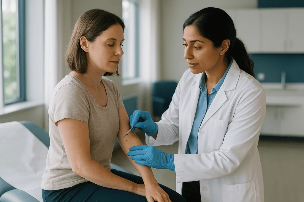
If removal is planned, the area is cleaned and marked. A tiny needle delivers local anesthetic; the sting lasts seconds, and the skin goes numb within a minute or two. Many people say the injection is the only uncomfortable part.
For a shave, the clinician holds the blade almost parallel to the skin and draws it across the base of the mole in a single sweep or a few passes. Bleeding is controlled with pressure, gentle heat or a styptic solution. A small dressing goes on, and you can walk out within minutes.
For an excision, an oval is drawn around the mole, roughly three times as long as it is wide so the skin closes flat without puckering. The scalpel follows the line through the skin’s full thickness, the specimen is lifted free and placed in a labeled container, and the wound is closed in one or two layers. Dissolving stitches may sit below the surface, with removable ones on top.
Either way, the mole leaves the building in a jar. That part is not optional in good practice, and the reason deserves its own section.
Why the mole decides the method: depth, pigment and suspicion
Think of a mole as an iceberg. Some sit almost entirely above the waterline: soft, skin-colored or lightly tan, often with a few hairs, raised like a tiny dome. These intradermal moles have most of their cells in a shallow layer, and a blade can remove nearly all of the visible lesion without going deep. They are classic shave candidates.
Others are flat or only slightly raised, darker, and extend further down. Here the shave would leave cells behind, which risks regrowth and, more importantly, gives the pathologist an incomplete specimen. If the lab cannot see the base of the lesion, it cannot measure how deep any abnormal cells reach.
That measurement is the crux. When melanoma is a possibility, thickness is one of the most important findings in the report and guides every decision afterward. The NHS and Mayo Clinic both describe the ABCDE features that raise concern: asymmetry, an irregular border, more than one color, a diameter larger than about 6 millimeters, and evolution or change over time. A mole with any of those features is normally excised whole rather than shaved, precisely so nothing about its depth is lost.
Location plays a part too. Thin skin over the shin or the back of the hand heals slowly after a deep cut, which can tip a borderline decision toward a shave for a clearly benign lesion. Cosmetic priorities matter on the face, where a fine linear scar may be preferred over a round, pale shave mark or vice versa, depending on the exact spot.
Clinicians weigh all of this in seconds, but the logic is consistent: the less certain the diagnosis, the more complete the removal.
Who is usually offered a shave removal, and who is usually asked to wait
Shave removal is typically offered when the mole looks reassuring under the dermatoscope and the main complaints are practical: it rubs on a bra strap, it bleeds when nicked while shaving, or it simply bothers the person who owns it. Raised, evenly colored, long-standing moles with a soft texture fit this description well.

Certain situations push a clinician toward waiting, choosing a different method, or investigating first.
- Any feature that raises suspicion of melanoma, in which case full excision, not a shave, is the standard route.
- A mole that has visibly changed in recent months, which usually earns an examination and possibly a biopsy before any cosmetic decision.
- Active skin infection at the site, which raises the chance of a wound complication and is normally treated first.
- A tendency to form keloids, the thick, raised scars that spread beyond the original wound; the care team may discuss scar risk before proceeding on the chest, shoulders or jawline.
- Pregnancy, when moles can darken or enlarge naturally; many clinicians prefer to reassess after delivery unless there is a medical reason to act.
- People taking medicines that thin the blood, who are not excluded but usually need a plan for bleeding control agreed with the prescribing clinician. Never alter such medicines on your own.
Children can have moles removed when there is a medical reason, though the pathway leans on preparation, comfort and keeping the visit calm rather than on speed. A cooperative older child may manage local anesthetic in clinic; a younger one may be scheduled differently. None of these factors are refusals; they are reasons the method or timing may change, and the final call rests with the treating team.
When surgical excision is usually the method of choice
Excision is the workhorse whenever certainty matters more than a minimal scar. The clearest case is a mole with ABCDE features, a new mole appearing in adulthood that looks unlike the others, or one that has started to itch, crust or bleed without an obvious injury. Removing the whole lesion with a margin gives the pathologist an intact specimen, top to bottom and edge to edge.
It is also preferred for flat moles of any color, because a shave cannot easily remove a lesion that has no height. Moles with hair-bearing roots that sit deep, large moles where partial removal would leave an uneven patch, and moles that have already regrown after a previous shave often fall into the excision column as well.
If a shave specimen comes back from the lab with a note that abnormal cells extend to the deep edge, a second procedure, usually an excision of the scar and surrounding skin, may be recommended. This is not a failure of the first procedure; it is the system working as designed, and it is one reason clinicians try to match the method to the mole from the start.
The trade-offs are real. Excision takes longer, requires stitches and a return visit to remove them, and leaves a linear scar that is longer than the mole itself because of the oval shape needed for flat closure. Bleeding and infection are uncommon but more possible with a deeper wound, as MedlinePlus notes in its guidance on surgical wound care. For many moles, that is a fair price for a complete answer. For an obviously benign bump, it may be more than the situation needs. The judgment belongs to the clinician holding the dermatoscope.
Shave vs excision mole removal at a glance
Laid side by side, the two methods are easier to compare than they sound. The table below summarizes what mainstream sources such as Cleveland Clinic and MedlinePlus describe; the ranges are typical, not promises, and your own experience will depend on the site, your skin and the lab result.
| Feature | Shave removal | Surgical excision |
|---|---|---|
| Best suited to | Raised, evenly colored, clinically benign moles | Flat, dark, changing or suspicious moles; deep or recurrent moles |
| Depth of removal | Upper skin layers, at or just below the surface | Full thickness with a margin of normal skin |
| Anesthetic | Local injection | Local injection |
| Stitches | None | Yes, surface and often deeper dissolving sutures |
| Typical time in the chair | Minutes | Longer, with marking and layered closure |
| Specimen for pathology | Yes, but base may be incomplete | Yes, complete lesion with margins |
| Wound healing | Shallow graze; around 2–3 weeks to close (Cleveland Clinic) | Sutured line; stitches usually out at a follow-up visit, scar maturing over months (Cleveland Clinic) |
| Scar type | Flat, often paler, roughly the mole’s footprint | Thin line, longer than the mole |
| Chance the mole regrows | Possible, since some cells may remain | Low once margins are clear |
| Follow-up visit | Usually only for results | Suture removal plus results |
One detail in that table carries more weight than the rest: the pathology row. A shave gives a good answer for a mole that was never in doubt. An excision gives a complete answer for a mole that was. Everything else, from scar shape to the number of visits, follows from that distinction.
Is it safe to get moles removed from the face?
Facial mole removal is routine, and both methods are used on the face every day. The real question is which approach leaves the least visible mark in a particular spot, and that depends on anatomy as much as on the mole.
Faces heal well because the skin has a rich blood supply, which supports faster closure and lower infection rates than the lower legs. The flip side is that any scar sits where people look. Clinicians therefore plan with the skin’s natural lines in mind: the fine creases that run horizontally across the forehead, diagonally beside the eyes, and vertically down the cheeks. An excision scar aligned with these lines tends to fade into them; one that crosses them can stand out.
Shave removal on the face suits raised moles on the cheek or nose where a small, flat, slightly paler patch may be less noticeable than a line. The nose deserves special mention: its skin is thick and oily around the tip, and shave wounds there can heal with a subtle dip or a visible pale spot. A clinician will often talk through that possibility before proceeding.
Near the eyelid, lips and the corner of the nose, excision needs careful planning so closing the wound does not pull on a delicate edge. Those areas are sometimes handled by clinicians with additional training in facial repair.
Safety, in the medical sense, is high with both methods when performed by a qualified clinician using sterile technique and local anesthetic. The more useful conversation is about appearance: what the scar will likely look like at two weeks, two months and a year. Mayo Clinic notes that scars from mole removal are permanent even when they fade well, so an honest preview matters more on the face than anywhere else.
Why every removed mole should go to the lab
Histopathology is the examination of tissue under a microscope by a pathologist, and it is the quiet safeguard behind every mole removal. A dermatoscope is a powerful tool, but the eye cannot see individual cells. The microscope can. The National Cancer Institute explains that a common mole and a melanoma can look similar at the surface, which is why a tissue diagnosis, not a visual impression, is the standard of care.
This has a practical consequence for the method debate. Techniques that destroy the mole in place, such as laser ablation or freezing, leave nothing to send. That is why mainstream guidance, including the NHS, is cautious about laser treatment of pigmented moles: a lesion vaporized today cannot be examined tomorrow, and a darkening scar at the site years later is harder to assess because the original tissue was never studied. Laser has its uses for some flat, clearly benign spots, but for a true mole, shave or excision remains the mainstream route precisely because both produce a specimen.
Results typically take days to a couple of weeks depending on the lab. The report describes what the lesion was, whether its edges were clear, and whether any features need follow-up. Most reports for clinically benign moles come back benign. Some describe a dysplastic nevus, a mole with atypical features that is not melanoma but may prompt closer surveillance or a wider excision depending on the degree of atypia.
If you ever hear a mole will be removed without being sent anywhere, it is reasonable to ask why. In most settings the answer should be that it is going to pathology as a matter of routine.
How long does it take for a shaved-off mole to heal, and when do excision stitches come out?
A shave site behaves like a deep graze. For the first day or two it weeps a little clear or pink fluid and feels tender if pressed. Over the next week the raw surface fills in from the edges and the base. Cleveland Clinic describes mole removal wounds as generally taking around two to three weeks to close over, with shave sites on the face often at the faster end and sites on the lower legs at the slower end because circulation there is poorer.
What you see at three weeks is new pink skin, slightly shiny and a little lower or higher than its surroundings. That pink fades over the following months. Sun exposure during this window can darken the new skin permanently, so covering the site or using sunscreen once it has fully closed is standard advice.
An excision follows a different rhythm. The line is closed on day one, so there is no open wound to watch. Mild swelling and bruising are common for a few days. Surface stitches are generally removed at a follow-up visit on a timetable your clinician sets; Cleveland Clinic notes that this varies with the body site, with facial sutures often removed sooner and those on the back or limbs left longer because the skin there is under more tension. Dissolving sutures beneath the surface take care of themselves.
After the stitches are out, the scar goes through its own slow maturation. It may look red and slightly raised for weeks, then gradually flatten and pale over many months. Strenuous stretching of a fresh excision line can widen it, so most clinicians advise easing back into heavy exercise, especially for wounds on the shoulders, back or over joints. Ask for site-specific guidance rather than a general rule.
Why put petroleum jelly on a mole removal?
The instruction surprises people who grew up being told to let a wound dry out and form a good scab. Modern wound care says the opposite, and the reason is cellular. Skin cells migrate across a wound to close it, and they move faster across a moist surface than under a dry crust. A scab is essentially a dried barrier the new cells have to burrow beneath, which slows closure and tends to leave a more noticeable mark.
A thin layer of plain petroleum jelly keeps the surface moist, prevents the dressing from sticking, and seals out debris. MedlinePlus, in its guidance on caring for surgical wounds, describes this moist, covered approach as the standard way to support healing. Cleveland Clinic gives the same advice for mole removal sites specifically.
The routine is simple and worth doing properly for the first one to two weeks or until the site has fully closed, whichever your clinician specifies:
- Wash hands, then gently clean the area with mild soap and water once or twice a day; no scrubbing, no harsh antiseptics unless instructed.
- Pat dry, apply a thin smear of petroleum jelly, and cover with a fresh non-stick dressing.
- Leave any forming scab alone; picking it off restarts the clock.
Plain petroleum jelly is usually preferred over antibiotic ointments for routine care because the ointments offer little extra benefit on a clean wound and can trigger contact allergy, which shows up as new redness and itch that is easily mistaken for infection. If an antibiotic cream has been prescribed for a specific reason, follow that prescription rather than this general note.
The same principle applies to excision lines once the dressing comes off: moist, covered and protected from sun is the formula for the least conspicuous scar.
Can a mole grow back after shave removal, and what scar should you expect?
Yes, a shaved mole can return, and this is the method’s best-known limitation. Because the blade removes the upper portion of the lesion, pigment cells that sat deeper can repopulate the site, sometimes within months, sometimes years later. The regrowth is usually flat and darker than the original, confined to the scar. Pathologists have a name for this, recurrent nevus, and it deserves a careful look because its microscopic appearance can mimic melanoma. That is a strong argument for keeping records: tell any future clinician that the spot was previously shaved and ask whether the original report can be retrieved.
Regrowth after full excision with clear margins is uncommon, since the entire lesion was removed. When something reappears along an excision scar, it is examined promptly for the same reason.
Scars differ in shape more than in severity. A shave leaves a round or oval patch roughly the size of the mole, typically flat and paler than the surrounding skin, occasionally slightly depressed. On darker skin it may instead heal darker than its surroundings for a period, a change called post-inflammatory hyperpigmentation that often fades with time and sun protection. An excision leaves a straight line, longer than the mole, that starts pink and settles toward a thin pale thread.
Two scar types need mention. A hypertrophic scar is thick and raised but stays within the wound’s borders; a keloid grows beyond them. Both are more common on the chest, shoulders, upper back and jaw, and in people who have formed them before. Mayo Clinic notes that scars from mole removal are permanent even when faint. If scar appearance is your main concern, raise it before the procedure, when the method and the wound orientation can still be adjusted.
What people often get wrong about mole removal
Myths cluster around moles, partly because almost everyone has them and partly because advice from decades ago still circulates. A few deserve direct correction.
Cutting a mole makes it spread. There is no evidence that removing a mole, or taking a biopsy from one, causes cancer or spreads an existing cancer. The opposite is true: tissue sampling is how melanoma is diagnosed early. This myth stops people from getting changing moles examined, which is the real harm.
Hair in a mole means it is dangerous. Hair growth usually indicates a mature, deep-seated benign mole with intact follicles. It is not a warning sign on its own.
Home removal creams and kits are a reasonable shortcut. The products sold online typically contain caustic chemicals that burn the skin unpredictably. Mayo Clinic warns against them: they leave scars, can mask a melanoma by destroying the surface while abnormal cells remain beneath, and provide no specimen. No mainstream guideline supports them.
Laser is a scar-free upgrade. Laser can lighten some superficial pigmented spots, but it leaves nothing for the laboratory and may not reach deeper cells. For moles, shave and excision remain the standard because they allow diagnosis.
Excision is always the safer choice. Excision is more complete, but for a clinically benign raised mole it trades a small flat mark for a longer linear scar without adding diagnostic value. Matching the method to the mole is the point.
A shaved mole that comes back means the first clinician made a mistake. Regrowth is a known property of the method, discussed in advance, and the returning pigment is examined rather than ignored.
Once removed, you are done. People with many moles, a history of atypical moles, or significant sun damage usually benefit from ongoing skin checks regardless of how one particular mole was handled, as the NHS and Mayo Clinic both advise.
Questions to ask your care team
A good consultation leaves you able to explain, in your own words, why one method was chosen over the other. These questions tend to draw out the reasoning. Bring them written down; appointments move quickly.
- Based on the dermatoscope view, do you consider this mole clearly benign, uncertain or suspicious, and how does that drive the choice between shave and excision?
- If you plan a shave, what happens if the lab finds abnormal cells at the deep edge? Would a second procedure be needed?
- Will the specimen be sent to pathology, and how will I receive the result?
- What will the scar most likely look like at this specific site in two weeks, three months and a year? Can you show me examples of each scar type on similar skin?
- Are there features of my skin or history, such as previous keloids, that change your recommendation?
- Which direction will the excision line run, and why?
- How should I care for the wound day by day, and when exactly should I stop dressing it?
- When can I shower, swim, exercise, shave the area or wear makeup over it?
- If I take medicines that affect bleeding, what is the plan, and who should I speak to before changing anything?
- If this mole comes back or a new spot appears in the scar, what should I do?
- Do I need a full-body skin check now or on a regular schedule?
Notice that none of these questions ask the clinician to promise an outcome. They ask for reasoning, likelihoods and a plan. That is the conversation worth having, and a clinician who answers it clearly has already told you most of what you need to know about their approach.
When to call your doctor
Most shave and excision sites heal uneventfully, and a little redness, tenderness and clear weeping in the first days are expected. Certain signs are not, and they warrant a call to the clinic the same day rather than waiting for the scheduled follow-up. MedlinePlus lists the classic warning signs of a wound problem, and they apply here.
- Bleeding that soaks through a dressing and does not stop after 10–15 minutes of firm, continuous pressure.
- Spreading redness, warmth or swelling beyond the immediate wound edge, especially if it is enlarging day by day.
- Thick yellow or green discharge, a foul smell, or a wound that reopens.
- Pain that worsens after the first two days instead of easing.
- Fever or chills in the days after the procedure.
- Red streaks tracking away from the wound, or a tender lump in the nearby lymph node area such as the armpit, groin or neck.
- Stitches that come loose early, or a wound edge that gapes.
- Numbness, tingling or weakness beyond the small numbed patch, which can rarely indicate a nerve near the site was affected.
Separately from wound healing, there are mole-related reasons to seek care at any time, before or long after a removal. The NHS and Mayo Clinic both advise prompt assessment for a mole that is changing in size, shape or color, has an irregular or blurred border, shows several shades, measures more than about 6 millimeters across, or starts to itch, crust, bleed or feel different. A new pigmented spot within an old removal scar belongs on that list as well.
Severe allergic reactions to local anesthetic are rare, but difficulty breathing, facial or throat swelling, or faintness during or shortly after the procedure is an emergency. Every decision about reassessment, re-excision or surveillance rests with your treating team; your part is to report what you see without delay.
Frequently asked questions
How long does it take for a shaved-off mole to heal?
A shave site usually closes over in roughly two to three weeks, according to Cleveland Clinic, with the new skin staying pink for several months afterward. Facial sites often heal toward the faster end because of better blood supply, while sites on the lower legs can take longer. Keeping the wound moist, covered and protected from sun supports the best result.
Why put petroleum jelly on a mole removal?
Petroleum jelly keeps the wound surface moist, and skin cells travel across a moist surface faster than under a dry scab. MedlinePlus describes this moist, covered approach as standard care for surgical wounds. It also stops the dressing sticking and seals out debris. Plain petroleum jelly is generally preferred over antibiotic ointments for routine care because those can trigger contact allergy that mimics infection.
Is it safe to get moles removed from the face?
Yes, both shave and excision are routinely performed on the face under local anesthetic, and facial skin tends to heal well because of its rich blood supply. The more pressing consideration is scar appearance, which depends on the exact site and how the wound is oriented along natural skin lines. Ask your clinician what the mark is likely to look like over the first year.
How do doctors remove moles?
After examining the mole with a dermatoscope, the clinician numbs the skin with a local anesthetic injection and either shaves the raised mole level with the skin using a blade, or cuts out the whole mole with a margin using a scalpel and closes the wound with stitches. In both cases the tissue is sent to a pathologist for microscopic examination.
What is the shave excision mole healing time compared with a full excision?
A shave heals as an open graze over about two to three weeks, per Cleveland Clinic, whereas an excision is closed on the day with sutures that are removed at a follow-up visit on a timetable set by your clinician. The excision scar then matures over months, starting red and slightly raised before flattening and fading.
When are mole excision stitches removed?
Surface stitches are taken out at a follow-up appointment, and the timing depends on the body site. Cleveland Clinic notes that facial sutures are generally removed sooner, while those on the back or limbs stay in longer because the skin there is under more tension. Dissolving sutures placed beneath the surface do not need removal.
Can a mole grow back after shave removal?
It can. Because a shave removes the upper part of the lesion, pigment cells left deeper may repopulate the scar, usually as a flat, darker patch. This recurrent nevus can look worrying under the microscope, so it should be examined and the original pathology report retrieved. Regrowth after a complete excision with clear margins is uncommon.
Does mole removal on face always leave a scar?
Every mole removal leaves some mark, and Mayo Clinic notes these scars are permanent even when they fade well. A shave leaves a flat patch about the size of the mole; an excision leaves a thin line longer than the mole. Aligning the wound with natural skin creases, moist wound care and sun protection help the scar settle as inconspicuously as possible.
Why is laser not usually used for mole removal?
Laser destroys the pigment in place, so there is no tissue to send to a pathologist and no way to confirm what the lesion was. It may also leave deeper cells behind. The NHS and other mainstream sources are cautious about laser for pigmented moles for this reason, reserving shave or excision as the standard methods that allow diagnosis.
What happens if the lab finds something unexpected after a shave?
If the report describes atypical cells or notes that the lesion reaches the deep edge of the specimen, your clinician may recommend a second procedure, typically an excision of the scar and surrounding skin, so the complete lesion can be assessed. This is a planned safety step rather than a sign the first procedure went wrong, and your treating team will explain the options.
References
This article is for general information only and is not a substitute for professional medical advice. Please consult a qualified doctor about your individual situation.

More from the Blog
Before Acne Scar Treatment: Sun, Exfoliants and the Products Your Dermatologist Pauses
Preparing for acne scar treatment usually means getting active acne under control first, protecting the skin from sun for several weeks beforehand, and pausing…
Supporting Someone After a Skin Graft: Protecting the Site, Positioning and the First Weeks
Caring for someone after a skin graft means keeping the first dressing dry and undisturbed until the care team removes it, keeping the grafted…
Who Suits a Suture Suspension Lift? Mild Sagging, Skin Quality and Realistic Goals
Thread lift candidates are usually adults with mild to moderate sagging of the cheeks, jawline or brow, reasonably good skin thickness and elasticity, and…
How Many Microneedling Sessions Does Skin Usually Need and How They Are Spaced
Most people are offered a course rather than a single microneedling treatment. Published dermatology reviews describe courses of roughly three to six sessions, spaced…

How Tissue Expansion Works: Expander Placement, Gradual Saline Fills and the Second Surgery
A tissue expander is a deflatable silicone balloon placed under skin or muscle during a first operation, then slowly filled with saline through a…
Laser Treatments for the Eye: Refractive, Retinal and Glaucoma Procedures Explained
Laser treatment for eye conditions is not one procedure but three families. Refractive lasers reshape the cornea to reduce dependence on glasses; retinal lasers…