

Laser Treatments for the Eye: Refractive, Retinal and Glaucoma Procedures Explained
Key Takeaways
- Refractive lasers reshape the cornea to reduce reliance on glasses; retinal and glaucoma lasers protect sight rather than improving it, so expectations should match the row you are in.
- In the LiGHT trial, 74.2 percent of people treated with selective laser trabeculoplasty first were at target eye pressure without drops at three years, though the effect can fade and repeat treatment is sometimes needed.
- Panretinal photocoagulation deliberately sacrifices some peripheral and night vision to stop fragile new vessels bleeding, and the NHS puts a session at roughly 20 to 40 minutes.
- After LASIK, the Mayo Clinic notes vision takes about two to three months to stabilize and eyes commonly feel dry for the first six months or so.
- No laser treats dry macular degeneration, and wet macular degeneration is now managed mainly with anti-VEGF injections rather than thermal laser.
- A sudden curtain across vision, a burst of floaters, or severe pain with nausea after any eye laser needs same-day assessment, because retinal detachment and acute pressure rises are emergencies.
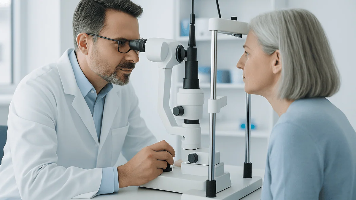
Laser treatment for eye conditions is not one procedure but three families. Refractive lasers reshape the cornea to reduce dependence on glasses; retinal lasers seal leaking or torn tissue, mainly in diabetic eye disease and retinal tears; glaucoma lasers improve fluid drainage or lower pressure. Each takes minutes as an outpatient, carries its own risks, and suits different eyes, so the choice rests with an ophthalmologist.
The letter arrives after a routine diabetic eye screening, or an optometrist pauses a little too long over a pressure reading, or a thirty-year-old simply tires of fogged glasses on a cold morning. Different people, same word on the next appointment slip: laser. It sounds high-tech and slightly ominous at once, and most people walk in with a mental picture borrowed from a friend’s refractive surgery, which may have nothing to do with what they are about to have.
That confusion is understandable. Laser treatment for eye conditions covers procedures that share a light source and almost nothing else. One reshapes the front window of the eye in under a minute of laser time. Another places hundreds of tiny burns on the back wall to starve abnormal blood vessels. A third nudges a drainage meshwork into working better. Different tissue, different wavelength, different goal.
This explainer walks through all three groups, what actually happens in the chair, who tends to be offered each one, what the evidence shows about how long results hold, and the warning signs that should prompt a phone call rather than patience.
What does laser treatment for eye conditions actually mean?
A laser is simply light of a single wavelength, focused so precisely that it can act on a spot thinner than a human hair. In ophthalmology that precision is used in three quite separate ways, and it helps to sort any proposed procedure into one of them before worrying about the details.
The first group is refractive. The cornea, the clear dome at the front of the eye, does most of the eye’s focusing. Shave or reshape a few microns of it and the eye’s focal point moves, which is how LASIK, PRK and small-incision lenticule extraction (SMILE) reduce short-sightedness, long-sightedness and astigmatism. These are elective procedures for people who can already see well with glasses or contact lenses.
The second group is retinal. The retina is the light-sensing layer lining the back of the eye, and when it leaks, tears or grows fragile new blood vessels, a thermal laser can seal, weld or quieten it. Diabetic retinopathy and retinal tears are the common reasons; these lasers aim to protect sight rather than sharpen it.
The third group targets glaucoma, a condition in which pressure inside the eye damages the optic nerve. Here the laser either stimulates the eye’s natural drainage filter, opens a bypass for fluid, or reduces how much fluid is made.
Why does the sorting matter? Because almost every question people bring to the consultation (will it hurt, how long will it last, could I go blind, do I still need drops) has a different honest answer in each group. A single phrase such as ‘eye laser’ hides that, and clinic leaflets rarely spell it out.
How does a laser change eye tissue without cutting it?
Think of the difference between sunlight through a magnifying glass, a hammer blow and a very fine chisel. Eye lasers borrow from all three mechanisms, and the one chosen explains most of what you will feel and see afterwards.
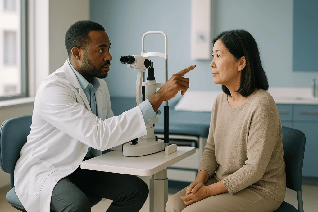
Photoablation is the chisel. The excimer laser used for refractive surgery emits ultraviolet light that breaks molecular bonds in corneal tissue, vaporizing a layer roughly a quarter of a micron deep with each pulse and leaving the surrounding tissue cool. That is why the cornea does not scar in the way a burn would, and why a computer map of your prescription can be translated so directly into a reshaping pattern.
Photocoagulation is the magnifying glass. Retinal lasers deliver visible light that the dark pigment layer under the retina absorbs and converts to heat. Each pulse makes a tiny controlled burn that scars down leaky vessels or glues retina to the wall beneath it. Patients often see a flash and may feel a dull ache, because the heat is real even if the spot is minuscule.
Photodisruption is the hammer. A neodymium:YAG laser focuses a pulse so intense that it creates a microscopic shockwave at its focal point. That shockwave can punch a hole in a clouded membrane behind a lens implant or in the edge of the iris, without the beam harming tissue in front of or behind the focal point.
Selective laser trabeculoplasty uses a fourth trick: a very short, low-energy pulse absorbed only by pigmented cells in the eye’s drainage meshwork. Those cells release signals that appear to prompt the meshwork to clear itself, with no visible burn at all. Same word, laser, four very different conversations with the tissue.
Refractive laser surgery: LASIK, PRK and SMILE in plain language
All three refractive procedures end in the same place, a cornea reshaped by an excimer or femtosecond laser so that light focuses on the retina instead of in front of or behind it. They differ in how the surgeon gets to the tissue.
In LASIK, a thin hinged flap is created in the cornea’s surface, either with a blade or a femtosecond laser, folded back, and the excimer laser reshapes the exposed bed. The flap is laid back without stitches. The Mayo Clinic notes the whole procedure usually takes 30 minutes or less, with the laser itself working for well under a minute per eye.
PRK, and its close cousin LASEK, skip the flap. The surgeon removes or loosens the cornea’s outer skin layer, the epithelium, and the laser works on the surface. The epithelium grows back over several days under a bandage contact lens. Recovery is slower and more uncomfortable in the first week, but there is no flap to dislodge, which is why PRK is often discussed for people with thinner corneas or contact-sport lifestyles.
SMILE uses only a femtosecond laser to carve a lens-shaped sliver of tissue inside the cornea, then removes it through a small keyhole incision. It currently treats short-sightedness and astigmatism rather than long-sightedness.
Each method carries trade-offs rather than a winner. Flap procedures tend to give faster visual recovery; surface procedures avoid flap complications; SMILE leaves more of the surface nerves intact, which may matter for dry eye, though long-term comparative evidence is still maturing. A surgeon’s recommendation should rest on your corneal thickness and shape, tear film, prescription and occupation, not on which machine the clinic recently bought.
Who is usually offered refractive laser, and who is asked to wait
The honest starting point: refractive surgery is for people whose eyes are healthy and whose vision is already correctable with glasses. It treats inconvenience, not disease, so the bar for safety is deliberately high.

The NHS guidance on laser eye surgery describes it as suitable for most adults over 18, provided the prescription has been stable, and advises that it is not usually recommended for people whose eyesight is still changing, who have certain eye diseases, or who have conditions that affect healing. Surgeons typically want a prescription unchanged for at least a year or two, because reshaping a cornea that is still drifting locks in yesterday’s number.
Common reasons to be asked to wait or to consider alternatives include:
- A cornea that is too thin, or has an irregular shape on mapping, raising the risk of post-operative bulging (ectasia).
- Significant dry eye, which laser surgery often worsens for months.
- Pregnancy or breastfeeding, when hormonal shifts can temporarily change the prescription.
- Uncontrolled diabetes, autoimmune disease or medicines that impair healing.
- Active eye infection, uncontrolled glaucoma or early cataract.
- Prescriptions outside the range the laser can safely correct.
People over roughly 40 face a different conversation. The lens inside the eye stiffens with age, a process called presbyopia, and no corneal laser reverses it. Some are offered monovision, one eye set for distance and one for near, which suits some brains and not others; many are better served by lens-based surgery instead. A good consultation spends as much time explaining why someone might not be a candidate as it does describing the procedure, and that reluctance is a sign of quality, not salesmanship.
What is laser eye surgery for glaucoma?
Glaucoma lasers do not improve vision. They protect what remains by lowering eye pressure, the only treatable risk factor for a disease that damages the optic nerve silently and permanently. Three procedures cover most situations, and which one you are offered depends on the type of glaucoma.
Selective laser trabeculoplasty (SLT) is for open-angle glaucoma and ocular hypertension, where the drainage angle is open but the trabecular meshwork, the sponge-like filter fluid drains through, is underperforming. Low-energy pulses are aimed at pigmented cells in the meshwork through a contact lens placed on the numbed eye. The NHS describes it as an outpatient procedure that takes a few minutes, after which you may need fewer or no eye drops, though pressure may rise again over time and the treatment can sometimes be repeated.
Laser peripheral iridotomy (LPI) is for angle-closure glaucoma, where the iris physically blocks the drain. A YAG laser makes a pinpoint hole near the edge of the iris, creating a second route for fluid and letting the iris fall back. It is also used preventively in eyes judged at risk of sudden closure.
Cyclophotocoagulation (often called cyclodiode) takes the opposite approach, treating the ciliary body, the ring of tissue that manufactures eye fluid, to reduce production. It is usually reserved for glaucoma that has resisted other treatments, because the effect is less predictable.
UK guidance from NICE, reflected in NHS pages, now positions SLT as a first-line option for many people with newly diagnosed open-angle glaucoma, alongside or instead of starting drops. Whether that applies to a given eye is a decision for the ophthalmologist, who weighs angle anatomy, pressure target and how much damage already exists.
What the evidence shows about laser versus eye drops for glaucoma
For decades the sequence was fixed: drops first, laser if drops failed, surgery if both did. One large randomized trial changed the conversation, and it is worth knowing what it did and did not show.
The LiGHT trial, published in The Lancet, randomized 718 people with newly diagnosed open-angle glaucoma or ocular hypertension to either SLT first or eye drops first. At three years, 74.2 percent of the laser-first group were at their target pressure without needing any drops, and disease progression was less frequent in that group than in those started on drops. No sight-threatening complications were attributed to the laser. These are trial figures, not promises for an individual eye, and they came from a specific population treated under protocol in UK hospital clinics.
Why might a laser perform at least as well as medicines? Partly because drops only work when they land in the eye on schedule, and real-world adherence is imperfect; a laser’s effect does not depend on remembering. Partly because preservatives in drops can irritate the eye surface over years. The flip side is that SLT’s effect fades in a proportion of eyes, repeat treatment is sometimes needed, and some eyes simply do not respond.
What the evidence does not support is the idea that laser means glaucoma is dealt with. Monitoring continues for life. Pressure checks, visual field tests and optic nerve imaging carry on exactly as they would on drops, because the question is never whether the pressure fell for a season but whether the nerve stays stable for decades.
Someone already on drops should not read any of this as a reason to stop them. Whether to switch, add or hold is a conversation with the prescribing clinician, informed by that person’s own pressure readings and field tests.
What is a focal laser? Retinal laser treatment for diabetic eye disease
Diabetes damages the retina’s small blood vessels in two broad ways, and the laser answer to each is different. Understanding which pattern you have explains why one person leaves with a handful of laser spots and another with more than a thousand.
Focal laser targets specific leaking points. When damaged vessels ooze fluid into the macula, the central patch of retina responsible for reading and faces, the result is diabetic macular edema, a swelling that blurs central vision. Using a dye-based photograph or an optical scan as a map, the surgeon places a small number of gentle burns on the leaking microaneurysms, or a light grid over a diffusely swollen area, to seal them and encourage the retina to reabsorb fluid. Modern practice often combines or replaces focal laser with injections of anti-VEGF medicines, drugs that block the growth signal driving leakage, particularly when swelling involves the very center; the retina specialist chooses based on where and how much fluid there is.
Panretinal photocoagulation (PRP) treats proliferative retinopathy, the stage where the oxygen-starved retina grows fragile new vessels that bleed and scar. Rather than targeting individual vessels, PRP scatters hundreds to thousands of burns across the peripheral retina, deliberately sacrificing some side and night vision to reduce the retina’s oxygen demand and switch off the growth signal. The NHS describes it as an outpatient procedure of around 20 to 40 minutes, sometimes spread over more than one visit, and notes that blurred vision for a few hours and sensitivity to light afterwards are expected.
Neither laser restores vision already lost. Both aim to stop the next bleed, and both work best alongside good control of blood glucose, blood pressure and lipids, which is why the eye team and the diabetes team should be reading from the same notes.
Lasers for retinal tears, and what laser cannot do for macular degeneration
A sudden shower of floaters or a flicker of lightning at the edge of vision sends many people to an eye emergency service, and a proportion of them leave having had a laser the same afternoon. This is retinal laser treatment at its most straightforward.
When the vitreous gel inside the eye shrinks with age, it can tug a small tear in the retina. Fluid then seeps through the tear and lifts the retina off the wall, a retinal detachment, which MedlinePlus describes as a medical emergency that can cause permanent vision loss if untreated. Caught at the tear stage, before significant lifting, a surgeon can ring the tear with rows of laser burns through a contact lens or headset. Over the following one to two weeks those burns scar into a seal, like spot-welding a loose edge of wallpaper before it peels. The procedure itself takes minutes and the retina stays where it is. Once detachment has happened, laser alone is rarely enough and operating-room surgery is usually needed.
Age-related macular degeneration (AMD) is a different story, and a common source of disappointment. In the dry form, where the macula thins gradually, no laser has been shown to slow vision loss, and a historical attempt to laser drusen deposits did not help. In the wet form, where abnormal vessels grow under the macula, direct thermal laser was once standard but has been almost entirely replaced by anti-VEGF injections, because a burn to the center of the macula destroys the very cells you are trying to save. Photodynamic therapy, a cold laser activated by an injected light-sensitive dye, is still used for a few specific vessel patterns. Anyone promised a laser that reverses macular degeneration should ask for the published evidence.
YAG capsulotomy: the laser for a cloudy lens implant
Months or years after successful cataract surgery, vision can quietly mist over again, and people often assume the cataract has returned. It has not; a cataract cannot grow back once the natural lens is removed. What has happened is posterior capsule opacification, in which the thin membrane left behind to hold the new lens implant becomes hazy as residual cells spread across it.
The fix is a YAG laser capsulotomy. Sitting at a slit-lamp machine with a numbed eye and usually a contact lens on the surface, the patient hears a series of clicks while the laser’s shockwave opens a small central window in the clouded membrane. Light passes through the clear implant again. The treatment itself typically lasts a few minutes, and most people notice improvement within a day or two as any debris settles, though floaters from membrane fragments are common for a while.
Because it is a photodisruption procedure, there is no burn and no healing of corneal tissue to wait for. The risks are small but real: a temporary rise in eye pressure, which is why many clinics check pressure before you leave and may use a pressure-lowering drop around the procedure; swelling of the macula in a small proportion of eyes; and a modest increase in the risk of retinal tear or detachment, particularly in highly short-sighted eyes. Those are the reasons it is done when haze genuinely affects function rather than at the first sign of film.
People sometimes ask whether it needs repeating. The window, once opened, does not usually close, so a single treatment generally suffices. If vision clouds again later, the cause is more likely to be something else, such as changes in the retina, and warrants a fresh examination.
Which eye laser treats what: a side-by-side summary
Procedures that share a name rarely share a purpose. The table gathers the ones discussed so far by what they target, what they aim to achieve and the kind of eye they are usually offered to. Durations are typical ranges reported by the cited health services, not commitments.
| Procedure | Tissue treated | Goal | Usually offered for | Typical chair time |
|---|---|---|---|---|
| LASIK / PRK / SMILE | Cornea | Reduce need for glasses | Healthy adult eyes, stable prescription | Under 30 minutes for both eyes (Mayo Clinic) |
| Selective laser trabeculoplasty | Trabecular meshwork | Lower eye pressure | Open-angle glaucoma, ocular hypertension | A few minutes per eye (NHS) |
| Laser peripheral iridotomy | Iris edge | Relieve or prevent angle closure | Narrow or closed drainage angles | Minutes |
| Cyclophotocoagulation | Ciliary body | Reduce fluid production | Glaucoma resistant to other treatment | Minutes; often under local anesthetic block |
| Focal or grid laser | Macula (leaking vessels) | Reduce swelling, stabilize vision | Diabetic macular edema not involving the very center | Minutes |
| Panretinal photocoagulation | Peripheral retina | Stop new vessel growth and bleeding | Proliferative diabetic retinopathy | 20 to 40 minutes, sometimes over several visits (NHS) |
| Laser retinopexy | Retina around a tear | Seal tear, prevent detachment | Fresh retinal tear without detachment | Minutes, often same day |
| YAG capsulotomy | Posterior capsule behind lens implant | Clear clouded membrane | Posterior capsule opacification after cataract surgery | Minutes |
Two patterns stand out. Only the first row is about seeing better than you do now; every other row is about preventing loss or restoring what a complication took. And nothing in the table treats dry macular degeneration, presbyopia on its own, or an optic nerve already damaged by glaucoma. Knowing which row you are in reframes the whole conversation.
What the first days and weeks after eye laser usually look like
Recovery depends almost entirely on which row of that table you sat in. Grouping by mechanism gives a realistic picture.
After refractive surgery, most people have blurry, watery, light-sensitive eyes for the first day and are asked to rest with eyes closed and avoid rubbing. Following LASIK, functional vision often returns within a day or two; the Mayo Clinic notes that it takes about two to three months for the eye to fully heal and vision to stabilize, and that eyes commonly feel unusually dry for the first six months or so. PRK is slower: the surface layer regrows over several days, the bandage lens comes out at a follow-up visit, and sharp vision may take weeks. Surgeons generally restrict swimming, eye makeup and contact sports for a period, and night-time halos and glare are expected early on and usually fade.
After glaucoma lasers, the eye may ache and look red for a day or two, and vision can be mildly blurred from the gel and contact lens used during treatment. Many services check eye pressure an hour or so afterwards to catch a temporary spike, then review again within weeks to see whether the target has been reached. Existing drops are usually continued until that review; stopping them is the clinician’s call, never the patient’s assumption.
After retinal laser, the NHS advises that blurred vision and light sensitivity for a few hours are normal and that driving home is not advised. Panretinal treatment can bring a dull headache-type ache and, in the following weeks, a noticeable reduction in night vision or peripheral awareness that may be permanent; this trade is explained in advance because it is intentional. Retinal tear lasers need roughly one to two weeks to form a firm seal, so heavy lifting and vigorous activity are often limited during that window.
Is eye laser good or bad? Risks and alternatives, stated plainly
The question people type into search engines deserves a direct answer: eye lasers are neither good nor bad in the abstract. Each is a tool with a known risk profile that is justified in some eyes and not in others. What matters is whether the risk of the procedure is smaller than the risk of the condition it treats, and that calculation flips depending on the diagnosis.
For refractive surgery, the condition being treated is dependence on glasses, which carries no medical risk at all. The NHS lists dry eyes, glare and halos, and seeing less well than hoped as possible side effects, usually improving over months, and notes that serious complications are uncommon. Rare but serious problems include corneal infection, flap complications, ectasia, and persistent dry eye or visual disturbance that does not settle. The alternatives are glasses, contact lenses and, in some eyes, implantable lenses or lens exchange. Because the baseline risk is zero, every complication is weighed against convenience rather than against blindness.
For glaucoma and retinal lasers, the comparison is different. The condition left untreated leads toward permanent sight loss, so a procedure with a small chance of pressure spike, inflammation, macular swelling or lost peripheral vision becomes reasonable. Alternatives exist and should be discussed: drops or drainage surgery for glaucoma; anti-VEGF injections or vitrectomy surgery for retinal disease; observation for a stable retinal tear with no symptoms.
A useful habit is to ask two questions in any consultation. What happens to this eye over the next five years if we do nothing? And what is the most likely bad outcome of the laser, and how often does it occur in published series? An ophthalmologist who answers both without flinching is giving you the information you need.
How long does laser eye treatment last?
Durability is the question most often asked and most often answered badly, because the honest answer is three different answers.
Refractive lasers permanently remove corneal tissue; the cornea does not regrow its shape. The NHS describes the results as usually permanent. What changes is the rest of the eye. Presbyopia arrives in the forties regardless of surgery, so reading glasses become likely later. Natural lens changes and, eventually, cataract alter the prescription. A minority of eyes regress slightly in the first year as the cornea heals, and some people choose an enhancement procedure if enough tissue remains. So the laser’s effect is lasting; the eye’s refraction is not frozen.
Glaucoma lasers behave more like a medicine with a long half-life. The NHS notes that SLT’s pressure-lowering effect may wear off over time, and the LiGHT trial counted repeat treatments within its three-year protocol. Peripheral iridotomy, by contrast, creates an anatomical opening that generally stays open, though it may not prevent all future pressure problems. Cyclophotocoagulation sometimes needs repeating because the ciliary body partially recovers.
Retinal lasers create scars, and scars are permanent. A sealed retinal tear stays sealed; panretinal photocoagulation’s effect on new vessel growth is lasting in many eyes, although fresh vessels can appear elsewhere if diabetes control slips or the disease remains active, which is why further sessions are sometimes added. Focal laser for macular edema may need supplementing with injections as new leaks develop. A YAG capsulotomy window does not close.
Across all three families, the one thing that lasts without question is the need for review. Lasers alter tissue; they do not alter the underlying tendency of a short-sighted cornea to age, a glaucomatous drain to clog, or diabetic vessels to leak.
What people often get wrong about laser treatment for eye conditions
Some of these myths are harmless. A few lead people to delay sight-saving care or to expect what no laser can deliver.
Myth: ‘My aunt had laser, so I can have the same.’ Her procedure may have been for glaucoma, yours for a retinal tear. The word means nothing until you know which tissue is being treated and why.
Myth: ‘Glaucoma laser will fix my vision.’ Glaucoma lasers lower pressure to protect the optic nerve. Vision already lost does not return, and the procedure does nothing for a glasses prescription.
Myth: ‘After glaucoma laser I can stop my drops.’ Sometimes fewer drops are needed, sometimes none, sometimes all of them. Only the clinician, with pressure readings in hand, can make that call, and stopping early risks silent damage.
Myth: ‘Laser can treat my macular degeneration.’ Dry AMD has no laser treatment, and wet AMD is managed mainly with injections. Claims otherwise are not supported by mainstream evidence.
Myth: ‘Retinal laser will make my diabetic eyes see better.’ It is designed to prevent the next bleed, and panretinal treatment deliberately trades peripheral and night vision for that protection.
Myth: ‘Refractive surgery means I will never wear glasses again.’ Presbyopia still arrives, and some people need a small correction for night driving or fine print.
Myth: ‘The cataract grew back, so I need surgery again.’ It is the capsule behind the implant that has clouded, and a brief YAG laser treats it.
Myth: ‘I’ll be blinded if I move during the laser.’ Modern refractive lasers track eye movement and pause if it exceeds limits; retinal and glaucoma lasers are delivered through a contact lens that steadies the eye. Staying as still as you can helps, but a blink or twitch is anticipated.
Questions to ask your care team before any eye laser
Consultations run on a clock, and the questions you most need answered are rarely the ones a leaflet anticipates. Taking a short list in, and writing the answers down, tends to change the quality of the conversation.
- Which of the three families does this procedure belong to, and what exactly is it meant to achieve for my eye: better vision, stable vision or lower pressure?
- What is likely to happen to this eye over the next five years without the laser, and how confident is that prediction?
- What alternatives exist, including doing nothing for now, and why is the laser preferred in my case?
- What are the most common side effects I should expect, and which rare complications would you want me to understand before consenting?
- Will I still need my current drops, injections or glasses afterwards, and who decides when that changes?
- How many sessions might be needed, and what would prompt a repeat treatment?
- What will I be able to do in the first 24 hours, the first week and the first month? Can I drive home? When can I work, exercise, swim or fly?
- Which measurements will you use at follow-up to judge whether it worked, and when is that visit?
- If something feels wrong at night or on a weekend, whom do I call and how quickly should I be seen?
- For refractive surgery specifically: is my prescription stable, how thick and regular is my cornea, how dry are my eyes, and would you have this procedure yourself with my numbers?
You are entitled to answers that mention probabilities rather than reassurance alone, and to time to think before signing. For elective refractive surgery in particular, a clinic that pushes a same-day decision is telling you something about its priorities. For urgent retinal problems the timeline is shorter for good reason, and the surgeon will say so.
When to call your doctor after eye laser treatment
Most discomfort after an eye laser is expected and settles within hours to days. A smaller set of symptoms signals a complication that needs assessment the same day, and the general rule is simple: new or worsening symptoms after an initial improvement are never normal.
Seek urgent care, through the number your clinic gave you or an emergency eye service, if you notice any of the following:
- A sudden drop in vision, a dark curtain or shadow spreading across part of your sight, or a new burst of floaters or flashing lights; these can indicate retinal detachment, which MedlinePlus classes as an emergency.
- Severe or escalating eye pain, especially with nausea, vomiting or a headache over the brow, which can accompany a sharp rise in eye pressure after glaucoma or YAG lasers.
- Increasing redness, thick discharge, or pain and light sensitivity that worsens after the second day following refractive surgery, which may point to infection or inflammation of the cornea.
- A sensation that something has shifted in the eye after LASIK, particularly if you were struck or rubbed the eye; a displaced flap needs prompt repositioning.
- Haloes with a steamy, misty cornea and a hard, tender eye, a pattern seen in acute angle closure.
- Vision that was improving and then becomes blurred, distorted or wavy in the center, which can suggest macular swelling.
Call during working hours, without alarm but without delay, for dryness that is not easing after several weeks, persistent glare that interferes with driving, double vision in one eye, or any uncertainty about whether to continue a drop. Nobody at an eye clinic minds a phone call that turns out to be nothing; what they dread is the call that comes two days late. Every decision about continuing, changing or adding treatment remains with the team who examined your eye, and they can only make it if they know what you are experiencing.
Frequently asked questions
What is eye laser surgery for glaucoma?
It is a group of procedures that lower eye pressure to protect the optic nerve, not to improve vision. Selective laser trabeculoplasty stimulates the eye’s drainage filter in open-angle glaucoma, laser peripheral iridotomy opens a tiny hole in the iris for angle closure, and cyclophotocoagulation reduces fluid production in harder-to-control cases. Each takes minutes as an outpatient, and lifelong monitoring continues afterwards.
What is a focal laser treatment?
Focal laser is a retinal treatment that places a small number of gentle burns on specific leaking blood vessels near the macula, the center of the retina, to reduce swelling from diabetic macular edema. Unlike panretinal laser, which scatters hundreds of spots across the periphery, focal laser is targeted using a dye photograph or scan as a map. It is often combined with or replaced by anti-VEGF injections, depending on where fluid sits.
Is eye laser good or bad?
Neither in the abstract; each laser is justified when its risk is smaller than the risk of the condition it treats. For glaucoma and retinal disease, where untreated progression threatens sight, the balance usually favors treatment. For refractive surgery, which treats dependence on glasses, every risk is weighed against convenience alone, so candidacy criteria are strict and the decision should never feel rushed.
How long does eye laser treatment last?
It depends on the procedure. Refractive laser results are described by the NHS as usually permanent, although age-related changes such as presbyopia and cataract still alter vision later. Glaucoma laser effects can wear off over years and may be repeated. Retinal laser scars are permanent, but new leaks or vessels can develop elsewhere if the underlying disease remains active, so review continues regardless.
What is retinal laser treatment used for?
Mainly to seal retinal tears before they become detachments, to shut down abnormal new blood vessels in proliferative diabetic retinopathy, and to reduce leakage causing diabetic macular edema. In each case the goal is to prevent further loss of sight rather than restore what has already gone. Historical use for wet macular degeneration has largely given way to injections.
Does laser eye surgery hurt?
Numbing drops mean most people feel pressure rather than pain during refractive, glaucoma and YAG lasers, though the contact lens placed on the eye can feel odd. Retinal photocoagulation can cause a dull ache or sharp twinges because the laser creates heat, and longer panretinal sessions are sometimes done with additional local anesthesia. Soreness, grittiness and light sensitivity in the first day or two are common.
Can laser eye surgery help macular degeneration?
For the dry form, no laser has been shown to slow vision loss. For the wet form, direct thermal laser has been almost entirely replaced by anti-VEGF injections because a burn at the macula’s center damages the cells responsible for detailed vision. Photodynamic therapy, a cold laser activated by a light-sensitive dye, remains an option for a few specific vessel patterns chosen by a retina specialist.
Will I still need glasses after refractive laser surgery?
Possibly, at some stage. The laser corrects the prescription you have at the time, but the eye’s lens stiffens in the forties, a process called presbyopia, and reading glasses commonly become necessary. Some people also need a small correction for night driving or fine detail. Surgeons may discuss monovision or lens-based alternatives for people over 40 for this reason.
Can I drive home after an eye laser procedure?
Usually not. Vision is typically blurred for several hours after refractive, retinal and glaucoma lasers because of dilating drops, the gel used with the contact lens, and the treatment itself, and the NHS specifically advises against driving after retinal laser. Arranging a lift or an escort is standard, and your clinic will tell you when driving is reasonable again at follow-up.
Does a cataract grow back and need laser?
A cataract cannot return once the natural lens is removed. What can happen is clouding of the thin capsule left behind to hold the lens implant, called posterior capsule opacification. A YAG laser capsulotomy opens a small window in that membrane in a few minutes, and the opening does not usually close, so a single treatment generally suffices.
References
- NHS: Laser eye surgery and lens surgery
- NHS: Diabetic retinopathy, treatment
- MedlinePlus: Retinal detachment
This article is for general information only and is not a substitute for professional medical advice. Please consult a qualified doctor about your individual situation.

More from the Blog

Questions to Ask Your Reconstructive Surgeon Before Agreeing to a Staged Plan
Before agreeing to a staged reconstructive plan, ask your surgeon why staging is needed, how many stages are expected, what each one aims to…
Cryotherapy Results: How Treated Skin Looks Once Healed and Why Repeat Sessions Happen
Cryotherapy results depend on what was frozen. For warts, sun-damaged patches and other skin growths, the treated area blisters, crusts and sheds within roughly…
Cryotherapy vs Surgical Removal for Skin Lesions: Why Pathology Sometimes Wins
Cryotherapy freezes a skin lesion with liquid nitrogen and leaves nothing for a laboratory to examine, while surgical excision removes the tissue intact so…
Dermabrasion Recovery: The Raw Days, Ointments and When Redness Finally Fades
Dermabrasion recovery time typically runs in stages: the treated skin is raw, red and weeping for the first days, new skin usually covers the…
Sun Protection After Dermabrasion: Why Strict Cover Matters for Months, Not Days
Sun protection after dermabrasion needs to continue for months, not days, because the newly grown skin is thin and its pigment cells are easily…
What Happens During an IPL Photorejuvenation Session: Cooling Gel, Flashes and Timing
During an IPL photorejuvenation session, a clinician cleanses the skin, applies a cool clear gel, gives you protective eyewear, and passes a handpiece across…