

Cryotherapy Results: How Treated Skin Looks Once Healed and Why Repeat Sessions Happen
Key Takeaways
- Liquid nitrogen boils at about minus 196 degrees Celsius, and clinical lesion freezing works by killing cells with ice crystals and then collapsing the small blood vessels that fed them during the thaw.
- Mayo Clinic describes the dead wart tissue sloughing off within a week or so after freezing, but the pink spot left behind keeps changing color and texture for months.
- Pigment cells are more cold-sensitive than the surrounding skin, which is why a healed cryotherapy spot often ends up paler and why clinicians freeze more lightly on the face and on darker skin.
- Repeat sessions for warts are the planned trade-off for lighter freezing on the face, and NHS guidance notes warts can still recur after apparent clearance.
- A pigmented mole should never be frozen without a diagnosis because cryotherapy destroys the tissue a pathologist would need to examine.
- Whole-body cold chambers and cryofacials cool the skin surface for a few minutes without injuring it, so they have no plausible mechanism for lasting skin tightening and no controlled evidence for large calorie burn.
Cryotherapy results depend on what was frozen. For warts, sun-damaged patches and other skin growths, the treated area blisters, crusts and sheds within roughly one to two weeks, then heals to skin that may stay slightly pale or pink for months. Repeat sessions are common because a single freeze often cannot destroy every affected cell without damaging the surrounding skin. Whole-body cold chambers and cryofacials have no comparable evidence base.
The mirror check happens on day three. A woman in her fifties leans in under the bathroom light, tilts her chin, and studies the small dome of fluid where a rough, sun-damaged patch used to sit on her cheekbone. The dermatologist warned her about the blister. Nobody warned her how much she would want to poke it.
Across town, a college student is on his fourth visit for the same stubborn wart beside his nostril. He is polite about it, but the question is written on his face: why does a treatment that takes twenty seconds need to be repeated so many times?
Both of them are asking about cryotherapy results, and both deserve a more honest answer than the glossy ones online. Freezing skin is old, cheap in effort, and genuinely effective for the right lesion. It is also messier, slower and more repetitive than most people expect, and the phrase now gets borrowed by cold chambers and “ice facials” that share the name but not the science.
What cryotherapy results actually mean: two very different treatments share one name
Cryotherapy simply means treatment with cold. Under that umbrella sit two practices that have almost nothing in common apart from a low thermometer reading, and confusing them is the root of most disappointment about cryotherapy results.
The first is medical cryotherapy of skin lesions. A lesion is any abnormal patch or growth on the skin. A clinician applies liquid nitrogen, a cryogen (a substance used to produce extreme cold) that boils at about minus 196 degrees Celsius, to a wart, an actinic keratosis, a seborrheic keratosis or a skin tag. The aim is destruction: the frozen cells die and the body clears them. Cleveland Clinic lists this among the most common in-office procedures in dermatology, and it has decades of clinical use behind it.
The second is what a gym, spa or wellness studio calls cryotherapy: a whole-body chamber cooled with nitrogen vapor or refrigerated air, or a handheld wand waved over the face for a “cryofacial.” These are marketed for recovery, calorie burn, mood and skin tightening. The temperatures sound dramatic, but nothing is frozen and no tissue is meant to die. The skin surface cools for a few minutes and rewarms. Rigorous, controlled evidence for lasting effects is thin, and this article will keep saying so wherever the marketing gets ahead of the data.
So when someone asks what cryotherapy results look like, the first question back should be: which cryotherapy? The rest of this explainer takes the medical version seriously, because that is where real, visible, sometimes permanent skin changes happen, and where the question of repeat sessions has a clear biological answer.
How cryotherapy works on skin: what actually happens during the freeze
Watch closely during a lesion freeze and the whole event lasts less time than it takes to read this paragraph. The clinician sprays or dabs liquid nitrogen onto the growth until a white, frosted halo forms and extends a millimeter or so beyond its edge. Then they stop and let it thaw. That pause is the treatment.
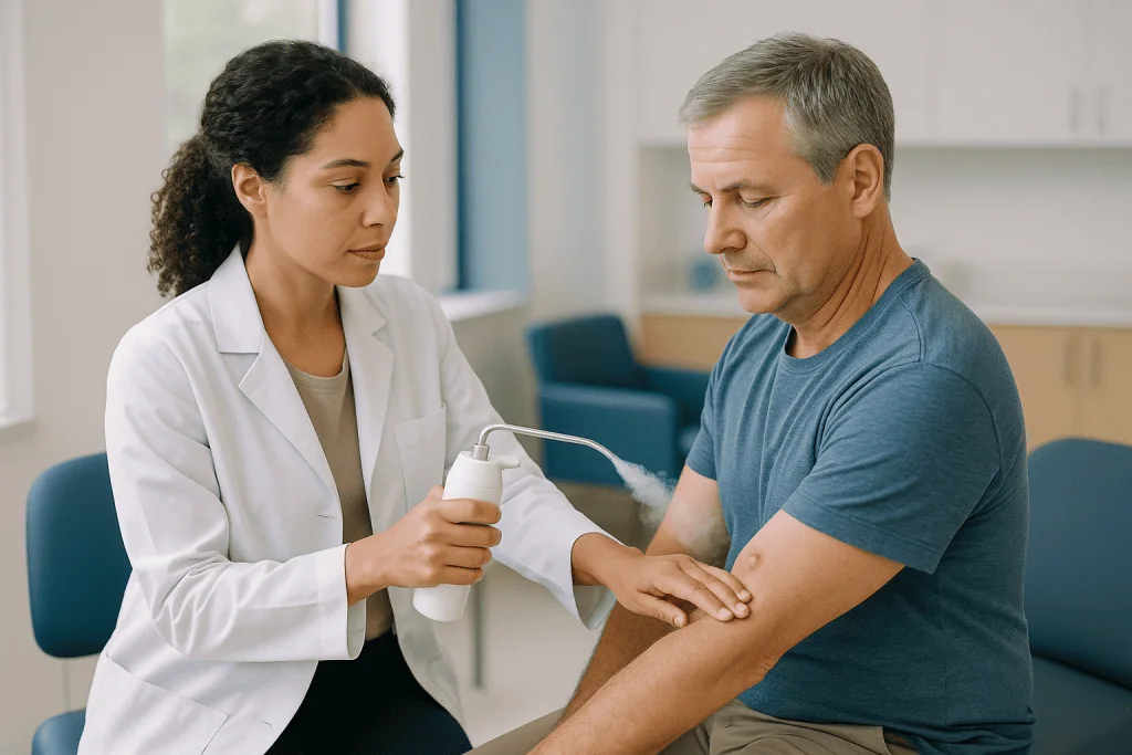
Freezing kills cells in two overlapping ways. Ice crystals form inside and between cells, tearing membranes and pulling water out so that salts concentrate to toxic levels. Then, as the tissue thaws, the tiny blood vessels that fed the lesion clot and collapse, starving whatever survived the ice. Mayo Clinic describes the practical outcome for warts: a blister forms under and around the wart, and the dead tissue sloughs off within a week or so.
The thaw matters as much as the freeze. A slow thaw causes more damage than a fast one, which is why clinicians let the frost fade on its own rather than warming the spot. For thicker or more resistant lesions, they may repeat the freeze-thaw cycle once during the same visit.
Pigment cells, called melanocytes, are more sensitive to cold than the surrounding keratinocytes that make up most of the outer skin. That single fact explains the most common long-term cosmetic result: a treated spot that heals paler than its neighbors. It also explains why clinicians freeze conservatively on the face and on darker skin, accepting a higher chance of needing a second session in exchange for a lower chance of a permanent light patch.
Mayo Clinic notes one more mechanism for warts specifically: the local injury may prompt the immune system to recognize and attack the wart virus. That is a bonus, not a guarantee, and it is part of why identical warts on two people can respond so differently.
Who cryotherapy is usually for, and who is usually asked to wait
The ideal candidate for lesion cryotherapy has a growth that a clinician has looked at and confidently identified as benign or premalignant, that is small and well defined, and that sits on skin where a pale mark would not be a major concern. Warts, actinic keratoses (rough, scaly patches caused by years of sun exposure that can occasionally progress to skin cancer), seborrheic keratoses (harmless, waxy, stuck-on growths that appear with age) and skin tags make up most of the list. MedlinePlus and NHS both describe freezing as a standard option for actinic keratoses and warts.
Some people are asked to wait, or to consider a different route, and the reasons are usually about diagnosis or skin type rather than about the freeze itself.
- An undiagnosed pigmented spot. Freezing a mole or anything that could be melanoma destroys the tissue a pathologist would need to examine. Clinicians biopsy first, freeze later, or not at all.
- Deeply pigmented skin. The risk of long-lasting or permanent hypopigmentation (loss of skin color) is higher, so the treating team may favor lighter freezing, or alternatives such as prescription creams or curettage (scraping under local anesthetic).
- Cold-sensitivity conditions. Cold urticaria (hives triggered by cold), Raynaud phenomenon and cryoglobulinemia, a rare blood-protein disorder worsened by cold, make freezing a poor choice.
- Lesions over nerves or on thin skin. Fingers, the side of the nose and the lower eyelid need extra care because nerve injury and scarring are more likely.
- Poor healing. Uncontrolled diabetes, circulation problems or medicines that suppress immunity can slow the healing that cryotherapy relies on.
None of these is an absolute wall. They are conversations. The person who decides is the clinician who has examined the lesion and knows the patient’s history, which is why this article never substitutes for that appointment.
What the first two weeks look like after cryotherapy on the face
Facial skin is thin, richly supplied with blood and constantly on display, so the healing sequence is both quicker and more visible than on a knee or a shin. Here is the arc most people can expect, drawn from Cleveland Clinic and Mayo Clinic descriptions of the procedure.
The first hours bring redness and a stinging ache, often described as a bee-sting throb that fades over the evening. Swelling can be surprising near the eyes, where loose tissue pools fluid overnight.
Between day one and day three, a blister usually appears. It may be clear, or it may fill with blood and look alarming; a dark blister is common on the face and is not by itself a sign of trouble. Mayo Clinic lists blistering as an expected part of freezing therapy rather than a complication.
By around day three to five, the blister flattens or breaks and a crust forms. This is the stage where the temptation to pick is strongest and where the cosmetic outcome is most easily damaged. The crust is a biological bandage; under it, new skin is migrating across the wound.
Somewhere in the second week, the crust releases on its own. Mayo Clinic’s “within a week or so” for sloughing applies to the dead lesion tissue; the surrounding skin may take a little longer to settle. What is revealed is a pink, shiny, slightly indented spot. That pink is not the final result. It is the start of a months-long remodeling.
Practical care during these two weeks is simple. Gentle washing, a thin layer of plain petroleum-based ointment if the clinician suggests it, sun protection over the area, and no picking, scrubbing or exfoliating acids on the spot. Makeup is usually fine once the crust is intact and not weeping, but the treating team’s instructions override any general advice here.
How long does cryotherapy take to heal, and when do results show?
The honest answer splits into three timelines, and mixing them up is why “how long does cryotherapy take to show results” gets such different replies.
The wound timeline is the shortest. On the face, the crust from a small lesion typically separates within about one to two weeks, in line with Mayo Clinic’s description of dead tissue sloughing within a week or so. Lower legs, where circulation is slower, can take considerably longer, and many people are surprised that a shin wart heals more slowly than one on the cheek.
The lesion timeline is what most people mean by results, and it depends on what was frozen. A thin actinic keratosis or a small seborrheic keratosis is often gone once the crust falls away. A wart is different. The visible bump may be gone, but wart virus can persist in cells that look normal, and the growth can re-emerge from the edges. NHS advice is blunt that warts frequently need several freezing sessions and may still come back, which is why the clinician often books a review a few weeks later rather than declaring victory at the first visit.
The cosmetic timeline is the longest and the least discussed. The pink, shiny spot that appears after the crust falls gradually fades over several months as collagen reorganizes and pigment cells, if they survived, slowly repopulate the area. Skin-color changes after cryotherapy can persist for months and are sometimes permanent, a risk Mayo Clinic lists explicitly for actinic keratosis treatment.
So: wound closed in a couple of weeks, lesion outcome judged over a couple of months, final color known perhaps a year later. Anyone promising a flawless spot at the two-week mark is describing the first timeline and selling it as the third.
Cryotherapy before and after: how treated skin looks once healed
Set aside the airbrushed side-by-side photos and picture the actual healed spot at, say, three months. In most cases it is flat, smooth and slightly shinier than its surroundings because the new outer layer has fewer of the fine lines and pores that texture older skin. On close inspection it is often a shade lighter. In fair skin that difference can be nearly invisible. In olive, brown or Black skin it can be a distinct pale coin that lasts years or never fully recolors, because the melanocytes were killed alongside the lesion.
Less often, the opposite happens: the spot heals darker. Post-inflammatory hyperpigmentation (extra pigment laid down after any skin injury) is more likely if the area was exposed to sun while healing or if the person is prone to dark marks after acne or cuts. Strict sun protection over the treated spot for several months is the one cosmetic intervention with solid logic behind it.
True scarring, meaning a raised or pitted mark, is uncommon after appropriately light freezing of small lesions but becomes more likely with deep freezes, large areas, repeated aggressive sessions on the same spot, or infection during healing. Cleveland Clinic and Mayo Clinic both list scarring and skin-color changes among recognized risks, which is why conservative treatment on the face is the norm.
Hair does not regrow in a deeply frozen patch, because hair follicles sit in the same layer as the pigment cells. On a cheek or brow edge this matters; on a bald scalp it does not.
What healed skin should not show: persistent scale, a growing edge, bleeding, or a bump that returns to its old size. Those are signals that the lesion is still there or was something other than expected, and they belong in the clinician’s chair, not in a skincare routine.
Why repeat sessions happen: margins, wart biology and the immune system
The student on his fourth visit is not a treatment failure. He is experiencing the ordinary arithmetic of cryotherapy, which trades depth of destruction against damage to normal skin.
Every freeze has to stop somewhere. Frost spreads outward and downward from the point of contact, and the clinician chooses how far to let it go. Push far enough to guarantee every lesion cell is dead and the halo of injured healthy skin widens, raising the odds of a pale scar or a nerve injury. Stop early and some lesion cells survive at the base or the edges. On the face, nearly every clinician chooses the second problem over the first, planning from the outset that one or two more light sessions may be needed.
Warts add biology to the geometry. They are caused by human papillomavirus living inside skin cells, and the infected zone usually extends beyond the visible bump. Mayo Clinic notes that repeat treatments are commonly needed for warts, and NHS guidance describes freezing as something that may have to be repeated several times, with no guarantee the wart will not return. The immune-stimulation effect described earlier is real but unpredictable; some people clear a wart after one freeze because their immune system joins in, while others need a series.
Thickness matters too. A wart with a callused cap or a raised seborrheic keratosis insulates its own base; the frost may not reach deep enough on the first pass. Clinicians sometimes pare down thick lesions before freezing for exactly this reason.
Spacing between sessions is usually a few weeks, long enough for the previous wound to heal so the clinician can see what remains. The number of sessions is not fixed in advance. It is a review-and-decide process, and the treating team is the right party to make that call.
Cryotherapy for warts results: why the face needs a lighter touch
A wart on the heel and a wart beside the nose are the same virus, but they are treated like different patients. On the sole, thick skin protects deeper structures and a pale patch is nobody’s concern, so freezing can be firmer. On the face, three factors change the calculation.
Skin thickness comes first. Facial skin, especially on the eyelids and the sides of the nose, is a fraction of the thickness of the sole. A freeze that would barely blister a heel can produce a deep wound on a cheek. Clinicians shorten freeze times and often use a cotton-tip applicator rather than a spray for precision near the eyes.
Nerves and cartilage come second. The nose, ears and the area in front of the ear carry superficial nerves and, in the ear and nose, cartilage that heals poorly. Over-freezing here can cause numbness or a notched contour, which Cleveland Clinic lists among possible complications.
Cosmetic tolerance comes third and is the reason most facial warts are treated in a series. Accepting three or four light sessions in exchange for a near-invisible result is a reasonable trade that many people, once it is explained, are happy to make.
Flat warts, the small smooth-topped kind that cluster on the forehead and jawline, respond less reliably to freezing than raised common warts and can spread along shaving lines. For these, the treating team may prefer a topical approach or a combination, and freezing may be reserved for a few stubborn ones.
Filiform warts, the thread-like growths that project from the eyelid or lip margin, are the facial exception that often does well with a single quick freeze or snip because there is so little base to treat.
Whatever the type, NHS and Mayo Clinic agree on the two points that shape expectations: warts often need repeat treatment, and they can recur even after apparent clearance. Anyone quoting a single-session success rate is not quoting either source.
Typical cryotherapy results by lesion type: a comparison table
The table below summarizes what mainstream sources describe for the lesions most often frozen on the face and neck. It shows patterns, not promises; the treating clinician’s assessment of the individual lesion always governs.
| Lesion | What it is | Usual healing pattern | Repeat sessions | Cosmetic notes |
|---|---|---|---|---|
| Common or filiform wart | Skin growth caused by human papillomavirus | Blister, crust, shedding within about a week or so of dead tissue (Mayo Clinic) | Frequently needed; recurrence possible (NHS, Mayo Clinic) | Light freezing preferred; pale mark possible |
| Actinic keratosis | Rough, scaly sun-damage patch with small potential to progress to skin cancer | Similar blister and crust cycle; new smooth skin beneath | Sometimes, for thick or incompletely cleared patches; new patches develop on sun-damaged skin | Skin-color change and scarring listed as risks (Mayo Clinic) |
| Seborrheic keratosis | Benign, waxy, age-related growth | Crust lifts off, often taking the growth with it | Usually one to two sessions for small lesions; thick ones may need more | Higher hypopigmentation risk in darker skin |
| Skin tag | Soft, harmless flap of skin on a stalk | Darkens, shrivels and drops off | Rarely | Small pale dot possible |
| Pigmented mole | Cluster of pigment cells | Not appropriate for freezing | Not applicable | Must be examined and, if needed, biopsied instead |
Two things stand out. First, actinic keratoses are a field problem: the freeze treats one patch, but the sun-damaged skin around it keeps producing new ones, which is why MedlinePlus and NHS stress ongoing sun protection and periodic skin checks after treatment. Second, the mole row is not a footnote. Destroying a pigmented lesion without a diagnosis is the one cryotherapy outcome no one can reverse.
Does cryotherapy tighten skin? What cryofacials and cold wands can and cannot do
The claim runs like this: cold makes blood vessels constrict, they then dilate as the skin rewarms, circulation surges, pores shrink, collagen is stimulated, and the face looks lifted. The first two steps are real physiology. The rest is a leap.
Vasoconstriction followed by rebound flushing does happen after a few minutes of cold air or a chilled roller. The skin looks briefly tighter and brighter for the same reason it does after a cold shower: mild swelling of the surface and a temporary reduction in redness. That effect wears off within hours. There is no plausible mechanism by which a few minutes of surface cooling, with no injury to the dermis, tells fibroblasts to lay down new collagen. The treatments that do stimulate collagen, such as resurfacing lasers or microneedling, work precisely by creating controlled injury and a healing response, which a cryofacial is designed not to do.
Medical cryotherapy of lesions does provoke collagen remodeling, but as part of wound healing in a small destroyed spot. Nobody would freeze an entire face to chase that effect; the pigment loss and scarring risk would be unacceptable.
Where does that leave the question “does cryotherapy tighten skin”? The evidence-graded answer is that no controlled trial in the mainstream medical literature demonstrates lasting tightening, wrinkle reduction or pore shrinkage from cold facials or whole-body chambers. A cryofacial may feel pleasant and reduce puffiness for an afternoon, which is a fair reason to enjoy one. Presenting it as a tightening treatment is not supported.
People with rosacea, cold urticaria or very reactive skin should also know that rapid temperature swings can trigger flares. A treatment with no proven benefit and a real chance of a rebound flush is a poor bargain for those complexions.
Does cryo really burn 500 calories? Whole-body cryotherapy claims versus evidence
The number is everywhere on studio websites, usually rounded and rarely sourced. Here is the reasoning behind it and why it does not hold up.
Cold exposure does raise energy expenditure. Shivering burns fuel, and the body activates brown fat, a heat-generating tissue, when core temperature is threatened. Those are real, well-studied responses. The problem is scale and duration. A whole-body cryotherapy session lasts two to four minutes. Core temperature barely moves in that window; the skin cools sharply, then rewarms once the person steps out. Any extra energy spent rewarming is modest and short-lived, and no controlled study in the mainstream medical literature has measured anything close to the figures in the marketing.
The comparison that helps: a brisk half-hour walk expends a few hundred calories in most adults according to standard exercise tables, and it requires thirty minutes of continuous muscle work. The idea that standing still in cold vapor for three minutes matches or beats that is not physiologically coherent.
Safety deserves a sentence. Cold chambers have caused frostbite, cold burns and, in rare cases involving unsupervised use, asphyxiation from nitrogen displacing oxygen. Cleveland Clinic describes whole-body cryotherapy as lacking rigorous evidence for most claimed benefits and notes it is not appropriate for people with certain heart, circulation and cold-sensitivity conditions. Anyone with high blood pressure, heart disease, a pacemaker, Raynaud phenomenon, cold urticaria or pregnancy should discuss it with their clinician before trying it, and should treat any studio that waves those concerns away as a red flag in itself.
Where does that leave the calorie question? Cold exposure is an interesting area of research, particularly around brown fat and metabolic health. Whole-body cryotherapy as sold today is not a weight-loss tool, and the evidence does not support the number.
Is cryotherapy actually worth it? Weighing risks and alternatives
For a clearly diagnosed wart, actinic keratosis or seborrheic keratosis, the case for freezing is strong and boring, which is the best kind of medical case. It takes seconds, needs no injection of anesthetic for small lesions, uses no incision, and its main downsides are temporary discomfort and a possible pale mark. Mayo Clinic describes it as the most common treatment for actinic keratoses, and it remains a first-line office option for warts across NHS and MedlinePlus guidance.
Worth it compared with what? The alternatives each carry their own trade-offs, and the treating team weighs them against the person and the lesion.
- Watchful waiting. NHS notes that many warts clear on their own, though this can take months or years. For a harmless seborrheic keratosis that does not bother the person, doing nothing is entirely reasonable.
- Topical treatments. Salicylic acid preparations for warts, and prescription creams for actinic keratoses, work over weeks rather than seconds and cause their own redness and peeling. They spare pigment cells better than freezing, which can matter on darker skin.
- Curettage and cautery. Scraping and heat-sealing under local anesthetic gives a tissue sample and a single-visit result but leaves a small scar.
- Laser or photodynamic therapy. Useful for widespread actinic damage; more equipment, more visits, more expense in time.
The strongest argument against cryotherapy is not that it fails. It is that it can be used on the wrong lesion. Freezing a spot that turns out to be an early skin cancer delays diagnosis and destroys the evidence. That risk is managed by one thing: a clinician examining the lesion first and, when in doubt, sampling it.
For cold chambers and cryofacials, “worth it” is a personal question about how much a pleasant, temporary sensation is worth to you. Framed that way, honestly, most people can decide for themselves.
What people often get wrong about cryotherapy results
Some of these myths come from the internet. Several come from well-meaning friends who had a wart frozen once, years ago, and remember only the ending.
“If it needed a second session, the first one failed.” On the face, a planned series of light freezes is the intended strategy, not a fallback. Mayo Clinic and NHS both describe repeat treatment as routine for warts.
“The blister means something went wrong.” Blistering is the expected mechanism. A blood-filled blister looks dramatic and is common. Spreading redness, pus or fever are different matters, covered in the red-flag section below.
“Popping the blister speeds healing.” It does the opposite, opening the wound to bacteria and disturbing the new skin forming underneath. If a blister is painfully tense, the clinician can drain it cleanly; kitchen-needle surgery is not the same thing.
“Once the crust falls off, that is the final look.” The pink spot underneath changes for months. Judging the cosmetic result at two weeks is like judging a paint job while it is still wet.
“A pale spot means the clinician froze too hard.” Sometimes, but pigment cells are simply more cold-sensitive than the rest of the skin. Some lightening can follow even a careful, appropriate freeze, particularly in deeper skin tones.
“Cryotherapy is cryotherapy.” A liquid nitrogen freeze that destroys a lesion and a three-minute chill in a spa chamber share a word and nothing else. Evidence for one does not transfer to the other.
“You can freeze off a mole at home.” Over-the-counter freezing kits reach far milder temperatures than clinical liquid nitrogen and are labeled for warts only. Using any freezing method on a pigmented spot without a diagnosis is the single most consequential mistake in this entire topic.
Questions to ask your care team before a cryotherapy session
A good consultation before a freeze takes only a few minutes, but the right questions turn a quick procedure into an informed one. Bring these, and expect straight answers.
- What exactly is this lesion, and how confident are you in the diagnosis without a biopsy?
- Why cryotherapy rather than a cream, curettage or simply leaving it alone? What would you choose if it were on your own face?
- How many sessions do you anticipate for this type of lesion, and how far apart would they be?
- Given my skin tone, how likely is a lasting pale or dark mark here, and would a gentler option protect pigment better?
- What will the area look like over the first two weeks, and what care do you want me to do at home?
- Which signs would you want a phone call about, and which are normal?
- How will we know the lesion is fully gone, and when should I come back for a check?
- If it recurs, what is the next step?
- For actinic keratoses: how often should my skin be checked from now on, and what sun protection do you recommend for the treated area?
Two questions deserve a slower conversation. The first is about pigment. If the answer to “how likely is a pale mark” is vague, ask again; on the face, this is the outcome most people care about a year later. The second is about diagnosis. A clinician who says “I would rather biopsy this first” is not being cautious for the sake of it. That sentence protects the one thing cryotherapy cannot give back, which is the tissue itself.
Write the answers down or ask for them in the visit summary. Healing instructions given in the thirty seconds after a stinging freeze are easy to forget by the time the blister appears.
When to call your doctor: red-flag signs after cryotherapy
Most cryotherapy wounds heal without a hitch and never need a second thought. A small number do not, and because the face heals in plain view, early problems are usually easy to spot. Contact the treating clinic, or seek urgent care if it is closed, for any of the following.
- Redness that spreads outward from the treated spot after the first two days, especially with warmth, increasing pain or swelling. Early redness is normal; widening redness is not.
- Thick yellow or green discharge, a foul smell, or a crust that keeps re-forming with pus beneath it. These suggest infection, which Mayo Clinic lists as a recognized complication of freezing.
- Fever or feeling generally unwell in the days after treatment.
- Severe or worsening pain that is not easing with the simple measures the clinic advised, or pain that seems out of proportion to the size of the wound.
- Bleeding that does not stop with ten minutes of firm pressure.
- New numbness, tingling or weakness near the site, particularly on the side of the nose, the ear or the temple, which may indicate nerve irritation.
- A blister that is unusually large, very tense, or near the eye and interfering with vision or eyelid closure.
- Any change in vision or eye pain after treatment close to the eyelid.
Later signs matter too. A spot that has healed but starts to grow again, develops a raised pearly edge, bleeds without being knocked, or produces persistent scale should be reviewed rather than re-frozen on assumption. Actinic keratoses in particular sit on skin that can produce other lesions, and MedlinePlus advises ongoing checks after treatment.
For anyone who has used a whole-body cold chamber: numb, white or waxy patches of skin that do not recolor within minutes, blistering after the session, chest pain, breathlessness or fainting all need urgent medical attention.
None of this is meant to frighten. It is a short list so that the many normal, ugly-but-healthy stages of a healing freeze can be left alone with confidence, and the few genuine problems get seen quickly. The treating team makes every decision about next steps; this list only helps decide when to pick up the phone.
Frequently asked questions
How long does cryotherapy take to show results?
For skin lesions, the visible growth usually blisters, crusts and sheds within about one to two weeks on the face, with Mayo Clinic describing dead wart tissue coming away within a week or so. Whether the lesion is fully gone is judged a few weeks later at review, and the final skin color may take months to settle. Warts often need more than one session before the result is clear.
What does cryotherapy before and after skin actually look like?
Before treatment there is a raised, rough or waxy growth. Immediately after, the spot is red and stinging, then blisters within a day or two, crusts, and sheds. The healed spot is typically flat, smooth, slightly shiny and often a shade paler than the surrounding skin. In darker skin tones the pale patch can be more noticeable and may persist for a long time.
How long does cryotherapy take to heal on the face?
The open-wound phase on the face usually lasts about one to two weeks, from freeze to crust separation, because facial skin has good blood supply. Lesions on the lower legs can take longer. The pink new skin that appears afterward continues to remodel for several months. Individual healing varies, and the treating clinician’s aftercare instructions and review schedule are the guide.
Why does cryotherapy for warts need so many sessions?
Wart virus lives in skin cells that extend beyond the visible bump, and a freeze deep enough to destroy every infected cell in one go would damage surrounding skin. Clinicians, particularly on the face, choose lighter repeated freezes instead. Mayo Clinic and NHS both describe repeat treatment as common for warts, with sessions typically spaced a few weeks apart so the previous wound can heal.
Does cryotherapy really tighten skin?
No controlled evidence in the mainstream medical literature shows lasting skin tightening from cryofacials or whole-body cold chambers. Brief cooling constricts blood vessels and causes a temporary flush that can make skin look brighter for a few hours, but there is no injury to the deeper skin that would prompt new collagen. Treatments that do stimulate collagen work through controlled injury, which cold facials are designed to avoid.
Does cryo really burn 500 calories?
Cold exposure does raise energy use through shivering and brown fat activity, but a two-to-four-minute chamber session barely changes core temperature and no controlled study supports the large calorie figures in marketing. Whole-body cryotherapy is not an established weight-loss method. People with heart or circulation conditions, cold-sensitivity disorders or pregnancy should discuss any cold-chamber use with their clinician first.
Is cryotherapy actually worth it for a facial lesion?
For a clearly diagnosed wart, actinic keratosis or seborrheic keratosis, freezing is a well-established, quick office treatment with a long track record described by Mayo Clinic, NHS and MedlinePlus. Its main downsides are temporary discomfort and a possible pale mark. Whether it beats a cream, curettage or watchful waiting depends on the lesion, skin tone and personal priorities, which is a conversation for the treating team.
Will the pale spot after cryotherapy go away?
Often it fades over months as surviving pigment cells slowly repopulate the area, but Mayo Clinic lists lasting skin-color change as a recognized risk, and in some people, especially with deeper skin tones or after firmer freezing, the light patch is permanent. Strict sun protection of the healing area reduces the risk of the opposite problem, a dark post-inflammatory mark.
Can I use makeup or skincare on the treated area while it heals?
Gentle cleansing and, if the clinician advises, a thin layer of plain ointment are usually all that is needed. Exfoliating acids, retinoids and scrubs should stay off the spot until it has fully healed. Makeup over an intact, dry crust is often acceptable, but the specific instructions given at the visit take priority over any general guidance.
Can a mole be removed with cryotherapy?
Pigmented moles are not appropriate for freezing. Cryotherapy destroys the tissue, so if the spot were an early melanoma there would be nothing left for a pathologist to examine and the diagnosis would be delayed. Clinicians examine pigmented lesions first and, when there is any doubt, remove them by a method that preserves a sample. Home freezing kits are labeled for warts only.
References
This article is for general information only and is not a substitute for professional medical advice. Please consult a qualified doctor about your individual situation.

More from the Blog
Cryotherapy vs Surgical Removal for Skin Lesions: Why Pathology Sometimes Wins
Cryotherapy freezes a skin lesion with liquid nitrogen and leaves nothing for a laboratory to examine, while surgical excision removes the tissue intact so…
Dermabrasion Recovery: The Raw Days, Ointments and When Redness Finally Fades
Dermabrasion recovery time typically runs in stages: the treated skin is raw, red and weeping for the first days, new skin usually covers the…
Sun Protection After Dermabrasion: Why Strict Cover Matters for Months, Not Days
Sun protection after dermabrasion needs to continue for months, not days, because the newly grown skin is thin and its pigment cells are easily…
What Happens During an IPL Photorejuvenation Session: Cooling Gel, Flashes and Timing
During an IPL photorejuvenation session, a clinician cleanses the skin, applies a cool clear gel, gives you protective eyewear, and passes a handpiece across…

Why Your Cold Sore History Matters Before Laser Resurfacing and How Doctors Prepare for It
Your cold sore history matters before laser resurfacing because the herpes simplex virus that causes cold sores stays dormant in facial nerves and can…

What to Stop Before a Chemical Peel: Retinoids, Scrubs, Waxing and Sun Exposure
Before a chemical peel, people are usually asked to pause topical retinoids and exfoliating acids, stop facial scrubs, masks and exfoliating devices, avoid waxing,…