

How Deformity Correction Works: Osteotomy, Fixation and Gradual Realignment Explained
Key Takeaways
- Every deformity correction involves a deliberate cut through bone called an osteotomy; adult bone cannot be bent back into shape.
- In gradual correction, the cut bone is moved about one millimeter per day so new bone forms in the gap, and the device stays on for months while it hardens (MedlinePlus).
- External frames remain the most adjustable fixation for large or multiplane deformities because the correction can be fine-tuned against follow-up X-rays after surgery.
- Mayo Clinic describes bracing for growing children with scoliosis curves of roughly 25 to 40 degrees and surgery generally considered above about 45 to 50 degrees.
- Realigning a bone changes how a joint is loaded but cannot regrow cartilage that is already lost, which is why joint replacement is sometimes discussed instead.
- Slow correction is a safety measure for nerves and blood vessels, not a limitation of technique, and stretching neighboring muscles during lengthening is what keeps joints mobile.
Deformity correction surgery realigns a bone that has grown, healed or worn into the wrong shape. A surgeon cuts the bone at a planned point (an osteotomy), moves the pieces into better alignment either all at once or gradually over weeks, and holds them with plates, rods, nails or an external frame while new bone heals across the gap. Recovery is measured in months, and the plan is individual.
The X-ray goes up on the screen and the line the surgeon draws down the middle of the leg does not pass through the middle of the knee. It drifts inward, toward the other leg, by a couple of centimeters. The patient, a runner in her thirties who has spent two years blaming her shoes, sees it before anyone says a word.
That drawn line is the whole story of how deformity correction surgery works. It is geometry first, carpentry second and biology last, and each of those three steps can be done well or badly. Understanding them is the difference between a patient who feels processed and one who can ask the right question at the right moment.
This explainer walks through the cut, the hardware and the slow, deliberate realignment that some corrections need, and is honest about what the evidence does and does not say.
How deformity correction surgery works, in plain language
A deformity, in orthopedic terms, is a bone or joint that sits outside the range of shape and alignment the body was built to load. Bones can be too short, bent in a curve, twisted along their length, or angled at a joint so that weight lands on one side of the cartilage instead of spreading across it. The bone itself is often perfectly healthy; it is simply pointing the wrong way.
Correction rests on a biological fact that surprises many patients: bone is the only adult tissue that heals without a scar. Cut it cleanly, hold the ends still in a new position, and cells at the cut edges lay down fresh bone that eventually looks like it was always there. Surgeons borrow this behavior. They deliberately break the bone where the plan says it should bend, reposition the fragments, and stabilize them until healing locks in the new shape.
There are three moving parts to how deformity correction surgery works, and this article takes them in order. The osteotomy is the cut. Fixation is whatever holds the pieces in place, from a small plate to a large external frame. Realignment is the act of moving the fragments, either in one step on the operating table or in tiny daily increments over weeks. Which combination a patient receives depends less on the diagnosis than on the size of the correction, the age of the bone and the soft tissues that must stretch to follow it.
One more idea runs through everything below. The goal is rarely a perfectly straight limb on a radiograph. It is a limb, foot or spine that loads its joints evenly enough to protect them, and the treating team decides how much correction is worth the risk of achieving it.
What is osteotomy surgery?
An osteotomy is a surgical cut through bone, made on purpose and in a planned place. The word comes from Greek roots meaning bone and cutting, and it describes a technique rather than a specific operation: the same cut principle is used around the knee, in the foot, in the forearm and along the spine.
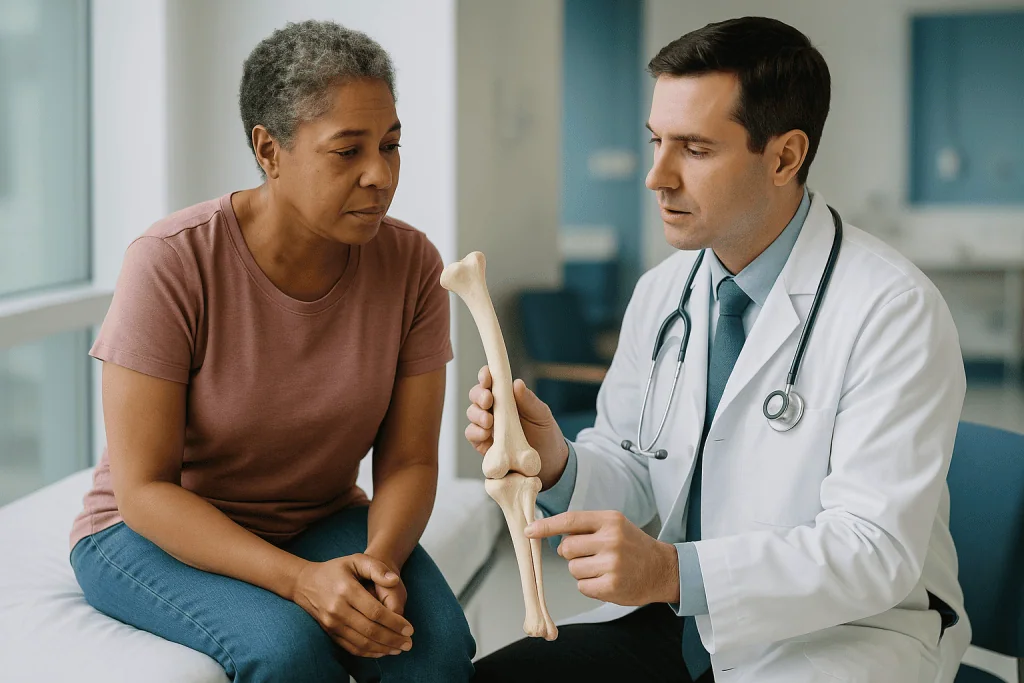
The location of the cut is chosen from measurements, not by eye. Before the operation, the surgeon marks the point where the bone actually bends, often called the apex of the deformity, on long-leg or full-spine radiographs. Cutting exactly at the apex lets the bone hinge into alignment with minimal shift. Cutting away from it, which is sometimes unavoidable because of a joint, a growth plate or hardware from an earlier operation, means the bone must both angle and slide, and the plan has to account for that.
The shape of the cut also varies. A wedge can be removed so the bone closes down onto itself, which shortens it slightly but heals with bone touching bone. A wedge can instead be opened and the gap filled with graft, which preserves or adds length but asks the body to bridge a space. A dome-shaped cut lets fragments rotate around one another. Each choice trades stability against length against healing time, and reasonable surgeons weigh those trades differently.
Most osteotomies are done with a saw or a series of drill holes joined with a fine chisel, the second method being kinder to the blood supply that new bone depends on. The cut itself usually takes minutes; the planning and the fixation that follow are where the operating time goes. Patients often imagine the osteotomy as the dramatic part of the day. To the surgical team it is the most rehearsed.
Why the plan matters more than the hardware
Ask an orthopedic surgeon what makes deformity correction succeed or fail and the answer is rarely the implant. It is the measurement done beforehand. Imaging is taken with the patient standing, because a bone that looks straight lying down can load very differently under body weight. The team draws lines along the shafts and across the joints and reads the angles where they meet, then compares each angle with published normal ranges.
From those numbers comes a target. For a bowed leg, the target might be a weight-bearing line that passes through or slightly outside the knee’s center rather than inside it. For a foot, it might be a first metatarsal that points forward instead of sideways. For a spine, it is usually a trunk that sits balanced over the pelvis so muscles are not working all day to hold the head upright.
Three-dimensional imaging has changed this step for complex cases. Twisting deformities, where one end of a bone is rotated relative to the other, are hard to see on flat films and easy to miss; a CT scan can measure the twist in degrees. Some teams now print a plastic model of the bone and rehearse the cut on it, or use a guide that fits the bone and shows where the saw should sit. These are planning aids, not proof of a better result, and the evidence comparing them with conventional planning is still developing.
What patients can take from this section is a question to bring to their consultation: what is my target, and how did you arrive at it? A team that can show the drawn lines and the angle they are aiming for has done the part of the operation that matters most, before anyone is asleep.
Fixation explained: plates, nails and frames
Once the bone is cut and moved, something must hold it there. Fixation is the collective name for that hardware, and it splits into two families: internal, which sits under the skin and stays in during healing, and external, which connects to the bone through pins or wires and lives outside the limb.
Internal options include plates screwed to the bone surface, nails passed down the hollow center of a long bone, and screws or staples for small bones of the foot and hand. A plate works like a splint bolted to the outside of a fence post. A nail works like reinforcing rod inside concrete. Both hold a correction that has been completed on the operating table, and both are usually left in permanently unless they cause irritation.
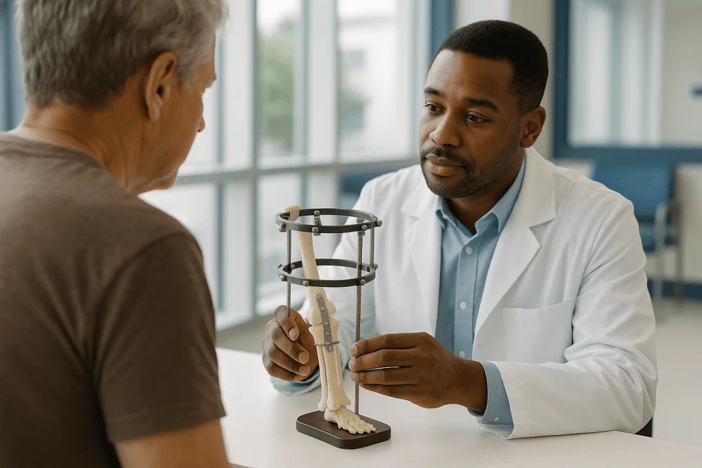
External fixation looks alarming and is the workhorse of gradual correction. Rings or bars sit around the limb, connected to the bone by thin tensioned wires or thicker pins. Struts between the rings can be lengthened or shortened by a small daily adjustment, moving the bone fragments in three dimensions over weeks. Because the frame can be adjusted after surgery, the correction does not have to be perfect on day one, which is exactly why surgeons choose it for large or complex deformities. The trade-off is pin sites that need daily care and carry an ongoing infection risk, and a device that is worn for months.
Newer internal nails can lengthen from inside the bone using a magnet held against the skin, avoiding an external frame for some straightforward lengthenings. Which fixation is appropriate is a judgment about the size of the correction, the quality of the soft tissue, the patient’s ability to manage a device, and the surgeon’s experience with each system.
Gradual realignment: how limb lengthening and deformity correction use time
Bone can be persuaded to grow across a gap if the gap is opened slowly enough. This is the principle behind distraction osteogenesis, a term that simply means making new bone by gradually pulling cut ends apart. It was refined for limb lengthening and deformity correction and remains the only reliable way to add several centimeters of length to a bone.
The sequence has three phases. After the osteotomy comes a short latency period, typically a few days to a week, while the body starts the early healing response. Then distraction begins: the frame or internal nail is adjusted in tiny steps, most commonly adding up to about one millimeter per day according to MedlinePlus, so that the soft callus forming in the gap is stretched rather than torn. The final and longest phase is consolidation, during which the new bone hardens; MedlinePlus notes the device stays in place for months while this happens, and the total time roughly scales with how much length was gained.
The same slow approach corrects angles and rotations, not just length. Muscles, nerves and blood vessels that have spent years adapted to a crooked bone cannot be stretched suddenly without risk of injury. Moving a few degrees a day gives them time to lengthen, which is why large angular corrections in adults and many corrections in children with growth remaining are done gradually rather than in a single step.
The cost of this biology is patience and daily attention. Adjustments are made at home, often by the patient or a family member, and progress is checked with regular X-rays. Physical therapy runs alongside to keep neighboring joints moving, because a joint that stiffens during lengthening is a common and avoidable problem.
Can bone deformity be fixed? What correction really means
Can bone deformity be fixed? In the mechanical sense, most can: a bent, short or twisted bone can be cut and realigned, and the tools described above are mature and widely used. What the question usually hides is a second one, about whether correcting the bone will also correct the pain, the limp or the wear that brought someone to the clinic. That answer is less certain and depends on how much damage the deformity has already caused.
Bone deformity has many origins, and the origin shapes what correction can do. Some people are born with a bone that grows at an angle or a leg that grows shorter than its partner. Some develop bowing during childhood from a growth plate that closes early on one side, from rickets, or from certain skeletal conditions. Many adult deformities are acquired: a fracture that healed at an angle, a joint that has worn down on one side so the limb has drifted, or a bone weakened by disease.
Where the joint surfaces are still healthy, realigning the bone changes how weight passes through them, which is the whole point of a knee osteotomy for early one-sided arthritis. Where the cartilage is already gone, straightening the bone will not bring it back, and the team may discuss joint replacement instead or alongside.
Correction also does not guarantee that a deformity will stay corrected. In a growing child the bone keeps growing, and if the underlying growth imbalance persists, the curve can return. That is why pediatric teams sometimes prefer to guide growth with a small plate across one side of a growth plate rather than cut the bone, and why follow-up continues until the skeleton matures. “Fixed” is a reasonable hope; “finished” is a separate question.
Can foot deformity be corrected?
The foot is where most people first meet the idea of deformity correction, usually through a bunion. A bunion is a bony bump at the base of the big toe that forms when the first metatarsal, the long bone behind the toe, drifts toward the midline while the toe angles the other way. Shoes do not cause it, though tight ones can make it hurt more; the NHS describes bunions as often running in families and worsening over time.
Correcting a bunion is deformity correction in miniature. The surgeon cuts the metatarsal, shifts the head of the bone back toward its neighbor, and fixes it with small screws or a plate. Larger angles may need a cut nearer the base of the bone or a fusion of the joint behind it. Recovery involves weeks in a stiff postoperative shoe; the NHS notes that a full return to normal footwear and activity commonly takes several weeks to a few months, and that surgery is generally considered only when pain limits daily life despite non-surgical measures such as wider shoes and padding.
Other foot deformities follow the same logic at different scales. A flatfoot in an adult may be treated with a cut through the heel bone that slides it sideways, combined with tendon work. A high-arched foot may need the opposite. Clubfoot in infants, where the foot points down and inward, is usually managed first with serial casting, a gradual non-surgical realignment that stretches soft tissue over weeks, with surgery reserved for what casting cannot achieve.
Toes and small bones heal on the same biology as large ones but tolerate less error, since a millimeter of shift changes which part of the foot strikes the ground. Foot correction is precise work, and swelling can persist for months after the bone itself has healed.
Spinal deformity correction surgery: can spinal deformities be corrected?
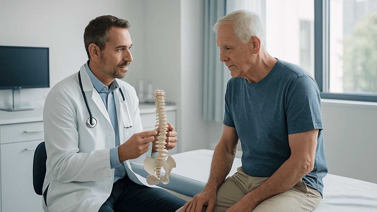
Spinal deformities can be corrected, and the principles carry over, but the spine adds two complications a leg does not have: the deformity is spread across many bones rather than one, and the spinal cord runs through the middle of them. Scoliosis, a sideways curve of the spine that also rotates the vertebrae, is the most common reason for spinal deformity correction surgery; kyphosis, an excessive forward rounding, is the other.
Most scoliosis is never operated on. Mayo Clinic describes bracing as the usual approach for a growing child whose curve measures roughly between 25 and 40 degrees, with the aim of stopping progression rather than straightening, and notes that surgery is generally considered when a curve exceeds about 45 to 50 degrees in a child who is still growing or when a curve in an adult causes pain or nerve symptoms. Smaller curves are watched with periodic X-rays.
The standard operation is a spinal fusion. Screws are placed into the vertebrae along the curve, rods are attached and used to pull and rotate the segments toward a straighter, balanced position, and bone graft is packed between the vertebrae so they knit into one solid segment. Correction is partial by design; the aim is a trunk balanced over the pelvis with the shoulders level, not a ruler-straight spine, and the fused section no longer bends. MedlinePlus notes that a hospital stay after spinal fusion is commonly several days, and the NHS advises that children typically return to school within weeks and to sports over subsequent months, with full bone fusion taking many months.
Younger children with severe curves may instead receive growing rods, which are lengthened periodically to let the chest keep developing before a final fusion. Nerve monitoring throughout surgery is standard, and the risk of neurological injury, while low, is the one every team discusses first.
Acute versus gradual correction: a side-by-side comparison
Patients are often told they will have either a “one-stage” correction or a frame, without much explanation of why. The table sets the two approaches beside each other. It generalizes, and an individual plan can blend elements of both, but the pattern of trade-offs is consistent across body regions.
| Feature | Acute (one-stage) correction | Gradual correction |
|---|---|---|
| What happens | Bone is cut, moved to final position and fixed during a single operation | Bone is cut, then moved in small daily steps over weeks using an adjustable device |
| Typical fixation | Plate and screws, or intramedullary nail | External ring frame or magnetically lengthened internal nail |
| Suited to | Smaller angular corrections; little or no lengthening needed; healthy soft tissue | Large angles, significant lengthening, multiplane deformities, tight or scarred soft tissue |
| Adjustability after surgery | Essentially none without reoperation | High; the plan can be fine-tuned against follow-up X-rays |
| Main risk profile | Nerve or vessel stretch if correction is large; non-union of an opened gap | Pin-site infection, joint stiffness, delayed consolidation, device wear over months |
| Patient workload | Weight-bearing limits and therapy for weeks | Daily adjustments and pin care plus therapy for months |
| Time to healed bone | Weeks to a few months | Months, scaling with the amount of length gained (MedlinePlus) |
Two points deserve emphasis. First, gradual correction is not a fallback for difficult cases; for many large deformities it is the safer first choice precisely because nerves and vessels are given time to adapt. Second, the acute approach is not the “easy” one; a plate that is fixed in a slightly wrong position cannot be nudged afterward, so it places more weight on the accuracy of the preoperative plan. The treating team chooses between them by measuring how far the bone must travel and how much the tissues around it can tolerate.
Who deformity correction surgery is usually for, and who is asked to wait
Surgery is generally offered when a deformity is causing a problem that non-surgical care has not controlled, or when it is expected to cause one that would be harder to treat later. In adults that typically means pain with weight-bearing, a limp or leg-length difference that is affecting the back and hips, early arthritis concentrated on one side of a joint, or a spinal curve that is progressing or pressing on nerves. In children it means a curve or length difference that is growing faster than the child, or one already large enough to affect walking or, in the spine, chest development.
Candidates are also assessed for their capacity to heal and to participate. Bone healing depends on blood supply and nutrition; teams look at smoking status, vitamin D levels, diabetes control and other factors that influence how well a gap fills with new bone. Gradual correction in particular asks the patient or family to perform daily adjustments and pin care, and a realistic conversation about that commitment is part of every assessment.
Several groups are commonly asked to wait or to consider alternatives. A growing child whose deformity is mild and not progressing is often watched, because growth itself may improve the alignment or because guided growth with a small plate is a smaller operation than an osteotomy. A person whose joint cartilage is already severely worn may be steered toward joint replacement rather than realignment. Someone with an active infection, uncontrolled medical illness or who smokes heavily is often asked to address that first, because each raises the risk of the bone failing to heal.
None of these are rules. They are the considerations a treating team weighs, and the decision belongs to that team together with the patient.
What the following days and weeks usually look like
The first days after surgery are about pain control, swelling and getting moving. Many limb corrections allow partial weight-bearing early with a walker or crutches; some require no weight for several weeks while the cut heals, depending on the bone, the fixation and the surgeon’s protocol. After spinal fusion, MedlinePlus describes a hospital stay of several days with early walking encouraged. Physical therapy typically begins within the first week for any of these procedures, aimed at keeping adjacent joints moving rather than building strength.
With a gradual correction, the distraction phase starts after a short latency period. Adjustments are made at home each day according to a written schedule, and X-rays every week or two confirm that the bone is moving as planned and that new bone is appearing in the gap. Pin sites are cleaned daily. Some discomfort during lengthening is expected and tends to be worse toward the end of each day’s distraction; therapy focuses hard on stretching muscles that are being asked to grow.
The consolidation phase follows, when the new bone hardens. The NHS and MedlinePlus both describe healing from major bone surgery in months rather than weeks, and for lengthening MedlinePlus notes the device remains until the new bone is strong enough to bear load, with total time scaling with the length gained. For bunion correction, the NHS describes several weeks in a protective shoe before normal footwear, and residual swelling that can last longer.
Return to work depends on the job. Desk-based tasks may resume within weeks of many procedures; standing or physical work waits for solid bone. Driving is generally not advised until the operated limb can perform an emergency stop and pain medicine that impairs alertness is no longer needed, a judgment the treating team makes rather than a fixed date.
Risks and alternatives, described neutrally
Every operation that cuts bone carries the risks of any surgery: bleeding, infection, blood clots and reaction to anesthesia. Deformity correction adds risks specific to the task. The cut bone may heal slowly or not at all, a problem called non-union, which is more common when a gap has been opened, in smokers, and in bones with poor blood supply. It may heal in a slightly wrong position, called malunion, requiring further adjustment. Nerves and vessels that run alongside the bone can be stretched or injured, particularly during large acute corrections.
Gradual correction trades some of those risks for others. Pins and wires pass through the skin for months and pin-site infection is common enough that most teams treat it as expected rather than exceptional; most cases settle with local care, while a smaller number need oral antibiotics or pin exchange. Joints near the lengthening segment can stiffen, and muscles may tighten faster than they stretch. Bone that forms in the gap can be slow to consolidate, extending time in the frame.
Spinal correction carries the additional possibility of neurological injury, which is why intraoperative nerve monitoring is standard, and of the fusion failing to knit or of hardware loosening. Adjacent segments of the spine take on more motion after a fusion and may wear faster over years.
Alternatives are real and often used first. Shoe inserts or a heel lift can compensate for modest leg-length differences. Bracing is the mainstay for moderate scoliosis in growing children. Wider footwear and padding manage many bunions. Guided growth is a smaller operation than osteotomy in children. For a worn joint, replacement is an alternative to realignment. Which path is appropriate is a decision between patient and treating team, informed by these trade-offs rather than by the promise of any single technique.
What people often get wrong about how deformity correction surgery works
The first and most persistent misunderstanding is that the bone is somehow bent back into shape. It is not. Adult bone does not bend; it breaks. Correction always involves a cut, even for what sounds like a small adjustment, and the new shape is made by how the pieces are positioned and healed, not by force applied to intact bone.
A second myth is that a straight X-ray equals a solved problem. Alignment is a means to an end. If the cartilage in a joint is already destroyed, realigning the bone above it will not regrow that cartilage. If a spinal curve has been corrected but the trunk still leans forward, the patient may feel no better. Good teams aim for balance and load distribution, and a modest correction that achieves those often serves a patient better than a dramatic one that overshoots.
Many people also believe external frames are an outdated or last-resort method. In practice they remain the most versatile tool for large, complex or multiplane deformities because they can be adjusted after surgery; internal nails that lengthen are a welcome addition for some cases, not a replacement for all of them.
A fourth misconception concerns speed. Patients sometimes ask why the surgeon cannot simply add the full length or angle on the day. The answer is soft tissue: nerves stretched quickly can stop working, and vessels can spasm or tear. Slow correction is a safety choice, not a limitation of technique.
Finally, there is a widespread assumption that once bone has healed the job is done. In children the skeleton continues to grow and the correction is monitored until maturity; in adults the joints above and below adapt to a new load pattern over a year or more, and therapy through that period matters as much as the operation.
Questions to ask your care team
A consultation about deformity correction can move quickly from anatomy to scheduling. Bringing written questions slows it down to a pace where understanding is possible. The following are the ones experienced patients most often wish they had asked earlier, grouped by the stage of care they concern.
- What exactly is my deformity, in degrees or millimeters, and what target alignment are you aiming for? Could you show me the lines on my X-ray?
- Will the correction be done in one stage or gradually, and what led you to that choice for me?
- Which type of fixation will you use, and does it stay in permanently or come out later?
- What is my role after surgery: daily adjustments, pin care, weight-bearing limits, therapy sessions?
- How long, as a typical range, until I can bear full weight, drive, return to my particular work, and stop attending regular follow-up?
- What are the specific complications you see most often with this operation, and how are they managed?
- What happens if the bone is slow to heal or the correction is not where we planned?
- Are there non-surgical options or a smaller operation that would be reasonable to try first?
- If I am still growing, or my child is, how will growth affect the result and how long will monitoring continue?
- Is there anything about my health, such as smoking, vitamin D, diabetes or weight, that you would want addressed before surgery?
Answers to these questions do not need to be memorized. They need to be heard, and ideally written down or recorded with permission, so that the decision to proceed is made with the plan understood rather than merely trusted. A team confident in its planning will welcome them.
When to call your doctor
Most recovery after deformity correction is uneventful, and the team will describe what normal discomfort and swelling look like for the specific operation. Certain signs, however, should prompt a call to the treating team the same day, or emergency care if the team cannot be reached.
Around the surgical site or pin sites, seek care for spreading redness, increasing warmth, pus or cloudy drainage, a foul smell, or a pin that has become loose or newly painful. A fever, especially with chills or feeling generally unwell, warrants a call even without an obvious wound problem, because deep infection around bone or hardware needs prompt assessment.
In the operated limb, new or worsening numbness, tingling, weakness, or an inability to move the toes or fingers can signal nerve stretch or compression and should not wait until the next appointment. Pain that is escalating rather than settling, is out of proportion to what was described, or is not relieved by rest and elevation also needs review, as does a limb that becomes pale, cold or unusually swollen and tight.
After spinal surgery, new weakness or numbness in the legs, new difficulty controlling the bladder or bowel, or severe headache when upright are red flags requiring urgent contact.
Anywhere in the body, calf pain or swelling, sudden breathlessness, chest pain or coughing up blood can indicate a blood clot and are reasons to seek emergency care immediately rather than waiting for a callback.
If something feels wrong and does not fit any of these descriptions, calling is still the right choice. Teams that perform these operations expect questions during recovery and would rather hear about a concern early than manage a complication late.
Frequently asked questions
What is the process of correcting a deformity?
The process has four stages: measuring the deformity on standing X-rays or CT to set a target, cutting the bone at the planned point (an osteotomy), moving the fragments into alignment either in one operation or gradually over weeks, and holding them with plates, nails or an external frame until new bone heals across the cut. Rehabilitation and follow-up imaging continue for months afterward.
Can bone deformity be fixed in adults, or only in children?
Adult bone deformities can be corrected; the biology of healing across a cut works at any age, though it is slower in adults and in smokers. Children have the extra option of guided growth, where a small plate slows one side of a growth plate so the bone straightens as it grows. Adults rely on osteotomy with fixation, sometimes with gradual correction for large deformities.
What is osteotomy surgery and does it always need a plate?
An osteotomy is a planned surgical cut through bone that lets a surgeon reposition it. Fixation is almost always needed so the pieces heal in the new position, but it does not have to be a plate. Nails inside the bone, screws, staples, or an external frame are all used, and the choice depends on the bone, the size of the correction and whether the correction will be gradual.
How does limb lengthening and deformity correction add length to a bone?
It uses a process called distraction osteogenesis. After the bone is cut, an adjustable frame or internal nail pulls the ends apart in tiny daily steps, commonly about one millimeter per day according to MedlinePlus, and new bone forms in the widening gap. The device then stays in place for months while that bone hardens; total time scales with the length gained.
Can spinal deformity correction surgery make the spine completely straight?
Rarely, and that is not the goal. Spinal fusion for scoliosis uses screws and rods to pull the curve toward a balanced position and then fuses those vertebrae so the correction holds. Surgeons aim for a trunk centered over the pelvis with level shoulders rather than a perfectly straight radiograph, partly because over-correction can stress the unfused segments above and below.
Can foot deformity be corrected without surgery?
Often the symptoms can be managed without surgery, even when the shape cannot be changed. The NHS recommends wider shoes, padding and pain relief for bunions before considering an operation, and clubfoot in infants is usually treated first with serial casting. Surgery is generally reserved for deformities that cause pain or function problems despite these measures.
How long does deformity correction surgery take to heal?
Healing is measured in months. MedlinePlus describes bone-lengthening devices remaining in place for months until new bone is strong enough to bear weight, with time scaling to the length gained. The NHS notes that children return to school within weeks of scoliosis fusion but to sports over months, and that bunion recovery involves several weeks in a protective shoe. Your team will give a range for your case.
Is deformity correction surgery painful?
Pain is expected after any bone surgery and is managed by the treating team with a plan tailored to the procedure. With gradual correction, discomfort often accompanies the daily adjustment and stretching of muscles, and physical therapy helps. Pain that escalates rather than settles, or that comes with numbness or weakness, is a reason to contact the team rather than something to tolerate.
Does the hardware from deformity correction stay in forever?
Internal plates and nails are frequently left in place permanently unless they irritate soft tissue or a later operation requires removal. External frames are always removed once the new bone has consolidated, usually in a short procedure. Magnetically lengthened internal nails are often removed after healing because they are not designed for lifelong load. Your surgeon will explain the plan for your specific implant.
What happens if the bone does not heal after an osteotomy?
Slow or failed healing, called delayed union or non-union, is a recognized complication and has established treatments. Options include extending the time in the device, adding bone graft to the gap, changing the fixation to provide more stability, or addressing factors such as smoking or low vitamin D that impair healing. The treating team monitors with regular X-rays so problems are caught early.
References
- MedlinePlus – Leg lengthening and shortening
- MedlinePlus – Spinal fusion
- NHS – Bunions
- NIH NIAMS – Scoliosis
This article is for general information only and is not a substitute for professional medical advice. Please consult a qualified doctor about your individual situation.

More from the Blog

What Happens During Knee Replacement Surgery: A Step-by-Step Look Inside the Operating Room
During knee replacement surgery, a surgeon opens the front of the knee, removes the worn cartilage and a thin layer of damaged bone from…

Hip, Knee or Shoulder Replacement: How Recovery Differs Joint by Joint
Recovery after hip, knee and shoulder replacement follows the same broad arc but differs in detail. Hip replacement usually allows walking within a day…

Wound Care After Knee Replacement: What Healing Looks Like and When to Call Your Team
Wound care after knee replacement means keeping the incision clean, dry and covered as your surgical team instructs, changing dressings only when told, and…

How Is Pain Managed After Hand Surgery? Elevation, Splints and Safe Relief
Pain after hand surgery is usually managed with a combination of measures: keeping the hand raised above heart level to limit swelling, wearing the…

Glucosamine, Collagen and the Arthritis Diet: What Trials Show for Joint Pain
Glucosamine is a natural building block of cartilage sold as a supplement for osteoarthritis. Large randomized trials show it works no better than placebo…
Kinesiology Tape: What the Studies Show Behind the Colorful Strips
Kinesiology tape is a stretchy cotton strip applied over muscles and joints. Randomized trials and systematic reviews show it produces, at best, small and…