

How Doctors Check Whether CAR-T Is Working: Scans, Blood Tests and the Waiting Period

Key Takeaways
- Response after CAR-T is assigned a formal category, complete, partial, stable or progressive, on a scheduled test, and most centers perform that first assessment around one month after infusion (Cleveland Clinic).
- Blood tests in the first two weeks, including CRP, ferritin and blood counts, track the safety reactions CRS and ICANS and say almost nothing about whether the cancer is responding (Lee et al.).
- For lymphoma the key response test is a PET/CT read on the Deauville five-point scale, where uptake no brighter than the liver is generally treated as complete metabolic response (MedlinePlus; Lugano criteria).
- For leukemia and myeloma, bone marrow sampling and measurable residual disease testing can detect one abnormal cell among ten thousand or more, far below what any scan can see (MedlinePlus).
- Disappearance of normal B cells after CD19-targeted CAR-T is an indirect sign that engineered cells are still active, and their return often signals the cells have faded (NCI).
- Inflammation caused by active CAR-T cells can make an early scan look worse than the disease actually is, so a repeat scan or biopsy is sometimes needed before anyone declares the treatment has not worked.
Doctors judge whether CAR-T has worked mainly with imaging and laboratory tests done on a schedule, not in the first days after infusion. For lymphoma, a PET/CT scan is usually the key test; for leukemia and myeloma, bone marrow samples and blood markers matter more. Early blood tests track safety and CAR-T cell activity rather than proving a response, and the treating team interprets results over weeks to months.
The infusion itself is oddly brief. A small bag, a line already in place, and in well under an hour the cells are in. Then comes the part nobody warns you about properly: the silence. No scan the next morning, no verdict on the ward round. Just temperature checks, blood draws before breakfast, and a nurse asking you to write your name and the date on a whiteboard, again.
People facing this stretch almost always ask the same thing, sometimes on day three: how to know if CAR-T worked. It is a fair question with a frustrating answer, because the tests that can actually answer it are deliberately scheduled weeks away, while the tests happening every morning are answering something else entirely.
This explainer walks through what each test measures, when it is usually done, and what it can and cannot tell you. The point is not to turn you into your own hematologist. It is to let you read the waiting period for what it is: a plan, not a gap.
How to know if CAR-T worked: what "worked" actually means
The question sounds simple, but “worked” means different things to different people in the room. To a patient, it usually means the cancer is gone. To a hematologist, it means the disease has met a defined response category on a defined test at a defined time, and that category has a name.
Response is graded, not felt. A complete response means no detectable disease on the agreed test: no active lymphoma on a PET/CT scan, or a bone marrow sample without leukemia cells. A partial response means the disease has shrunk substantially but is still measurable. Stable disease means little change, and progression means growth or new sites. These definitions come from international response criteria for lymphoma and leukemia, which is why two hospitals on different continents can compare notes about the same scan.
Two further ideas matter. Depth of response describes how little disease remains; measurable residual disease testing, covered later, pushes that measurement far below what a scan can see. Durability describes how long a response lasts, and it can only be judged by time passing. A complete response at the first assessment is genuinely good news, yet the treating team will not call it durable until later checks agree.
So the honest answer is layered. There are early laboratory clues within weeks, a formal response assessment that most centers schedule around the one-month mark, and a series of confirmations over the following months (Cleveland Clinic). Each layer answers a slightly different question. Knowing which question is being answered on any given day spares a great deal of unnecessary worry, and it stops a routine blood count from being read as a verdict it was never designed to deliver.
What actually happens during CAR-T, from blood draw to infusion
CAR-T stands for chimeric antigen receptor T-cell therapy: a treatment that takes a patient’s own T cells, a type of white blood cell that hunts abnormal cells, and re-engineers them in a laboratory to recognize a target on the cancer. Understanding the steps explains why the checking happens when it does.
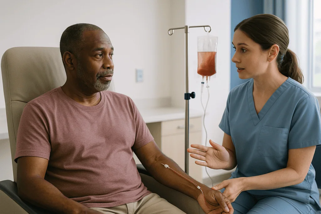
First comes collection, called leukapheresis. Blood flows out through a line, a machine separates white cells, and the rest returns to the body. This usually takes a few hours in one sitting. The collected cells are sent to a manufacturing facility, where a gene for the chimeric antigen receptor is inserted so the T cells display a new sensor on their surface. The cells are then multiplied into the hundreds of millions. Manufacturing generally takes several weeks (NCI).
Shortly before the cells return, patients receive lymphodepleting chemotherapy: a short course intended to lower the existing lymphocyte population so the engineered cells have room, and the right chemical signals, to expand. Choices about that chemotherapy sit entirely with the treating team.
The infusion itself is a single intravenous procedure. Afterwards, the engineered cells do something no ordinary medicine does: they multiply inside the body, often by orders of magnitude, seek out their target, and destroy cells carrying it. In many current treatments the target is CD19, a protein on B cells, or BCMA, a protein on myeloma cells (NCI).
That expansion phase is the reason for the waiting period. The drug is alive. Its peak activity, its side effects and its anti-cancer work all unfold across the first two to three weeks rather than at the moment of infusion. Assessing response before that process has run its course would be like judging a bread dough before it has proved.
Who CAR-T is usually for, and who is asked to wait
CAR-T is currently used for certain blood cancers, chiefly some forms of B-cell lymphoma, B-cell acute lymphoblastic leukemia and multiple myeloma, when the disease has come back after earlier treatment or has not responded to it (NHS England; NCI). Eligibility is a specialist decision made by a multidisciplinary team, and the checklist is longer than most people expect.
The team looks at the disease itself: its type, its target protein, how fast it is moving, and whether it can be held steady during the manufacturing weeks. They look at the patient: heart, lung, kidney and liver function, because the immune reaction after infusion places real demands on those organs. They look at recent infections, because an active infection during the period of low blood counts can be dangerous. And they look at practical matters, such as whether someone can stay close to the center and have a caregiver with them.
Being asked to wait is common and is not a refusal. Someone may be asked to wait while an infection is treated, while bridging therapy brings a rapidly growing lymphoma under short-term control, or while a specialist reviews cardiac or neurological findings. Others may be told that a different option, such as a stem cell transplant or a bispecific antibody (a laboratory-made protein that links a T cell to a cancer cell), is more suitable for their situation. Those alternatives carry their own risks and timelines, and weighing them is the team’s job.
This matters for the theme of this article, because the waiting begins before infusion, not after it. Patients who understand that manufacturing, bridging and safety checks are part of the treatment rather than delays to it tend to find the later waiting period less alarming.
Why nobody checks for a response in the first week
In the first week after infusion the daily attention is intense, and yet almost none of it is about whether the cancer is responding. It is about safety, and specifically about two reactions that come from the treatment doing exactly what it was designed to do.
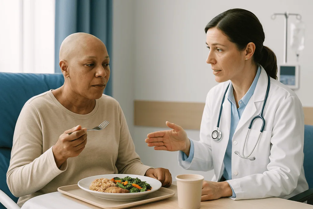
The first is cytokine release syndrome, or CRS: a flood of inflammatory signaling molecules released as activated T cells attack their targets. It typically shows up as fever, sometimes with low blood pressure or low oxygen, and it usually begins within the first days after infusion, though timing varies with the product and the disease (Lee et al., PubMed). The second is immune effector cell-associated neurotoxicity syndrome, or ICANS: inflammation affecting the brain that can cause confusion, word-finding difficulty, drowsiness or, rarely, seizures. Both are graded on a standardized international scale so that teams anywhere describe them in the same way (Lee et al.).
That is why you are asked to write a sentence or name objects each morning. It is a neurological screen, not a memory test, and a changed handwriting sample can prompt action hours before anything else would.
Hospitals and national health services typically ask patients to remain in or very near the treating center for around four weeks, with a caregiver present, and to avoid driving for a longer period afterwards (Cleveland Clinic; NHS England). The exact rules are set by the treating team and the product’s safety information.
None of this measures response. A fever is not proof the treatment is working, and the absence of fever is not proof it is failing. The cells may be expanding vigorously in someone who feels well. The first week answers the question “is this safe right now,” and it is meant to.
CAR-T blood tests in the first month: what counts, ferritin and CRP really show
The morning blood draw is the most misread event of the whole waiting period. It is worth knowing what each common test is for, so that a single number does not ruin a day.
The complete blood count measures red cells, white cells and platelets. After lymphodepleting chemotherapy these fall, sometimes steeply, and may stay low for weeks; prolonged low counts after CAR-T are well recognized (NCI). A low white cell count on day ten says nothing about the lymphoma. It says the chemotherapy did its job and the marrow is recovering on its own schedule.
C-reactive protein, or CRP, is a general marker of inflammation made by the liver. Ferritin is an iron-storage protein that also rises sharply during inflammation. Both climb during CRS and are used to track its course. A rising ferritin is a safety signal for the team, not a scorecard for the cancer.
Liver enzymes, kidney markers and electrolytes are checked because inflammation and supportive treatment can strain those organs. Clotting tests may be added when CRS is more severe.
Some centers also measure the engineered cells directly, using flow cytometry, a technique that counts cells by the proteins on their surface, or PCR, which detects the inserted gene. These show how much the CAR-T population has expanded and, later, whether it persists. Expansion is generally regarded as a good sign of activity, but it is not itself a response measurement, and it is not available everywhere.
A practical habit helps: ask the team which numbers they are watching this week and why. In the first month the answer is almost always “the safety ones.” Hearing that plainly makes the printout less frightening.
B-cell aplasia and CAR-T expansion: the indirect clues that the cells are active
Between the safety tests and the formal scan sits a category of evidence that is easy to overlook: indirect signs that the engineered cells are alive and doing their work.
For treatments that target CD19, the clearest sign is B-cell aplasia. Because CD19 sits on healthy B cells as well as cancerous ones, active CAR-T cells clear both. A blood test showing that normal B cells have disappeared, and stay absent, indicates that functional CAR-T cells are still on patrol (NCI). Clinicians sometimes describe this as an “on-target, off-tumor” effect: unwanted in principle, but informative in practice. When B cells eventually return, it often means the engineered cells have faded, which is one reason teams keep an eye on that count for a long time.
The consequence is a fall in immunoglobulins, the antibodies B cells produce. Low immunoglobulin G raises infection risk, and many centers monitor levels and may offer replacement therapy; whether and when is the treating team’s decision.
For myeloma treatments targeting BCMA, the analogous clue comes from disease markers rather than normal cells. Paraprotein, an abnormal antibody made by myeloma cells and measured by serum protein electrophoresis, and serum free light chains, fragments of antibody, usually begin to fall within weeks if the treatment is active. These are followed at each visit.
The intensity of CRS is sometimes read as a proxy for activity, since more T-cell activation tends to release more cytokines. Published experience shows the relationship is loose, and the international grading paper treats CRS strictly as a toxicity to be managed rather than a marker of benefit (Lee et al.). A mild course is not a bad omen. A stormy one is not a guarantee.
When is the first PET scan after CAR-T, and how is it read?
For lymphoma, the PET/CT scan is where the question in this article finally gets a direct answer. A PET scan uses a small amount of a radioactive sugar tracer that active cells, including many cancer cells, absorb more avidly than resting tissue; the scanner maps where the tracer collects (MedlinePlus). Combined with CT, which shows anatomy, it can distinguish a lymph node that is enlarged but quiet from one that is metabolically busy.
Most centers perform a first response PET/CT roughly one month after infusion, with a further scan around three months, then at intervals set by the team (Cleveland Clinic). Protocols differ, and the interval may be shortened if there is clinical concern or lengthened if a scan is clean.
Reading the scan uses the Deauville five-point scale, part of the internationally adopted Lugano response criteria. Each site is scored by comparing its brightness with two internal references: the mediastinal blood pool (the large vessels in the chest) and the liver. Scores of one to three, meaning no brighter than the liver, are generally treated as complete metabolic response. Scores of four and five, brighter than the liver or with new sites, indicate residual or progressive disease. The radiologist’s report will usually quote these numbers, and it is reasonable to ask what score each site received.
A few caveats keep the scan honest. The tracer is taken up by inflammation as well as by cancer, so recently treated tissue, infection, or even the immune activity of the CAR-T cells themselves can glow. Brown fat, muscle activity and recent growth-factor injections can add noise. That is why the team reads the scan alongside blood results and how you are doing, and why a single early image is rarely the final word.
MRD after CAR-T: bone marrow tests for leukemia and myeloma
Leukemia and myeloma live in the bone marrow, so a scan is the wrong instrument. Instead, the response assessment relies on a bone marrow sample and on the concept of measurable residual disease, usually shortened to MRD.
A bone marrow test involves numbing the skin over the back of the pelvis, drawing out a small amount of liquid marrow through a needle (an aspirate) and often removing a thin core of bone (a biopsy). The procedure typically takes a few minutes of actual sampling, with soreness for a day or two afterwards (MedlinePlus). For leukemia, the first sample is commonly taken around the one-month mark, mirroring the lymphoma scan schedule.
Under a standard microscope, a pathologist looks for leukemia cells among normal marrow cells. A marrow with fewer than a threshold percentage of blasts and recovering normal cells meets the conventional definition of remission. MRD testing goes far deeper. Using flow cytometry or molecular methods such as PCR or next-generation sequencing, laboratories can detect one abnormal cell among ten thousand, or even a million, normal cells. “MRD-negative” means none were found at that sensitivity; it does not mean none exist.
For myeloma, the picture is assembled from several sources: paraprotein and free light chains in blood and urine, a marrow sample, and sometimes imaging to check for bone lesions or soft-tissue disease that the marrow would miss.
Why does depth matter? Across blood cancers, deeper responses are generally associated with longer control, which is why teams track MRD over time rather than once. But a single MRD-negative result is a snapshot. The team will want to see it hold at later checks, and MRD status is one input into their thinking, not a decision made by the laboratory.
The waiting period: what the first weeks and months usually look like
Timelines vary by disease, by product and by how each person recovers, so the schedule below is a typical shape rather than a promise. Your team’s calendar overrides it.
| Period after infusion | What is usually being checked | What it can tell you about response |
|---|---|---|
| Days 0–14 | Temperature, blood pressure, oxygen, daily handwriting and orientation checks, blood counts, CRP, ferritin, organ function | Very little. This is the safety window for CRS and ICANS (Lee et al.) |
| Weeks 2–4 | Recovery of blood counts, B-cell count, immunoglobulins, CAR-T expansion where measured, myeloma markers | Indirect clues that the cells are active |
| Around 1 month | PET/CT for lymphoma; bone marrow and MRD for leukemia; marrow plus blood markers for myeloma | First formal response category (Cleveland Clinic) |
| Around 3 months | Repeat imaging or marrow, immune recovery, infection review | Confirmation and early sense of durability |
| 6–12 months and beyond | Periodic scans or marrow as directed, B cells, immunoglobulins, vaccination planning, late-effect monitoring | Durability, and whether engineered cells persist |
The first month is spent at or near the center, with a caregiver, because CRS and ICANS can appear with little warning and are treated far more easily when caught early (NHS England; Cleveland Clinic). Energy is often low. Appetite may be poor. Many people describe a fog that lifts unevenly.
After the first assessment, visits usually thin out. Blood counts may take months to normalize, and infection precautions continue while B cells and antibodies are low. Long-term follow-up is expected for years, both to watch the cancer and to monitor for late effects, and registries collect this information across countries so that experience is shared. Waiting, in other words, is not the absence of a plan. It is the plan, and every line of that table has a reason.
Why an early scan can look worse before it looks better
One of the harder conversations in a CAR-T clinic goes like this: the one-month scan shows a bright node, sometimes larger than before, and yet the team asks for patience. This is not evasion. It reflects a well-documented behavior of immune-based treatments.
When engineered T cells reach a tumor, they recruit other immune cells, release cytokines and set off local inflammation. Inflamed tissue takes up the PET tracer avidly and can swell. On the image, that looks exactly like growing cancer. The term for this is pseudoprogression: apparent worsening on imaging that is actually immune activity, followed by shrinkage on a later scan. It was first described with immune checkpoint drugs and has been reported after CAR-T as well.
Distinguishing pseudoprogression from real progression on a single image is often impossible. Clinicians therefore weigh other evidence: whether blood markers are falling, whether the patient is well, whether the pattern is confined to known sites or has appeared somewhere new, and whether CAR-T cells are still expanding. Sometimes the answer is a repeat scan in a few weeks. Sometimes it is a biopsy of the suspicious site, which can show inflammatory cells rather than lymphoma.
The mirror image also exists. A node can remain enlarged on CT while being metabolically silent on PET, because dead or scarred tissue does not shrink instantly. That is why the Deauville score, which measures activity rather than size, is the preferred yardstick, and why a report saying “residual soft tissue, no abnormal uptake” is usually reassuring.
What this means for you is simple to say and hard to do: try not to read the first image as the ending. The team is not withholding a verdict. The evidence needed for a verdict genuinely does not exist yet.
What if CAR-T doesn't work: partial response and relapse
Some people will hear that the assessment shows a partial response, or that disease has returned after an initial response. Both outcomes are real possibilities, and knowing how teams think about them removes some of the dread.
A partial response at one month is not a closed door. Because CAR-T cells can continue to act for weeks, some partial responses deepen into complete responses on the three-month assessment; others do not. The team may choose to watch and rescan rather than intervene immediately, depending on how the disease is behaving and how you are.
When disease progresses or relapses, the first question is why. Broadly, two mechanisms are recognized (NCI). In antigen-negative relapse, the cancer cells stop displaying the target protein, so the engineered cells can no longer see them; a biopsy or flow cytometry can show this. In antigen-positive relapse, the target is still there but the CAR-T cells have become exhausted, faded or been too few. The distinction matters because it shapes the next options.
Those options vary widely: other targeted or immune therapies, chemotherapy, clinical trials, a stem cell transplant for some, or in certain situations a second cellular therapy directed at a different target. Each carries its own risks and demands on the body, and the choice depends on the disease, prior treatments and personal priorities. Some people choose approaches focused on comfort and quality of life, and that too is a legitimate medical plan.
Nobody can promise which path leads where. What can be said, and is worth saying, is that a disappointing scan starts a new decision process rather than ending the conversation, and that the same team who planned the CAR-T will be the ones to lay out what comes next.
What people often get wrong about knowing if CAR-T worked
Waiting rooms generate their own folklore. A few of the most common misunderstandings deserve a correction grounded in what the evidence actually shows.
“If I don’t get a fever, it isn’t working.” Fever reflects cytokine release, which is a toxicity, not an efficacy readout. The international grading system treats CRS purely as something to manage (Lee et al.). Responses occur in people with little or no CRS.
“Feeling better means the cancer is gone.” Feeling better in week three usually means the chemotherapy and inflammation are wearing off. It is welcome, and it is not a response assessment.
“My white count is low, so the treatment has failed.” Low counts after lymphodepletion are expected and may persist for weeks or months (NCI). They describe the marrow’s recovery, not the lymphoma’s status.
“A clean one-month scan means I’m done.” It means complete response at that time point. Durability is judged over subsequent months, which is why the follow-up calendar continues.
“An MRD-negative result means zero cancer cells.” It means none were detected at the test’s sensitivity. That is meaningful, and it is different from zero.
“CAR-T works or fails immediately.” The cells expand over weeks, and partial responses can deepen. Early progression on imaging can also be inflammation, so a repeat scan is sometimes needed before anyone can say the treatment did not work.
“The nurse’s daily questions mean they think I’m confused.” They are a standardized neurological screen applied to everyone, and catching early ICANS is exactly why they exist.
Each of these misreadings comes from applying the wrong test to the question. Match the test to the question, and most of the false alarms disappear.
Questions to ask your care team
A good consultation is a two-way exchange, and the team will expect questions. Written down in advance, these tend to draw out the most useful answers about how your response will be judged.
- Which test will be used to assess my response, and roughly when is the first one planned?
- Which blood tests are you watching in the first month, and which of them are about safety rather than the cancer?
- Will you be measuring the CAR-T cells directly, or following indirect signs such as B-cell counts?
- How will you describe the result to me: as a response category, a Deauville score, an MRD level, or a change in paraprotein?
- If the first scan or marrow is unclear, what happens next, and how long might that take?
- What counts as a good result at one month, and what would prompt a change of plan?
- How long should I expect my blood counts and antibodies to stay low, and what infection precautions apply during that time?
- When are vaccinations reconsidered, and who coordinates that?
- How long do I need to stay nearby, and when is it safe to drive or return to work?
- Who do I call, at any hour, if I develop a fever or feel confused, and what information should I have ready?
- Will I be entered into a long-term follow-up registry, and what does that involve?
- If the treatment does not produce the response we hope for, what options would you want to discuss?
You will not need every answer at once. Many teams provide a written plan, and it is reasonable to ask for one. Bringing a caregiver to appointments helps too; the first month is tiring, and two sets of ears remember more than one. Whatever the answers, the decisions about tests, timing and next steps remain with the treating team, informed by what matters most to you.
When to call your doctor
The response tests can wait for their scheduled dates. Certain symptoms cannot, because they may signal CRS, ICANS, infection or bleeding at a time when blood counts are low, and early treatment changes outcomes for these complications (Lee et al.; NHS England). Every CAR-T center gives patients a card or wallet letter with a direct contact number. Use it without hesitation, at any hour, and show the card to any other clinician you meet.
Contact the team immediately, or seek emergency care, for any of the following:
- A temperature of 38 °C (100.4 °F) or higher, or shaking chills, even if you otherwise feel well
- New confusion, trouble finding words, difficulty writing a simple sentence, unusual drowsiness, or a seizure
- Dizziness, fainting, or a feeling that your heart is racing or pounding
- Shortness of breath, new cough, or chest pain
- Severe headache, stiff neck, or vision changes
- Bleeding that does not stop, black or bloody stools, or widespread bruising
- Persistent vomiting or inability to keep fluids down
- Any symptom that a caregiver notices as “not like you,” even if you feel fine
Caregivers carry particular responsibility here, because early ICANS can make a person unaware of their own confusion. If you are the caregiver and something seems off, call. No team will consider that an overreaction.
In the longer term, contact the team about fevers or repeated infections, since antibody levels can remain low for months; about new swellings, night sweats or unexplained weight loss between scheduled visits; and about anything that worries you, however small it seems. The follow-up schedule exists to catch problems, but it is built around the assumption that you will speak up between appointments. Treatment decisions, including whether any medicine should be started, adjusted or stopped, always rest with your treating team.
Frequently asked questions
How soon after CAR-T do you know if it worked?
The first formal response assessment is usually done around one month after infusion, with a confirmatory check around three months (Cleveland Clinic). Before that, daily tests focus on safety rather than response. Indirect clues, such as falling myeloma markers or disappearance of normal B cells, may appear within weeks, but the treating team will wait for the scheduled scan or marrow sample before assigning a response category, and for later checks before calling it durable.
What are the main CAR-T blood tests after infusion, and what do they show?
Early CAR-T blood tests include the complete blood count, CRP and ferritin to track inflammation, and liver, kidney and electrolyte panels. These monitor cytokine release syndrome and recovery from lymphodepleting chemotherapy, not the cancer. Later tests add B-cell counts, immunoglobulin levels, myeloma markers such as paraprotein and free light chains, and in some centers direct measurement of the engineered cells by flow cytometry or PCR.
Why do I need a PET scan after CAR-T if I feel fine?
Feeling well is welcome but it is not a measurement. Lymphoma can persist without symptoms, and the PET/CT scan detects metabolically active disease using a sugar tracer that busy cells absorb (MedlinePlus). The scan is read on the Deauville scale against the liver’s brightness, giving a standardized response category. It is the test the team uses to decide whether to continue observation or consider other options.
What does MRD after CAR-T mean, and is MRD-negative the same as no cancer?
MRD stands for measurable residual disease: cancer cells present at levels too low for a microscope or scan to find. Laboratories use flow cytometry or molecular methods to detect roughly one abnormal cell among ten thousand or more normal cells. MRD-negative means none were found at that sensitivity, not that none exist. Teams track MRD over time, since a response that stays negative at later checks is more informative than a single result.
Does a fever after CAR-T mean the treatment is working?
Not reliably. Fever after CAR-T usually reflects cytokine release syndrome, an inflammatory reaction that international guidance classifies as a toxicity to be graded and managed (Lee et al., PubMed). While more T-cell activation tends to produce more cytokines, responses occur in people with mild or no CRS, and severe CRS does not guarantee a response. Any fever should be reported to the team immediately because it needs assessment, whatever it means for the cancer.
What if CAR-T doesn't work at the first assessment?
A partial response at one month can still deepen, so the team may repeat imaging or marrow testing before changing course. If disease has progressed, they will usually try to establish why, for example whether the cancer has lost the target protein or the engineered cells have faded (NCI). Options afterwards range from other targeted or immune therapies to clinical trials, transplant for some, or comfort-focused care, and are weighed with you by the treating team.
Why are my B cells still low months after CAR-T, and is that a good sign?
With CD19-targeted CAR-T, the engineered cells remove healthy B cells as well as cancerous ones, so a persistently absent B-cell count suggests the cells are still active (NCI). Many clinicians read it as reassuring. The trade-off is lower antibody levels and higher infection risk, so the team monitors immunoglobulins and may discuss replacement therapy or vaccination timing. Those decisions rest with the treating clinicians.
Can the one-month scan after CAR-T be misleading?
Yes. Inflammation caused by immune cells attacking a tumor takes up the PET tracer and can make a site look larger or brighter, a phenomenon called pseudoprogression. Infection and recently treated tissue can do the same. Conversely, a shrunken but scarred node may stay enlarged on CT while being inactive on PET. That is why the team reads the scan alongside blood results and how you are, and sometimes repeats it or requests a biopsy.
How long do CAR-T cells stay in the body?
It varies widely between people and products. Some engineered cells persist for months or years and can be detected by molecular tests or inferred from continued B-cell aplasia; others fade within weeks after doing their work (NCI). Persistence is generally seen as helpful, but responses can last even after the cells decline. Your team may or may not measure the cells directly, and will explain what they are following in your case.
Do I need bone marrow tests after CAR-T if I have lymphoma?
Usually not as the main response test. For most lymphomas the PET/CT scan is the primary assessment, because the disease lives mainly in lymph nodes and organs. A bone marrow sample may be requested if the lymphoma involved the marrow before treatment, if blood counts stay low longer than expected, or if the team needs to rule out another cause for abnormal counts (MedlinePlus). The decision is individualized.
References
This article is for general information only and is not a substitute for professional medical advice. Please consult a qualified doctor about your individual situation.

More from the Blog

What a Melanoma Tumor Board Decides and Why Several Specialists Review Your Case
A melanoma tumor board is a scheduled meeting where dermatologists, surgeons, medical and radiation oncologists, pathologists and radiologists review one patient's slides, scans and…

Who Is a Candidate for CyberKnife? Tumor Size, Location and Prior Treatment Explained
A candidate for CyberKnife is usually someone with a small, well-defined tumor, benign or cancerous, that imaging can outline clearly, often sitting near sensitive…
Brachytherapy for Cervical Cancer: How Internal Radiation Sessions Are Delivered
Brachytherapy for cervical cancer delivers radiation from a sealed source placed inside the uterus and vagina through a hollow applicator. Each session usually involves…

Selective, Modified or Radical Neck Dissection: How Each Approach Is Performed
Neck dissection removes lymph nodes when cancer has spread to the neck or is likely to. A radical neck dissection takes all node groups…

What Happens on Bone Marrow Transplant Day: Why the Infusion Looks Like a Transfusion
On bone marrow transplant day, often called Day Zero, donated or previously collected blood-forming stem cells are given through a central line, much like…

Acute vs Chronic Graft-Versus-Host Disease: How Timing and Organs Involved Shape Treatment
Acute and chronic graft-versus-host disease are distinguished mainly by their clinical features, not just by timing. Acute GVHD usually appears within the first weeks…