

How Is a Mediastinoscopy Performed? Inside the Chest Lymph Node Biopsy Step by Step

Key Takeaways
- Mediastinoscopy is done through a single incision about an inch long at the base of the neck, not through the chest, and usually takes about 60 to 90 minutes under general anesthesia.
- The scope reaches the paratracheal and subcarinal node stations (2, 4 and 7) but cannot reach the nodes beside the aorta (5 and 6), which need a different approach.
- It is a sampling and staging procedure, not a treatment; nodes found to contain cancer are addressed through the treatment plan the team builds afterward.
- Needle biopsy through the airway (EBUS) is now usually the first test, with surgical mediastinoscopy reserved for larger samples or for confirming a negative needle result.
- The most commonly noticed complication is temporary hoarseness from irritation of the left recurrent laryngeal nerve; the most serious, major bleeding, is rare.
- Most people go home the same day and are back to light activities within a few days, though anesthesia effects rule out driving for the first 24 to 48 hours.
A mediastinoscopy is performed under general anesthesia. A surgeon makes a small incision just above the breastbone at the base of the neck, creates a narrow passage down behind the sternum, and slides a thin lighted scope alongside the windpipe to see and biopsy the lymph nodes in the central chest. The procedure usually takes about 60 to 90 minutes, and many people go home the same day.
The scan report arrives before the appointment does. Somewhere in the middle of it sits a phrase you have never had reason to look up: enlarged mediastinal lymph nodes. You search it once, close the tab, and then a surgeon you have met for twenty minutes is explaining that the next step is a small operation through the neck to take a piece of those nodes and find out what they are.
That is the moment most people first ask how is mediastinoscopy performed, and the honest answer is less alarming than the word suggests. It is a short procedure with a long track record, done through an incision roughly the width of a thumb, and its whole purpose is to replace a guess with a tissue diagnosis.
What follows walks through every stage: the preparation, the anesthesia, what the surgeon actually does behind the breastbone, what the days afterward look like, and where the real risks and limits lie. No hype, no reassurance you did not earn, just the evidence.
Where the mediastinum sits, and why lymph nodes there matter
The mediastinum is the central compartment of the chest, the space between the two lungs that holds the heart, the great vessels, the windpipe (trachea), the food pipe (esophagus) and a chain of lymph nodes. Lymph nodes are small bean-shaped filters in the body’s drainage system; they trap infection, inflammation and, sometimes, stray cancer cells carried from elsewhere.
Fluid from both lungs drains through these central nodes, which is exactly why they matter so much. If a lung tumor has spread, the mediastinal nodes are usually the first stop. Whether those nodes contain cancer often decides whether an operation to remove the tumor is likely to help, or whether treatment should start with chemotherapy, radiation or a combination instead (Mayo Clinic). Enlarged central nodes can also be the only visible sign of conditions that have nothing to do with lung cancer: sarcoidosis, an inflammatory condition that forms tiny clusters of immune cells; lymphoma, a cancer of the lymph system itself; or infections such as tuberculosis (MedlinePlus).
The problem is that a CT or PET scan shows size and metabolic activity, not cell type. A node can be large from old infection and perfectly benign, or normal-sized and still harbor cancer. Imaging narrows the question; only tissue answers it.
Reaching those nodes is the difficulty. They sit behind the breastbone, wrapped around the windpipe and pressed against some of the largest blood vessels in the body. Mediastinoscopy was designed as a direct route in, and understanding that anatomy makes every step of the procedure make sense.
How is mediastinoscopy performed? The procedure step by step
Once you are fully asleep under general anesthesia, the team places a breathing tube and positions you on your back with your neck gently extended, often with a small roll under the shoulders. The skin at the base of the neck is cleaned and covered with sterile drapes.

The surgeon makes a horizontal incision about an inch long just above the notch at the top of the breastbone (Johns Hopkins Medicine). Beneath the skin lie thin strap muscles that are parted, not cut, until the front wall of the trachea comes into view. Using a finger, the surgeon then creates a narrow tunnel downward along the trachea, behind the breastbone and in front of the great vessels. This part is done by touch, and it is where experience matters: the surgeon can feel the pulse of the arteries and the firmness of enlarged nodes before anything is seen.
The mediastinoscope, a slender metal tube with a light and, in most centers today, a video camera, is slid into that tunnel. On the monitor the surgeon identifies the node stations by their position relative to the trachea and its branching point, the carina. Each target node is freed a little with a blunt instrument; suspicious tissue is often first tested with a fine needle to confirm it is not a blood vessel, then biopsy forceps remove pieces for the pathologist.
Bleeding from small vessels is controlled with gentle pressure, packing or electrical sealing. When sampling is complete the scope is withdrawn, the tract is inspected, and the incision is closed in layers with dissolvable stitches and a small dressing. The whole operation typically takes about 60 to 90 minutes from start to finish (MedlinePlus).
Before the day: the tests and preparation that lead to a mediastinoscopy
Nobody arrives at a mediastinoscopy without a trail of results behind them. A chest CT maps the nodes and their relationship to the vessels. A PET scan, which highlights metabolically active tissue, often flags which stations look most suspicious. Many people will already have had a bronchoscopy or a needle biopsy that either did not reach the right node or did not yield enough tissue (Mayo Clinic).
The surgical consultation covers your medical history with particular attention to a few things. Previous neck or chest surgery, radiation to the neck, a very large thyroid or an aneurysm of the aorta can all make the passage behind the breastbone harder or riskier. The surgeon will also review any blood-thinning medicines, because the biopsy site sits beside major vessels; whether and when to pause an anticoagulant is a decision for the prescribing clinician and the surgical team together, never something to change on your own.
Preparation follows the usual rules for general anesthesia. You will be asked not to eat or drink for a set number of hours beforehand, because a full stomach raises the risk of stomach contents entering the lungs while asleep (NHS). Routine blood tests, and sometimes an ECG or lung function tests, check that you are fit for anesthesia.
Practical details matter too. Arrange for someone to drive you home and stay with you the first night. Bring a list of everything you take, including supplements. Wear something with a loose neckline. Ask which of your regular medicines to take on the morning of surgery; the answer varies, and only your team can give it.
Is a mediastinoscopy painful? What anesthesia does and what you will feel after
During the procedure itself you feel nothing, because mediastinoscopy is done under general anesthesia. General anesthesia is a controlled, medicine-induced state of unconsciousness in which the anesthetist manages your breathing, blood pressure and pain response throughout (NHS). This is not optional for this operation: the scope sits against major blood vessels, and any movement would be dangerous.
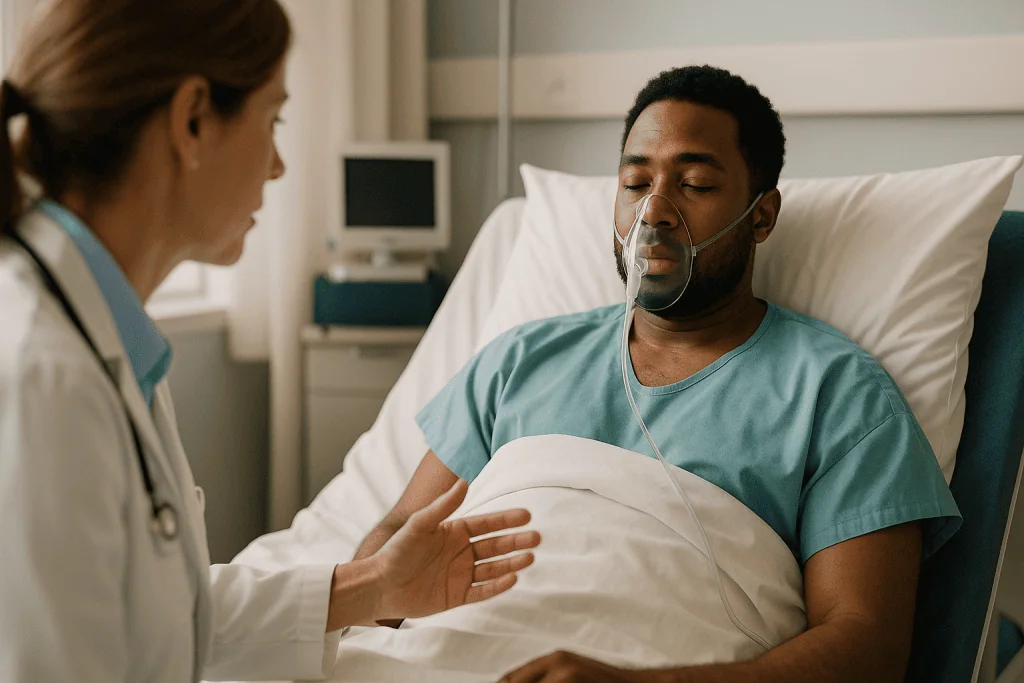
Afterward, most discomfort comes from two places rather than the chest. The first is the throat. A breathing tube passed between the vocal cords and a rigid scope pressed against the front of the windpipe leave many people with a sore throat and sometimes a hoarse voice for a few days (Cleveland Clinic). The second is the incision, which tends to feel like a bruise at the base of the neck, worse when swallowing or turning the head.
Pain scores after mediastinoscopy are generally lower than after operations that go through the ribs, simply because no bone is cut and no chest tube is usually needed. Most people manage with the pain relief plan their team prescribes and are surprised by how quickly it settles. What that plan contains, and for how long, is between you and your treating team; do not add over-the-counter products without asking, since some interfere with clotting.
Anesthesia itself carries familiar short-term effects: nausea, shivering, drowsiness, a fuzzy memory of the recovery room. Serious anesthetic complications are rare, and the pre-assessment exists to identify anyone at higher risk (NHS). If you have had a bad reaction to anesthesia before, or a family member has, say so early.
Which lymph nodes the scope can reach, and which it cannot
Lymph node stations are numbered zones agreed by thoracic specialists so that a report from one center means the same thing in another. Knowing which ones a cervical mediastinoscopy reaches explains why it is sometimes chosen and sometimes not.
Through the neck incision, the scope travels down the front and sides of the trachea. It gives good access to the upper and lower paratracheal stations on both sides (numbered 2 and 4), and to the subcarinal station (7), the node group tucked just beneath the point where the windpipe divides into the two main bronchi. These are the stations that most commonly matter for staging lung cancer, which is why the procedure has held its place for decades.
What it cannot reach is equally important:
- The aortopulmonary window and para-aortic stations (5 and 6), which sit to the left of the aorta and drain much of the left upper lobe. These usually need an anterior mediastinotomy, a small incision beside the breastbone, or a keyhole approach through the chest wall.
- The posterior stations along the lower esophagus (8 and 9), which are better sampled from inside the food pipe with endoscopic ultrasound.
- Nodes within the lung itself (stations 10 to 14), which belong to the surgeon at the time of any lung operation.
In practice, the surgeon plans the approach around where the suspicious nodes actually are on your scan. A left upper lobe tumor with a bright station 5 node may lead to a different procedure, or a combination, rather than a standard mediastinoscopy alone. That planning conversation is worth asking about explicitly.
Who is usually offered a mediastinoscopy, and who is usually asked to wait
The commonest reason is staging a known or suspected lung cancer: establishing whether central nodes are involved before anyone commits to a major lung operation (Mayo Clinic). Guideline bodies generally favor starting with a needle-based technique through the airway, and reserving surgical mediastinoscopy for situations where the needle result is negative but the scan remains worrying, where the needle sample was too small to analyze, or where local expertise and anatomy point that way.
The second group is people with enlarged nodes and no lung mass. Here the question is not stage but identity. Lymphoma in particular often needs a sizable intact piece of tissue so the pathologist can study its architecture, not just its cells, and a surgical biopsy can supply that where a fine needle cannot (MedlinePlus). Suspected sarcoidosis, certain infections and unexplained masses in the front of the chest fall into the same category.
Who is asked to wait, or offered something else? Someone whose scan already shows spread beyond the chest may not need surgical staging at all, because the treatment plan will not change. Someone with a previous mediastinoscopy has scar tissue in the exact tunnel the surgeon needs, which raises risk and lowers yield. A large thyroid extending into the chest, an aortic aneurysm, severe neck arthritis that prevents extension, or a previous tracheostomy can each tip the balance toward a needle approach or a keyhole operation instead.
Fitness for anesthesia is the final filter. The decision about which route suits you is made by the multidisciplinary team, weighing your scan, your other health conditions and what the answer will actually change.
Mediastinoscopy vs EBUS vs VATS: how the options compare
Three approaches can sample the same central nodes, and it helps to see them side by side. EBUS stands for endobronchial ultrasound: a flexible bronchoscope with an ultrasound probe at its tip passes through the mouth into the airways, and a fine needle is guided through the airway wall into the node. VATS, video-assisted thoracoscopic surgery, is keyhole chest surgery through small incisions between the ribs.
| Feature | Cervical mediastinoscopy | EBUS needle biopsy | VATS biopsy |
|---|---|---|---|
| Incision | One small cut at base of neck | None | One to three small cuts between ribs |
| Anesthesia | General | Sedation or general | General |
| Node stations reached | 2, 4, 7 | 2, 4, 7, 10, 11 | Any on one side, including 5 and 6 |
| Tissue size | Large pieces | Small cores or cells | Large pieces |
| Chest tube usually needed | No | No | Often |
| Typical setting | Day surgery | Outpatient | Overnight stay common |
The pattern is clear. EBUS is the least invasive and now usually the first test, especially when the scan shows a clear target. Mediastinoscopy earns its place when a larger sample is needed, when a negative needle result does not fit the picture, or when the surgeon wants direct visual confirmation before a major resection. VATS steps in for nodes on the left of the aorta and for masses that need a different angle entirely.
None of these is universally better. The right one depends on which stations look suspicious, what diagnosis is being considered and your fitness for anesthesia, and that judgment sits with your treating team (Mayo Clinic).
How long does it take to recover from a mediastinoscopy? Recovery time explained
Recovery is usually measured in days rather than weeks. Many people go home the same day once they are awake, breathing comfortably, passing urine and able to drink; some centers keep patients overnight, particularly if the procedure was longer or other conditions warrant observation (Cleveland Clinic).
The first 24 to 48 hours are dominated by anesthesia after-effects rather than the incision. Expect tiredness, a sore throat, possibly some hoarseness and a stiff neck. You should not drive, operate machinery, sign legal documents or drink alcohol during this window, and an adult should stay with you the first night (NHS).
By the third or fourth day most people are moving normally around the house, eating an ordinary diet and needing less pain relief. The dressing usually comes off within a few days; the stitches are commonly dissolvable and disappear on their own. Showering is generally allowed once the team says so, keeping the wound clean and patting it dry. The small scar sits in a natural skin crease and fades over months.
Returning to work depends on what you do. Desk work is often possible within a week; jobs involving heavy lifting or vigorous neck movement may need longer, and your surgeon will give a specific range. Light walking is encouraged from day one because it lowers the risk of blood clots and helps the lungs re-expand after anesthesia.
The part that often takes longest is not physical. Pathology results typically take several working days, and the waiting can feel heavier than the operation. Ask at discharge exactly how and when you will hear, so the silence has a shape.
What is the most common complication of a mediastinoscopy? Complications in honest terms
Mediastinoscopy is regarded as a low-risk operation, but it is performed millimeters from the aorta, the pulmonary artery, the azygos vein and the nerves that move the vocal cords, so the risk profile deserves plain description rather than reassurance.
The most common complication people notice is a hoarse voice. The left recurrent laryngeal nerve, which controls the left vocal cord, runs in the groove beside the trachea directly in the scope’s path. Stretching or bruising it during biopsy of left-sided nodes causes hoarseness that is usually temporary; permanent injury is uncommon but real (MedlinePlus). Sore throat and wound discomfort are near universal but are side effects, not complications.
The most serious complication is major bleeding from one of the great vessels. It is rare, but when it happens the surgeon may need to convert to an open operation through the breastbone to control it. This is the reason mediastinoscopy is done in an operating room with the ability to escalate, and why blood-thinning medicines are reviewed beforehand.
Other recognized risks (MedlinePlus, Cleveland Clinic) include:
- Pneumothorax, air leaking into the space around a lung, sometimes requiring a chest tube.
- Injury to the trachea or esophagus.
- Wound infection, or infection deeper in the mediastinum.
- Leak of lymphatic fluid (chyle) from a damaged lymph duct.
- Anesthetic reactions and blood clots, as with any general anesthetic.
There is also a diagnostic risk that does not appear on consent forms: a false negative, where nodes contain cancer that the biopsies happen to miss. No sampling technique is perfect, which is why surgeons often confirm the mediastinum again at the time of any lung resection.
What the results can tell you, and can cancerous mediastinal lymph nodes be removed?
The biopsies go to a pathologist, a doctor who examines tissue under the microscope. Sometimes a rapid frozen-section reading is available during the operation; the definitive report, with special stains and sometimes molecular tests, usually follows over several working days.
The result generally falls into one of a few categories. The nodes may show no cancer, which in a lung cancer workup shifts the conversation toward surgery or focused radiation. They may show lung cancer cells, which changes the stage and typically moves treatment toward chemotherapy, immunotherapy, radiation or a combination, with surgery considered later in selected situations (Mayo Clinic). Or they may reveal something else entirely: the granulomas of sarcoidosis, the specific cell patterns of a lymphoma, or evidence of infection.
This leads to a question people ask often: can cancerous mediastinal lymph nodes be removed? A standard mediastinoscopy is a sampling procedure, not a removal procedure. Its job is to take enough tissue to answer the question, then stop. When lung cancer is later treated with an operation to remove part of the lung, surgeons routinely remove or systematically sample the mediastinal nodes at the same time; this is called lymph node dissection and is part of accurate staging.
Whether removing involved nodes improves outcomes on its own is a different matter, and the evidence does not support node removal as a standalone treatment for cancer that has already reached them. Involved nodes signal that cancer cells have traveled through the lymph system, and treatment plans for that situation are built around therapies that act on the whole body or the whole region, decided by the multidisciplinary team. The biopsy is the map; it is not the journey.
How is mediastinoscopy performed today? Video-assisted scopes and other variations
The operation described by Carlens in the 1950s used a simple hollow tube and the surgeon’s naked eye. The modern version is recognizably the same route, refined in several ways.
Video-assisted mediastinoscopy replaces the eyepiece with a camera and a screen. The whole team sees what the surgeon sees, the image is magnified, and two instruments can sometimes be used at once, which allows more deliberate dissection and, when needed, more complete node removal. Most centers performing the procedure now use video equipment as standard.
A more extensive variation, sometimes called video-assisted mediastinoscopic lymphadenectomy, uses the same neck incision to remove the accessible node stations entirely rather than biopsy them. It is offered in selected centers for staging before lung surgery and is not the routine approach everywhere.
Anterior mediastinotomy, also known by its originator’s name Chamberlain, is a related operation for the stations mediastinoscopy cannot reach. A small incision is made beside the breastbone, usually on the left, through the space between the second and third ribs, giving direct access to the nodes beside the aorta. It is sometimes done in the same anesthetic as a cervical mediastinoscopy.
Increasingly, when the surgeon needs views of the left-sided stations or of the mass itself, keyhole chest surgery through the ribs is chosen instead, at the cost of a slightly longer recovery.
None of these variations changes the essentials for you as a patient: general anesthesia, a small incision, tissue to the pathologist, and a short recovery. What changes is which stations are reached and how much tissue comes out, and your surgeon should be able to explain why one variation suits your scan better than another.
What people often get wrong about mediastinoscopy
It is a treatment. It is not. Mediastinoscopy is a diagnostic and staging procedure. It removes tissue to answer a question; it does not treat cancer, sarcoidosis or anything else. The result guides treatment chosen afterward by the team.
Being offered it means the news is bad. Often the opposite. Surgeons frequently perform it because the scan is ambiguous and they want to prove that nodes are clear before committing to a lung operation. A negative result is a common and welcome outcome, though no single test can rule out microscopic spread entirely.
It is done through the chest. The standard approach is through a small incision at the base of the neck. Nothing passes between the ribs, no bone is divided, and a chest drain is rarely needed, which is why recovery is measured in days (Johns Hopkins Medicine).
A needle biopsy has made it obsolete. Needle techniques through the airway are now usually the first step, but they sample small amounts of tissue and can miss disease. Surgical biopsy remains the reference standard when a negative needle result does not fit the picture, or when a diagnosis such as lymphoma needs a larger, intact sample (MedlinePlus).
Hoarseness afterward means something went badly wrong. Mild, temporary hoarseness is a recognized effect of the breathing tube and of working beside the vocal cord nerve, and it usually settles. Hoarseness that persists or worsens should be reported, but its presence on day two is not in itself a sign of harm.
You will be back to everything in a day. Home the same day is common; fully back to normal is more often a matter of a week or so, with anesthesia effects, throat soreness and the wait for results all playing a part.
Questions to ask your care team before a mediastinoscopy
A good consultation leaves you knowing not just what will happen but why this procedure and not another. These questions tend to surface the answers that matter.
- Which node stations do you plan to sample, and why those? This tells you whether the scan findings and the procedure match.
- Why mediastinoscopy rather than a needle biopsy through the airway, or a keyhole operation through the chest? A clear reason should exist.
- Will you use a video scope, and might you also do an anterior mediastinotomy or another approach in the same anesthetic?
- What happens if the biopsy is negative but the scan still looks suspicious? Some teams plan to confirm again at the time of any lung surgery.
- How will the result change my treatment options? If the answer is that it will not, ask whether the procedure is still needed.
- What is your team’s experience with this operation, and what is your plan if bleeding occurs?
- Which of my medicines, including blood thinners and supplements, should I pause, and who will tell me when to restart them?
- Will I go home the same day, and who should be with me?
- When and how will I receive the pathology results, and who will explain them?
- What symptoms after discharge should prompt a call, and to whom?
Bring a written list and someone to take notes; most people retain only a fraction of a surgical consultation. If anything remains unclear, ask for a second conversation before the day itself. Consent that you do not fully understand is not really consent, and a good team will welcome the questions.
When to call your doctor after a mediastinoscopy: red-flag signs
Most recoveries are uneventful, but because the biopsy site lies beside major vessels, the airway and the space around the lungs, a small number of warning signs need prompt attention rather than watchful waiting.
Call emergency services or go to the nearest emergency department immediately if you experience:
- Sudden or worsening shortness of breath, or breathing that feels fast and shallow.
- Rapidly increasing swelling at the base of the neck, or a feeling of pressure in the neck or upper chest.
- Coughing or vomiting blood.
- Sharp chest pain, especially with breathing, or a racing heartbeat with light-headedness or fainting.
- Difficulty swallowing that is getting worse rather than better, or a crackling feeling under the skin of the neck or chest.
Contact your surgical team the same day if you notice fever or chills, spreading redness, warmth or discharge from the incision, hoarseness that is worsening or has not begun to improve after a week or two, or pain that is increasing rather than settling despite the plan you were given (Cleveland Clinic). Calf pain, swelling of one leg, or breathlessness after a period of inactivity can signal a blood clot and should also be reported promptly.
You should have been given a direct number for the ward or surgical team at discharge. Use it for anything that worries you, even if it is not on this list. Teams would far rather field a phone call about nothing than see a problem arrive late. And if you have not heard about your results within the timeframe you were promised, that too is a legitimate reason to call.
Frequently asked questions
Is a mediastinoscopy painful?
Not during the procedure, because it is performed under general anesthesia. Afterward, the main discomfort is usually a sore throat from the breathing tube and the scope, plus bruise-like soreness at the neck incision that is worse when swallowing or turning the head. Because no ribs are cut and no chest tube is usually needed, pain is generally less than after operations through the chest wall, and it typically settles within days on the pain plan your team provides.
How long does it take to recover from a mediastinoscopy?
Most people are home the same day or the next morning, moving around normally within a few days, and back to desk work within about a week. The first 24 to 48 hours are mostly about anesthesia effects, so you cannot drive or be alone that first night. Heavier physical work may need longer, and your surgeon will give a range specific to you. Hoarseness and throat soreness usually fade within days to a couple of weeks.
What is the most common complication of a mediastinoscopy?
Temporary hoarseness is the complication people most often notice. It comes from stretching or bruising the left recurrent laryngeal nerve, which runs beside the windpipe in the path of the scope and controls the left vocal cord. It usually improves on its own. Serious complications such as major bleeding from the great vessels, pneumothorax, or injury to the esophagus are uncommon but recognized, which is why the operation is performed in a fully equipped operating room.
Can cancerous mediastinal lymph nodes be removed?
Mediastinoscopy samples nodes rather than removing them; its purpose is diagnosis and staging. When lung cancer is treated with an operation, surgeons routinely remove or systematically sample the mediastinal nodes during the same procedure for accurate staging. Removing involved nodes on their own is not established as a treatment, because cancer in those nodes indicates spread through the lymph system, and the plan then centers on therapies chosen by the multidisciplinary team.
How is mediastinoscopy performed compared with EBUS?
Mediastinoscopy is an operation under general anesthesia through a small neck incision, giving the surgeon direct sight of the nodes and large tissue samples. EBUS is a flexible bronchoscope with an ultrasound tip passed through the mouth, usually under sedation, that guides a fine needle through the airway wall into the node with no incision. EBUS is now typically the first test; mediastinoscopy is used when more tissue is needed or a negative needle result does not fit the scan.
What does mediastinoscopy recovery time look like day by day?
Day one brings drowsiness, a sore throat and a stiff neck as the anesthesia wears off. By days two and three most people are eating normally and walking around the house. The dressing usually comes off within a few days and dissolvable stitches disappear on their own. Light activity is encouraged early to reduce clot risk. Pathology results typically arrive over several working days, and that wait is often the hardest part of recovery.
What are the main mediastinoscopy complications I should know about before consenting?
Recognized risks include hoarseness from vocal cord nerve irritation, bleeding from vessels beside the biopsy site, pneumothorax, injury to the windpipe or food pipe, wound or deep infection, lymphatic fluid leak, and the general risks of anesthesia and blood clots. Overall, serious complications are uncommon in experienced hands. A separate, non-surgical risk is a false negative, where nodes that contain disease are missed by the biopsies taken.
Why do I need a mediastinoscopy lymph node biopsy if I already had a PET scan?
Because PET and CT show size and metabolic activity, not cell type. Nodes can be bright from infection or inflammation and still be benign, or look normal and still contain cancer. A tissue diagnosis decides the stage of a lung cancer, and therefore the treatment path, and can identify conditions like lymphoma or sarcoidosis that imaging cannot distinguish. Surgeons often want proof before committing to a major lung operation.
Will I have a visible scar after mediastinoscopy?
Yes, but a small one. The incision is roughly an inch long and placed in a natural skin crease just above the top of the breastbone, so it tends to fade into the neck’s own lines over months. Keeping the wound clean, avoiding sun exposure on the fresh scar and following your team’s dressing advice all help. If you are prone to thick or raised scars, mention it beforehand so the closure can be planned accordingly.
Who should not have a mediastinoscopy?
It is usually avoided or reconsidered in people who have had a previous mediastinoscopy, because scar tissue fills the surgical path; in those with an aortic aneurysm, a large goiter extending into the chest, a previous tracheostomy or severe neck stiffness; and in anyone not fit for general anesthesia. If a scan already shows disease beyond the chest, staging the nodes may not change treatment. The decision always rests with the treating team.
References
This article is for general information only and is not a substitute for professional medical advice. Please consult a qualified doctor about your individual situation.

More from the Blog

How Long Does Lung Abscess Treatment Take? Why Antibiotic Courses Run for Weeks
Lung abscess treatment usually takes several weeks, not days. Published reviews describe antibiotic courses of roughly three to six weeks, sometimes extended to eight…

What Pulmonary Rehabilitation Offers People With Bronchiectasis: Realistic Expectations
Pulmonary rehabilitation for bronchiectasis is a supervised program of exercise training, airway clearance coaching, and education that usually runs for several weeks. Evidence from…

Daily Life With Pulmonary Disease: Air Quality, Vaccinations and Staying Active Under Treatment
Daily life with chronic lung disease goes more smoothly when three habits become routine: checking local air quality and adjusting outdoor plans on poor…

Inside the Operating Room During Robotic Thoracic Surgery: Ports, Docking and the Chest Tube
Robotic thoracic surgery procedure steps follow a set sequence: general anesthesia with one-lung ventilation, positioning on the side, three to five small incisions (ports)…

Pneumothorax in Young, Tall Adults and Athletes: Why It Happens and How Care Differs
Spontaneous pneumothorax, a collapsed lung with no obvious injury, occurs most often in tall, slim people in their late teens to thirties, usually because…

Preparing for a Pulmonology Consultation: Scans, Breathing Tests and Records to Bring
To prepare for a pulmonology appointment, gather your actual chest X-ray and CT images (not only the written reports), any previous spirometry or lung…