

How Periacetabular Osteotomy (PAO) Reorients a Shallow Hip Socket, Step by Step
Key Takeaways
- A periacetabular osteotomy frees the hip socket with several cuts through the pelvis and rotates it so the bony roof covers more of the femoral head, keeping the natural joint surfaces intact.
- The Bernese technique, first described by Ganz and colleagues, leaves the back column of the pelvis intact so the pelvic ring stays stable while the socket fragment is repositioned.
- PAO is generally offered to skeletally mature adolescents and adults with clear under-coverage and limited cartilage wear; hips with established arthritis are usually steered toward replacement instead.
- Recognized risks include outer-thigh numbness from a skin nerve, bleeding that may need transfusion, blood clots, delayed healing of a pelvic cut, and over- or under-correction of the socket.
- Weight through the operated leg is restricted until X-rays show bone healing across the cuts, and full return of strength is commonly described as taking six months to a year.
- Hip arthroscopy can repair a torn labrum but cannot rotate a shallow socket, so it is not a substitute for PAO when under-coverage is the underlying problem.
A periacetabular osteotomy (PAO) treats hip dysplasia by cutting the pelvic bone around the socket, rotating the freed socket so it covers more of the femoral head, and fixing it in place with screws. It is a major, planned operation usually offered to adolescents and adults with a shallow socket but little or no arthritis. Recovery typically spans months, and suitability is decided by an orthopedic team.
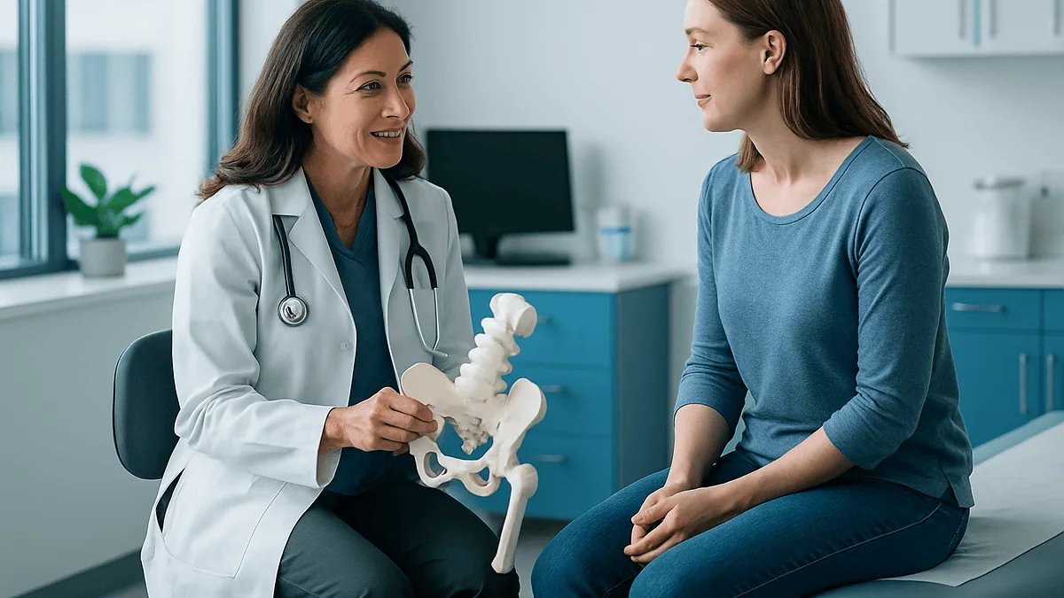
The X-ray goes up on the screen and the surgeon traces a line with one finger. On one side, the ball of the hip sits snugly under a roof of bone. On the other, the roof stops short, and the ball looks like it is peering out from under an awning that was built too small. For a 24-year-old who has spent three years being told her groin pain was a strained muscle, that image is the first thing that has made sense.
What follows is usually a conversation about the periacetabular osteotomy procedure, a mouthful of a name for an operation with a simple idea behind it: if the socket is pointing the wrong way, move the socket. Not replace it. Not smooth it. Reposition the bone you were born with so it does the job it was meant to do.
The idea is simple. The operation is not. This explainer walks through what actually happens, who tends to be offered it, what the weeks afterward look like, and where the honest uncertainties lie.
What is a periacetabular osteotomy procedure, in plain language?
Start with the words. The acetabulum is the cup-shaped socket in the pelvis that holds the ball of the thigh bone. An osteotomy is any operation in which bone is deliberately cut so it can be moved. Put them together and a periacetabular osteotomy is a set of cuts made around the socket that free it from the rest of the pelvis so a surgeon can turn it.
The operation is sometimes called a Bernese PAO because it was developed and first described by a team in Bern, Switzerland, in the late 1980s (Ganz and colleagues, PubMed). Their insight was that earlier pelvic osteotomies either could not swing the socket far enough or destabilized the pelvic ring. The Bernese version keeps the back column of the pelvis intact, so the pelvis stays stable while the socket fragment becomes free enough to rotate in three dimensions.
Why go to such lengths? In hip dysplasia, the socket is too shallow or angled too steeply, so the ball is not fully covered. The joint still works, often for decades, but the load that should spread across a wide dome of cartilage is concentrated on a narrow rim. Cartilage and the labrum, the ring of fibrous tissue around the socket edge, wear early (Mayo Clinic; Cleveland Clinic).
The PAO does not add tissue and does not replace anything. It takes the bone that already exists, turns it so the roof extends over the ball, and fixes it there with screws until it heals in the new position. The joint surfaces are the patient’s own. That single fact shapes almost everything else about who is offered the operation and why.
Why does a shallow hip socket matter if the joint still works?
Picture a golf ball resting in a shot glass instead of a cereal bowl. It stays put, but only a small ring of glass touches it, and every knock puts all the pressure on that ring. That is roughly what happens in a dysplastic hip. The ball is not dislocated and the person may run, dance and lift for years without a complaint.
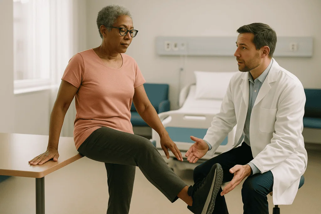
The trouble is cumulative. Cartilage tolerates load best when it is spread widely. When coverage is reduced, peak pressure along the socket edge climbs, and the labrum, which normally acts as a seal and shock absorber, is asked to carry weight it was never designed for. Over time it can fray or tear, and the cartilage beneath it thins. Mainstream medical sources identify hip dysplasia as a recognized cause of early osteoarthritis and of labral tears in younger adults (Mayo Clinic).
Symptoms, when they arrive, are often vague at first: a deep groin ache after long walks, a catch or click on getting out of a car, stiffness after sitting. Because the condition is not rare in babies (the NHS notes that roughly 1 or 2 in every 1,000 infants need treatment for developmental dysplasia of the hip), many adults were checked at birth and told all was well. Milder cases can be missed and declare themselves only in the twenties or thirties (NHS; MedlinePlus).
The logic of a PAO follows directly from this mechanism. If the underlying problem is load concentrated on too little surface, treating the tear alone leaves the cause untouched. Reorienting the socket aims at the load itself. Whether that translates into a healthier joint over a lifetime is a question the evidence answers only partially, and this article returns to it.
Who is PAO usually for, and who is usually asked to wait?
The ideal candidate is often described in three parts: a skeleton that has finished growing, a socket that is genuinely under-covering the ball on imaging, and a joint whose cartilage is still largely intact. Each part matters for a different reason.
Skeletal maturity matters because the cuts pass near growth areas; PAO is therefore generally reserved for adolescents near the end of growth and for adults, while younger children with dysplasia are treated with different operations (Mayo Clinic). Confirmed under-coverage matters because a PAO is a large intervention aimed at a specific mechanical fault; pain in a hip with normal coverage points elsewhere. Preserved cartilage matters most of all, because the operation redistributes load onto the existing joint surface. If that surface is already badly worn, moving it does not restore it, and the team may instead discuss joint replacement (Mayo Clinic; Cleveland Clinic).
Who tends to be asked to wait, or steered toward other options?
- People whose imaging shows moderate or advanced arthritis, for whom a reoriented but damaged socket is unlikely to help.
- Adolescents who are still growing, who may be monitored until the skeleton matures.
- People with mild dysplasia and few symptoms, where watchful waiting with activity adjustment and strengthening is reasonable.
- People whose hip pain has a different or additional cause, such as a spine problem, that needs to be sorted out first.
Age itself is not a hard barrier, but published series generally describe outcomes as more favorable in younger adults with less pre-existing wear, and surgeons weigh that heavily. None of these thresholds are rules a patient can apply alone. They are the questions an orthopedic team works through with imaging, examination and a frank conversation about goals.
How does the surgeon plan the reorientation before the first cut?
Much of a PAO happens before anyone enters an operating room. The planning phase is where the surgeon decides not just whether to move the socket but exactly how far, and in which directions.
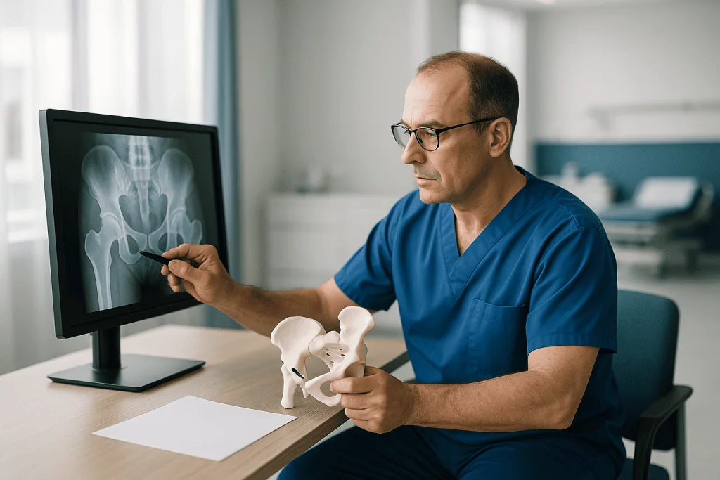
Plain X-rays of the pelvis, taken standing and in specific angled views, are the starting point. Surgeons measure how far the bony roof extends over the ball and how steeply the socket edge is tilted; these angles quantify what the eye sees as a shallow socket. Cross-sectional imaging, usually CT or MRI, adds the third dimension, showing whether coverage is lacking mainly at the front, the side, or the back, and whether the labrum and cartilage are intact (Mayo Clinic). Some teams also image the femur, because the ball and the top of the thigh bone can have their own shape variations that affect how the joint fits after the socket is turned.
The goal of planning is a corrected position that covers the ball adequately without over-correcting. Turn the socket too far and the front edge can pinch against the femoral neck when the hip flexes, trading one mechanical problem for another. Surgeons therefore aim for a defined target range of coverage rather than simply “as much as possible.”
Planning also includes the practical checklist that precedes any major operation: a review of medicines with the prescribing clinician, blood tests, an anesthesia consultation, and discussion of blood loss and the possibility of transfusion. Many programs ask patients to practice with crutches beforehand so that the first post-operative steps are not also the first lesson. Questions about which medicines to continue or pause should be answered by the treating and prescribing teams, not by a general article.
Step by step: what actually happens during the periacetabular osteotomy procedure
The operation is carried out under general anesthesia, often combined with a regional technique for pain control afterward. What follows is the sequence in broad strokes; individual surgeons vary the details.
The incision runs along the front of the hip and upper pelvis. Through it, the surgeon works between muscle groups to reach the pelvic bone around the socket while protecting the nerves and vessels that pass nearby. The Bernese technique, as originally described, is performed through a single approach and keeps the joint capsule closed unless a separate problem inside the joint needs attention (Ganz and colleagues, PubMed).
Then come the cuts. There are several, made in a set order with specialized saws and chisels, often with X-ray guidance:
- A cut through the ischium, the lower part of the pelvis you sit on, made partway and left incomplete for stability.
- A cut through the superior pubic ramus, the bar of bone at the front of the pelvis.
- A cut through the ilium, the broad wing of the pelvis above the socket.
- A connecting cut down the back, angled so the posterior column of the pelvis is left intact.
Once these cuts meet, the socket becomes a free fragment attached only by soft tissue. The surgeon grips it and rotates it, usually tilting the roof forward and outward so it covers more of the ball, then checks the new position with X-rays against the pre-operative plan.
When coverage looks right, the fragment is fixed with screws driven from the top of the pelvis into the repositioned socket. Bone from the cuts may be packed into gaps to encourage healing. The wound is closed in layers, and the patient wakes with the hip pointing, in bony terms, in a new direction. Several hours in the operating room is common for an operation of this scope.
How does PAO compare with hip arthroscopy, replacement and waiting?
People weighing a PAO are almost always weighing it against something. The table below sets the main options side by side in neutral terms; none is “better” in the abstract, and the right choice depends on the state of the joint, age, symptoms and goals as judged by the treating team (Mayo Clinic; Cleveland Clinic).
| Option | What it does | Typically considered when | Main trade-offs |
|---|---|---|---|
| Periacetabular osteotomy | Cuts and rotates the socket to improve coverage; keeps the natural joint | Skeleton mature, clear under-coverage, cartilage largely preserved | Major operation, months of recovery, does not repair existing wear |
| Hip arthroscopy | Keyhole surgery to repair the labrum or reshape bone at the joint edge | Labral or impingement problems with normal or near-normal coverage | Does not correct a shallow socket; may be combined with PAO in selected cases |
| Total hip replacement | Replaces ball and socket with implants | Established arthritis with persistent pain and loss of function | Reliable pain relief for arthritis, but implants have finite lifespans and revision may be needed |
| Non-surgical management | Activity modification, physical therapy, weight management, pain control | Mild symptoms, borderline dysplasia, or while decisions are being made | Does not change the socket; may slow but not reverse mechanical wear |
Two points from the table deserve emphasis. First, arthroscopy and PAO address different problems and are not interchangeable; a labral repair in a markedly shallow socket leaves the cause in place. Second, joint replacement in a young adult raises its own long-term question of implant wear and revision, which is one reason joint-preserving surgery exists at all. A candid team will explain which of these pathways applies to a particular hip and why.
Is PAO considered a major surgery, and is it high risk?
Yes to the first part, unambiguously. A PAO involves cutting through the pelvis in several places, a wide surgical exposure, meaningful blood loss and a hospital stay. It sits firmly in the category of major elective orthopedic surgery, and anyone told otherwise is being sold a softened version.
“High risk” is a different question, and the honest answer is that risk is real, specific, and usually manageable in experienced hands. The recognized complications of the procedure fall into a few groups:
- Nerve injury, most often to the lateral femoral cutaneous nerve that supplies skin sensation on the outer thigh, producing numbness that is frequently temporary but can persist. Injury to larger nerves controlling movement is much less common.
- Bleeding requiring transfusion, given the proximity of major vessels and the cut surfaces of pelvic bone.
- Blood clots in the leg veins, a risk shared by all major lower-limb surgery, which is why clot-prevention measures are standard.
- Delayed healing or non-union of one of the cuts, most often the pubic cut, which may or may not cause symptoms.
- Infection, wound problems, and irritation from screws that occasionally leads to their later removal.
- Over- or under-correction of the socket, which can cause pinching or leave coverage inadequate.
- Progression of arthritis despite a technically sound correction.
Rare but serious complications, including injury to major blood vessels or the joint surface itself, are described in the surgical literature. Numbers vary widely between series and depend on surgeon experience, patient factors and how complications are defined, which is why this article does not quote a single figure. The Bernese technique was designed in part to reduce the instability and vascular risk of earlier pelvic osteotomies (Ganz and colleagues, PubMed). Your own surgeon can tell you their approach to each of these risks and what their consent process covers.
How painful is a periacetabular osteotomy, honestly?
It hurts. Anyone who has had one will say so, and there is no value in pretending otherwise. Bone that has been cut and moved sends strong pain signals, and the muscles around the pelvis, which were retracted to reach the bone, ache and spasm afterward. The first few days are typically the hardest.
What has changed over the years is how that pain is managed. Modern programs generally use several methods at once, an approach often called multimodal analgesia. A regional anesthetic block or an epidural infusion may numb the area for the first day or two. Anti-inflammatory medicines, acetaminophen, and opioid medicines for breakthrough pain are commonly layered together so that no single class has to do all the work and side effects are spread thinner. Ice, positioning, and early gentle movement play a genuine role, not a decorative one.
Which medicines are used, in what combination and for how long, are decisions for the anesthesia and surgical teams, and they vary by hospital and by patient. If you take medicines regularly for other conditions, or have had problems with pain medicines in the past, tell the team well before surgery so the plan can be built around you.
Patients often describe a shift over the first two weeks from sharp, movement-triggered pain to a duller ache and a sense of deep bruising, followed by a longer stretch where fatigue and muscle weakness bother them more than pain does. Numbness on the outer thigh from the skin nerve mentioned earlier is common and distinct from pain. Persistent pain that is climbing rather than easing, or a new type of pain, is something to report rather than tolerate; the final section lists the signs that should prompt a call.
What do the first days in hospital look like after PAO surgery?
The first day is about waking up safely, controlling pain and watching the numbers. Nurses check blood pressure, oxygen levels, urine output and the wound. Blood counts are drawn because bleeding continues into the tissues for a day or so after the cuts are made, and some patients need a transfusion. A drain in the wound, if one was placed, is usually removed within the first couple of days.
Clot prevention starts immediately. Compression devices on the calves, early ankle and foot exercises and, very often, an anticoagulant medicine prescribed by the team are standard for major pelvic and hip surgery because immobility and the surgery itself raise the risk of clots forming in the leg veins. The choice and duration of any anticoagulant are set by the treating team.
Physical therapy begins early, usually on the first or second day. The initial goals are modest and specific: sitting at the edge of the bed, standing with support, learning to move the operated leg without letting it bear full weight, and practicing the crutch or walker pattern that will be used for weeks. Therapists also teach the transfers that matter most at home: bed to standing, toilet, a car seat, a flight of stairs if there is no way around them.
Bowel function, appetite and sleep are practical concerns in these days too. Pain medicines and reduced movement slow the gut, and most programs address this proactively. Hospital stays for PAO are typically a few days, with discharge depending on pain control, safe mobility with aids and stable blood results rather than on a fixed calendar. Some patients go home; others spend a short period in a rehabilitation setting if the home environment or support makes that safer. The team decides this with the patient, not for them.
PAO surgery recovery time: what the first twelve weeks usually hold
Recovery from a PAO is measured in months, and it moves in phases that track bone healing. Timelines below are typical ranges described in mainstream orthopedic patient information; they are not a schedule and a surgeon may lengthen or shorten each phase based on X-rays and progress (Mayo Clinic; Cleveland Clinic).
Weeks one to two. The focus is protection and basics. Weight through the operated leg is limited, usually to “toe-touch” or partial weight-bearing with two crutches or a walker, so the moved socket is not loaded before it knits. Pain and swelling are settling. Fatigue is pronounced; the body is spending its energy on healing bone. Wound checks and stitch or staple removal happen in this window.
Weeks two to six. Range-of-motion exercises expand, usually within limits set to avoid stressing the front of the hip. Many people can manage short outings on crutches. Sitting for long periods is often still uncomfortable. A follow-up X-ray around this time gives the surgeon a first look at early bone healing at the cut surfaces.
Weeks six to twelve. If imaging shows healing, weight-bearing is typically progressed in stages, and crutches are reduced from two to one and then set aside. Strengthening of the hip muscles becomes the main work of physical therapy. Walking pattern, which almost always develops a limp during the protected phase, is retrained deliberately.
Beyond twelve weeks, most programs continue therapy for several more months, with a gradual return to low-impact activities first. Full recovery of strength and stamina is commonly described as taking six months to a year. Return to running or impact sports, where permitted at all, is decided case by case. Anyone comparing their own progress against these ranges should remember that the range is wide precisely because bodies differ.
Walking after PAO surgery: crutches, weight-bearing, driving and work
The single question that dominates recovery is when it is safe to put weight through the leg, and the answer rests on one principle: the socket fragment is held by screws, but screws are a scaffold, not a weld. Bone has to grow across the cuts before the joint can take full load safely.
For that reason the early weeks involve strict weight-bearing limits, and the progression is driven by follow-up X-rays rather than by how good the hip feels. It is entirely possible to feel ready to walk unaided before the bone is ready to allow it. Surgeons are wary of that gap because loading a partly healed osteotomy risks shifting the fragment or breaking screws.
Driving depends on which hip was operated on, whether the car is automatic, and whether the person has stopped taking medicines that impair reaction time. Braking requires the right leg to move quickly and bear force. Most programs ask patients to wait until they are off opioid medicines, can bear weight comfortably, and can perform an emergency stop; the treating team confirms when that point is reached. Insurance and legal rules about driving after surgery vary by place and are worth checking.
Return to work follows the demands of the job. Desk-based work can sometimes resume, in part, within a few weeks if sitting tolerance allows, though fatigue may limit hours. Standing, walking or lifting jobs generally wait until weight-bearing is unrestricted and strength has returned, which can mean several months.
Practical preparation makes the difference between a frustrating recovery and a manageable one. A raised toilet seat, a shower chair, a firm chair with arms, a route through the home without loose rugs, and a plan for meals and groceries in the first weeks all pay off. So does a realistic conversation with family or friends about the help that will be needed and for roughly how long.
What does the evidence actually show about long-term results?
This is where honesty matters most, because the promise of joint preservation is easy to overstate. The original description of the Bernese osteotomy reported early results in a small group and was explicit that they were preliminary (Ganz and colleagues, PubMed). Since then, a substantial body of follow-up studies has accumulated in the orthopedic literature, some now extending beyond two decades.
Read carefully, that literature supports a few statements. Many reoriented hips remain in service without progression to joint replacement for long periods, and pain and function commonly improve compared with before surgery. At the same time, a meaningful proportion of hips do eventually develop arthritis and are converted to replacement, and the likelihood of that outcome is consistently linked to how much cartilage damage existed before surgery, to the patient’s age, and to how well the socket was positioned. Hips with little pre-operative wear and good correction fare best; hips with established arthritis fare worst, which is why the latter are usually steered toward replacement instead.
Specific percentages are not quoted here on purpose. Published figures for long-term survival of the native joint range widely because studies differ in patient selection, follow-up length, surgical era and how success is defined. Quoting one number risks implying a precision the evidence does not have.
Two further limitations deserve mention. Most studies are single-center series without a comparison group, so the natural history of untreated dysplasia in similar patients is inferred rather than measured directly. Reporting of complications and of patient-reported outcomes has also become more systematic over time, which makes older and newer series hard to compare. A surgeon who has performed many PAOs can share their own audited outcomes; asking for those, and for how they compare with published series, is a fair and useful question.
What people often get wrong about periacetabular osteotomy
Misunderstandings about PAO cluster around a handful of themes. Each is worth correcting plainly.
“It fixes the arthritis.” It does not. A PAO changes the direction of load on the joint. Cartilage that has already worn away does not grow back because the socket is turned. The operation is aimed at protecting the cartilage that remains, which is why it is offered mainly to hips with limited wear.
“It is the same as a hip replacement.” The two are almost opposites in intent. Replacement removes the joint surfaces and substitutes implants. PAO keeps every natural surface and moves the bone around them. A person who has had a PAO can still have a replacement later if needed; the reverse does not apply.
“Keyhole surgery would do the same thing with less recovery.” Arthroscopy can repair a torn labrum and reshape bone at the joint edge, but it cannot rotate the socket. In a genuinely shallow socket, repairing the labrum alone leaves the mechanical cause in place. Some surgeons combine arthroscopy with PAO to address both, but one does not substitute for the other.
“If it was missed at birth, it cannot be dysplasia.” Newborn screening detects many cases, but milder degrees of under-coverage can escape detection and present as adult hip pain (NHS; MedlinePlus).
“Recovery is a few weeks.” Protected weight-bearing alone typically lasts around six weeks or more, and return of full strength is measured in months (Mayo Clinic; Cleveland Clinic).
“The screws come out.” Usually they stay. Removal is considered only if they cause irritation once the bone has healed.
“Once corrected, the hip is normal.” Coverage can be made normal or near-normal; the joint’s history of altered loading cannot be erased, and long-term follow-up remains sensible.
Questions to ask your care team before agreeing to hip dysplasia surgery in adults
A good consultation leaves a person understanding not just what will be done but why this operation, why now, and what the alternatives would mean. The questions below are a starting point; write down the answers, because much is said in a short appointment.
- What do my X-rays and scans show about socket coverage, and how much cartilage wear is already present?
- Why are you recommending a PAO rather than arthroscopy alone, joint replacement, or continued non-surgical management for my hip?
- How will you decide how far to rotate the socket, and what is the target range you are aiming for?
- Will anything inside the joint, such as the labrum, be treated at the same time, and through what approach?
- How many of these operations do you perform, and what are your own complication and reoperation figures?
- What are the specific risks in my case, given my age, anatomy, weight, and other health conditions?
- How much blood loss do you expect, and what is the plan if transfusion is needed?
- What is your clot-prevention protocol, and how long will it continue after I go home?
- What weight-bearing limits will apply, for how long, and what will you use to decide when to progress?
- Which of my current medicines should be reviewed with my prescribing clinician before surgery?
- What does a realistic timeline look like for me to return to my particular job, driving and activities?
- If the hip develops arthritis in the future, does having had a PAO affect a later replacement?
- What symptoms after surgery should make me call you the same day?
It is reasonable to ask for a second opinion before major elective surgery, and reasonable surgeons expect it. It is also reasonable to ask whether waiting a year would change the picture, and what the team would watch for in the meantime. The decision belongs to the patient and the treating team together, informed by imaging, evidence and the life the person actually wants to lead.
When to call your doctor after a periacetabular osteotomy
Some discomfort, swelling, bruising and outer-thigh numbness are expected after a PAO. Certain signs, however, point to problems that need prompt assessment. Contact the surgical team the same day, or seek emergency care, if any of the following occur.
Seek emergency care immediately for:
- Sudden shortness of breath, chest pain, coughing up blood, or a racing heartbeat, which can signal a blood clot that has traveled to the lungs.
- Heavy bleeding from the wound that does not slow with firm pressure.
- A fall onto the operated hip, or a sudden “give” or shift in the hip accompanied by new severe pain.
- Fainting, confusion, or a leg that becomes cold, pale or pulseless.
Call the surgical team the same day for:
- New or worsening calf pain, swelling or tenderness in either leg, which may indicate a clot in a leg vein.
- Fever, chills, or a wound that becomes increasingly red, hot, swollen or leaks cloudy or foul-smelling fluid.
- Pain that is escalating rather than easing over days, or pain that is not controlled by the prescribed plan.
- New weakness in the leg or foot, such as difficulty lifting the foot, or numbness spreading beyond the outer thigh.
- Inability to pass urine, or persistent vomiting that prevents fluids or medicines being kept down.
- Any unexpected change in how the hip moves or bears weight.
Recovery is also a time when questions surface that are not emergencies but matter: whether a particular exercise is safe, whether a symptom is normal, when a follow-up X-ray is due. Programs generally provide a direct contact for exactly this purpose. Use it. Guidance from mainstream sources on hip dysplasia and its surgical treatment is consistent on one point: uncertainty after major surgery is a reason to ask, never a reason to wait and see (Mayo Clinic; Cleveland Clinic).
Frequently asked questions
How painful is a periacetabular osteotomy?
It is a painful operation, particularly in the first few days, because pelvic bone has been cut and moved and surrounding muscles have been retracted. Modern programs combine regional anesthesia, several classes of pain medicine and early gentle movement to keep pain manageable. Most people describe a shift from sharp movement-related pain to a duller ache over the first two weeks, with fatigue and weakness becoming the larger issue later. The pain plan is set by the anesthesia and surgical teams.
Is PAO one of the hardest orthopedic surgeries?
It is widely regarded as technically demanding. The cuts are made close to major nerves and blood vessels, the socket must be rotated to a precise target in three dimensions, and the exposure is deep. That complexity is one reason it is usually performed by surgeons with specific training and volume in hip preservation. Difficulty for the surgeon does not by itself mean poor outcomes for patients, but it is a fair reason to ask about a surgeon’s experience.
Is PAO considered a major surgery?
Yes. A periacetabular osteotomy involves multiple cuts through the pelvis, meaningful blood loss, general anesthesia, a hospital stay of several days and months of protected recovery. It belongs firmly in the category of major elective orthopedic surgery, comparable in scale to joint replacement, though different in purpose because it preserves rather than replaces the joint.
Is PAO a high risk surgery?
Risk is real and specific but generally manageable in experienced hands. Common complications include temporary or persistent numbness on the outer thigh, bleeding requiring transfusion, and blood clots; less common ones include delayed healing of a pelvic cut, infection, screw irritation and over- or under-correction. Serious injury to major vessels or nerves is rare but described. Published complication rates vary widely with surgeon experience and patient factors, so the treating surgeon is the best source for individualized figures.
What is the typical PAO surgery recovery time?
Recovery is measured in months. Protected weight-bearing with crutches or a walker typically lasts around six weeks or longer, progression is guided by follow-up X-rays showing bone healing, and strengthening continues for several months after that. Full return of strength and stamina is commonly described as taking six months to a year, according to patient information from mainstream medical centers. Individual timelines vary and are set by the surgical and therapy teams.
When can I start walking after PAO surgery without crutches?
Usually only after X-rays confirm that bone is healing across the pelvic cuts, which is commonly assessed around six weeks, though the exact timing varies. Before that, weight through the leg is limited so the repositioned socket is not loaded prematurely. Crutches are typically reduced in stages from two to one and then set aside as strength and walking pattern improve. Feeling ready is not the same as the bone being ready, so the surgeon’s clearance matters.
Does PAO stop hip arthritis from developing?
It cannot reverse wear that already exists, and the evidence does not show that it prevents arthritis in every case. Long-term studies report that many reoriented hips remain in service for decades, while a proportion progress to arthritis and eventual replacement, with outcomes strongly linked to how much cartilage damage was present before surgery and how well the socket was positioned. The aim is to reduce abnormal load on cartilage that is still healthy.
Can hip dysplasia surgery in adults be done with keyhole techniques instead?
Not for the socket reorientation itself. Hip arthroscopy, the keyhole approach, can repair a torn labrum or reshape bone at the joint edge, but it cannot cut and rotate the socket. When the underlying problem is a shallow socket, arthroscopy alone leaves the mechanical cause untouched. Some surgeons combine arthroscopy with PAO to address problems inside the joint at the same time, but one does not replace the other.
Will the screws from a PAO need to be removed later?
Usually not. The screws hold the socket fragment while bone heals across the cuts and are typically left in place permanently. Removal is considered only if a screw causes local irritation or pain after the bone has healed, which is a smaller, separate procedure. Screws generally do not interfere with a later hip replacement if one is ever needed, though the surgical team will review imaging before any future operation.
Can I still have a hip replacement after a periacetabular osteotomy?
Yes. A PAO preserves the natural joint, so if arthritis develops later, total hip replacement remains an option. Surgeons planning a replacement after a previous PAO review the altered pelvic anatomy and any retained screws beforehand, and the operation may require some additional planning. Preserving the option of a later replacement while potentially delaying the need for one is a central part of the rationale for joint-preserving surgery in younger adults.
References
This article is for general information only and is not a substitute for professional medical advice. Please consult a qualified doctor about your individual situation.

More from the Blog

How Long Does an Ankle Replacement Last? Honest Limits and What Affects Implant Wear
An ankle replacement does not last forever. Pooled data from a systematic review of total ankle replacement reported that close to nine in ten…

What Can a Knee Replacement Realistically Restore? Pain Relief, Bending, Kneeling and Sport
A knee replacement can realistically restore reliable, largely pain-free walking, stair climbing and sitting, and most people regain a bend of well over a…

Life After Shoulder Replacement: Reaching, Lifting Limits and Getting Back to Sport
Life after shoulder replacement usually means far less pain and a return to most everyday tasks, with reaching, gentle sport and light lifting typically…
Carpal Tunnel Surgery and Pregnancy: Why Many Teams Wait Until After the Birth
Carpal tunnel surgery during pregnancy is rarely performed because the condition is usually driven by pregnancy-related fluid retention and often eases after the birth.…

Private Hip Replacement: UK Costs, Waiting Times and the Abroad Option
A private hip replacement in the UK typically costs £11,000–£17,000, while NHS waits for the same operation can stretch well beyond the 18-week referral-to-treatment…
Limb Lengthening as an International Patient: Stay Length, Follow-Up X-Rays and Flying Home
Patients who travel for limb lengthening usually need to stay near the surgical team well beyond the operation itself, because the bone is lengthened…