

How Selective Dorsal Rhizotomy Works: Nerve Monitoring and Choosing Which Rootlets to Cut

Key Takeaways
- SDR reduces spasticity by cutting a portion of sensory (dorsal) rootlets in the lumbar and sacral spine; the ventral motor roots that move the legs are identified and left intact.
- During surgery, each rootlet is electrically stimulated while electrodes record leg muscle activity, and rootlets producing sustained or spreading responses are the ones divided.
- The NHS describes SDR as an option mainly for children with spastic diplegia, typically considered around ages 3 to 9, always followed by months of physiotherapy.
- Dystonia, underlying weakness and fixed contractures are the most common reasons a child is asked to wait or is offered a different treatment such as injections, a pump or orthopedic surgery.
- Unlike botulinum toxin injections or an intrathecal baclofen pump, SDR is permanent, which is why candidacy is assessed by a multidisciplinary team and reversible options are often tried first.
- Trials show SDR plus physiotherapy lowers leg tone more than physiotherapy alone with modest functional gains, but no single success percentage is supported by the evidence.
Selective dorsal rhizotomy (SDR) treats spasticity by cutting a portion of the sensory nerve rootlets in the lower spine that drive overactive stretch reflexes. During surgery, each rootlet is stimulated electrically while muscle activity is recorded; rootlets that produce abnormal, spreading responses are divided and the others are left intact. Movement nerves are not cut. It is usually considered for selected children with spastic cerebral palsy and is always followed by months of physiotherapy.
The referral letter arrives with a word most parents have never seen: rhizotomy. Underneath it, in smaller print, a note about a gait laboratory appointment and a request to bring the child’s usual splints. The mother reads it twice at the kitchen table, then types the term into her phone and finds videos of children walking on treadmills with reflective markers stuck to their knees. None of it explains what the surgeon actually does.
That gap is what this article fills. Understanding how SDR surgery works, from the anatomy of a stretch reflex to the electrodes taped over a child’s calf in the operating room, makes the conversation with the neurosurgeon far less intimidating. It also makes the hard questions easier to ask: why the sensory nerve and not the muscle, why only some rootlets, and what the months afterward demand of a family.
We will stick to what mainstream evidence supports and say plainly where it runs thin.
What spasticity is, and why a sensory nerve is the target
Spasticity is a velocity-dependent increase in muscle tone: the faster a joint is moved, the harder the muscle resists. A relaxed calf that stiffens the moment a physiotherapist bends the foot quickly is showing it. In cerebral palsy, the underlying injury sits in the brain, usually in the motor pathways near the fluid-filled ventricles, and it happens before, during or shortly after birth, as the Centers for Disease Control and Prevention describes.
So why operate on the spine? Because the brain does not create spasticity directly. It creates it by failing to quieten a loop that lives in the spinal cord. That loop is the stretch reflex. Inside every muscle sit tiny sensors called muscle spindles, which report how quickly the muscle is being lengthened. Their signal travels along sensory fibers into the spinal cord through the dorsal root, the sensory entry point on the back of the cord. There it connects to motor neurons that fire the same muscle, telling it to contract.
In a healthy nervous system, descending signals from the brain dampen this loop, so a brisk stretch produces a small, brief response. When those descending signals are damaged, the loop runs hot. Every stretch triggers an exaggerated contraction, and the child’s legs scissor, toes point and knees flex against their will.
Selective dorsal rhizotomy reduces the volume of sensory traffic entering that loop. Cutting some of the sensory rootlets means fewer spindle signals reach the motor neurons, so the reflex fires less forcefully. The motor nerves that carry commands from the cord to the muscle, which travel in the separate ventral root at the front, are not touched. This is the single most useful fact for understanding how SDR surgery works: it turns down an overactive input rather than switching off an output.
How SDR surgery works, step by step
The operation is done under general anesthesia with the child lying face down. A neurosurgeon opens a window in the lower back, over the lumbar spine, and reaches the dural sac, the tough membrane holding spinal fluid and the nerve roots. Below roughly the first or second lumbar vertebra, the spinal cord itself has ended; what remains is a bundle of nerve roots called the cauda equina, named for its resemblance to a horse’s tail.
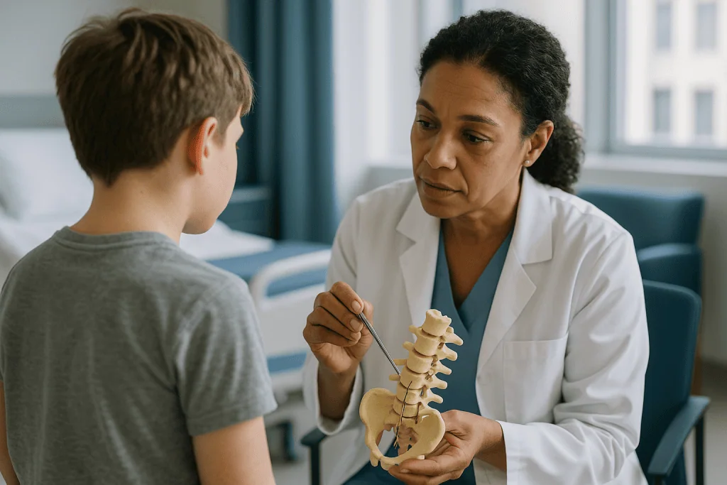
The surgeon identifies the dorsal (sensory) roots that serve the legs, typically from the second lumbar level down to the first or second sacral level. Each root is separated from its motor partner using anatomical landmarks and, when needed, gentle electrical stimulation: stimulating a motor root produces a strong muscle twitch at a very low current, while a sensory root does not.
Each dorsal root is then teased apart into several smaller strands called rootlets, often three to eight per root depending on the level. This is delicate work under an operating microscope. The rootlets are laid out, and one by one they are tested, which is the subject of the next two sections.
Rootlets judged to be feeding abnormal reflex activity are cut. Rootlets with normal responses are laid back in place. The dura is closed with fine sutures, sometimes reinforced with a sealant, and the muscle and skin are closed in layers. Depending on the technique, the operation commonly takes several hours, and the child wakes up on a ward or in a high-dependency unit with a drain or dressing over the incision.
Nothing about the brain injury changes. What changes is the amplification downstream, which is why physiotherapy afterward is not an optional extra but the second half of the treatment.
Why it is called selective: nerve monitoring inside the operating room
Before the child is positioned, a neurophysiology team places fine recording electrodes into or over muscles in both legs: typically the hip adductors, quadriceps, hamstrings, calf muscles and the small muscles of the foot. Some teams also record from the anal sphincter, because the sacral roots that serve the legs sit close to those controlling bladder and bowel.
This set-up is intraoperative electromyography, or EMG: a way of listening to muscles electrically while the patient is asleep. Each electrode feeds a channel on a monitor, so the team can see, in real time, which muscles respond when a particular rootlet is stimulated.
Anesthesia has to be planned around this. Muscle relaxants, which paralyze muscles during many operations, would silence the very signals the surgeon needs, so they are either avoided after the initial set-up or allowed to wear off before testing begins. The anesthetist keeps the child deeply asleep by other means and communicates constantly with the surgeon about timing.
Once a rootlet is isolated, the surgeon touches it with a tiny hooked electrode. A first, low-current stimulation confirms the rootlet is sensory rather than motor. A second stimulation, usually a short train of pulses, is used to provoke the reflex. The team then watches how the muscles answer.
Every aspect of this depends on teamwork. The surgeon cannot see the EMG screen while looking down the microscope, so the neurophysiologist calls out the grade of each response. It is slow, methodical and repetitive: dozens of rootlets, each tested, each graded, each decided. That is the meaning of selective, and it is why an SDR takes far longer than the size of the incision might suggest.
How surgeons choose which rootlets to cut
A normal sensory rootlet, when stimulated with a brief train of pulses, produces a short, contained response in the muscle it serves and then stops. An abnormal rootlet does something different. The response may continue after the stimulation ends, it may spread to muscles on the same leg that should not have been involved, or it may cross to the opposite leg entirely.
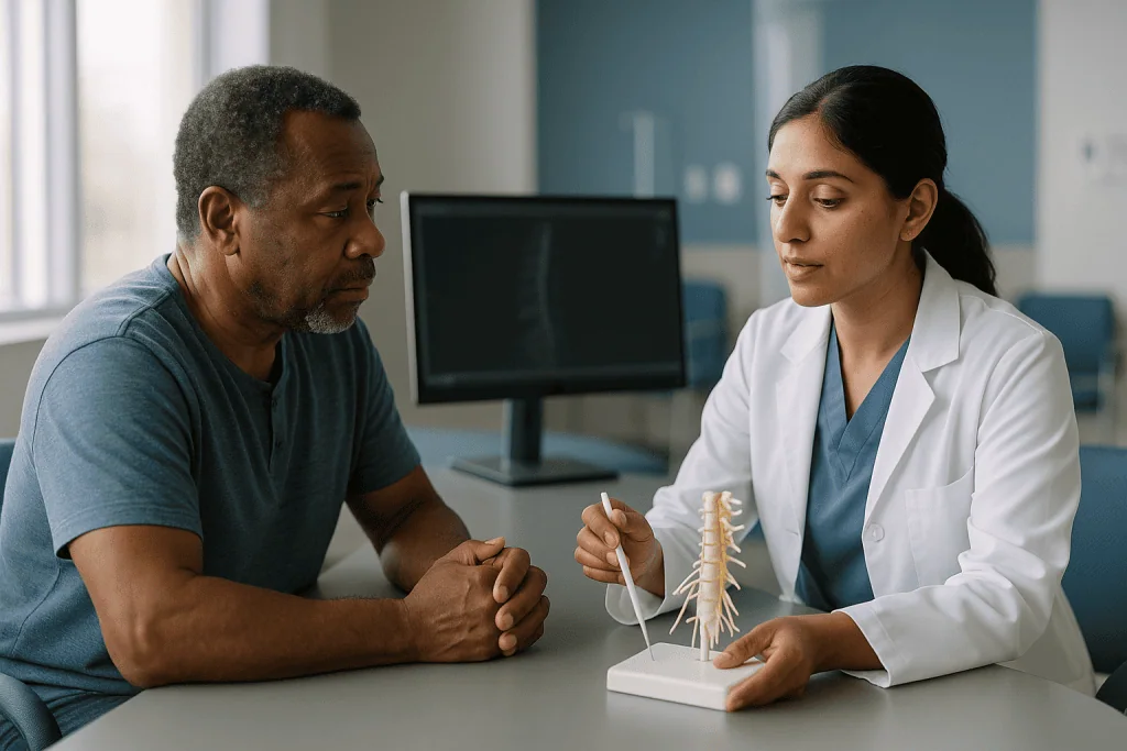
Teams typically grade these responses on a scale. Grading systems vary between centers, but the principle is shared: the more sustained and the more widespread the response, the more that rootlet is thought to be contributing to spasticity. Rootlets with the most abnormal grades are cut. Rootlets with normal or borderline responses are usually preserved, particularly at sacral levels where bladder and bowel fibers may be mixed in.
Surgeons also weigh the pattern of spasticity the child showed before surgery. If the hip adductors and calves were the biggest problem, rootlets driving those muscles receive closer attention. The surgical plan is therefore not a fixed recipe; it is shaped by the physical examination, the gait analysis and the intraoperative findings together.
An honest caveat belongs here. Some experienced surgeons argue that EMG grading is less predictive than once believed, because responses can be influenced by anesthetic depth, stimulation settings and electrode position. Others rely on it heavily. What both camps share is a commitment to cutting only a portion of the sensory rootlets at each level, never all of them, so that the child retains enough sensory input for balance, position sense and skin sensation.
Ask your surgeon which approach the team uses. Reasonable teams answer differently, and a clear explanation matters more than a particular number.
Single-level or multi-level: two ways to reach the same rootlets
Two broad surgical approaches exist, and the choice explains why one family hears about a small incision while another hears about several vertebrae.
The traditional route is a multi-level laminectomy or laminotomy. The surgeon removes or temporarily lifts the back portion of several lumbar vertebrae, exposing the dorsal roots where they leave the spinal canal at each level. Because each root is identified at its own exit, the surgeon knows with confidence which level it belongs to. The trade-off is a longer incision, more bone work and, over years of growth, a theoretical concern about spinal alignment.
The alternative is a single-level approach at the conus, the tapered end of the spinal cord. Here the surgeon opens one or two vertebrae and finds the dorsal roots where they emerge from the cord itself, before they fan out. All the target roots are reachable through one small window. The challenge is identification: roots at the conus are packed closely together and levels are less obvious, so stimulation and EMG mapping carry more weight.
Neither approach is proven superior in the mainstream literature, and both are performed by experienced teams. The ventral motor roots are avoided in each, and both rely on the same testing logic to choose rootlets.
What differs for the family is mostly practical. A single-level approach usually means a shorter scar and somewhat less bone disruption. A multi-level approach may allow a more anatomically certain map. Your surgeon should be able to explain which technique they use and why, and how it changes the expected hospital stay or restrictions on bending and twisting afterward.
Who qualifies for SDR surgery?
Selective dorsal rhizotomy is not a treatment for cerebral palsy in general. It is a treatment for one feature, spasticity, in one population where the balance of benefit and risk is best understood.
The NHS describes SDR as an option for children whose spasticity mainly affects the legs, the pattern called spastic diplegia, and notes that it is usually considered in childhood, with typical UK practice focused on children aged around 3 to 9 years. Many of these children were born preterm and have an MRI pattern of injury to the white matter near the ventricles.
Beyond diagnosis and age, teams look for a particular profile:
- Spasticity that is clearly the main obstacle to movement, rather than weakness, poor balance or a different tone disorder.
- Enough underlying leg strength to stand and take steps, since removing spasticity can reveal weakness that was hidden by stiffness.
- Reasonable selective motor control, meaning the child can move one joint without the whole leg following.
- Walking ability roughly in the middle of the Gross Motor Function Classification System, the five-level scale describing how a child with cerebral palsy moves; levels II and III are most often discussed.
- A family and child able to commit to intensive physiotherapy for months, as the NHS emphasizes.
Some centers also operate on children who do not walk, aiming at comfort, ease of care and positioning rather than gait, and a smaller number of centers offer SDR to adults. The evidence for these groups is thinner, and the goals are different, so the conversation should be too.
Eligibility is decided by a multidisciplinary team: neurosurgeon, pediatric neurologist or rehabilitation physician, physiotherapist, orthopedic surgeon and often a gait laboratory. No single clinician makes this call alone.
Who is usually asked to wait, or steered toward another option
Being told a child is not a candidate feels like a door closing. Often it is a door being held until the timing is right, or a redirection toward a treatment that fits the problem better.
Dystonia is the most common reason. Dystonia is involuntary, sustained muscle contraction that twists the limb into unusual postures, and it arises from different brain circuits than spasticity. Cutting sensory rootlets does little for it, and removing spasticity can occasionally make dystonic movements more visible. Children with mixed tone are therefore assessed carefully, sometimes over several visits.
Weakness is the second. If the stiffness is what allows a child to stand, taking it away without enough strength underneath may leave them less mobile, not more. Teams look for muscle power and trunk control before recommending surgery.
Fixed contractures, where a muscle has permanently shortened so the joint cannot be moved fully even when relaxed, are not helped by SDR. Contractures usually need orthopedic surgery, and the sequence matters: some teams do SDR first and address bones and tendons later, others reverse the order.
Other situations that typically prompt a pause or an alternative include previous spinal surgery, significant scoliosis or hip instability that needs attention first, poor general health, and family circumstances that make months of physiotherapy unrealistic. Very young children are sometimes asked to wait so that their movement pattern and strength can be assessed more reliably.
Waiting is not the same as being refused forever. Children change, and a reassessment after a year of therapy or after orthopedic treatment may reach a different conclusion. Ask the team what specifically would need to change for the answer to change.
SDR surgery for cerebral palsy compared with other spasticity treatments
Spasticity has several treatment routes, and most children with cerebral palsy use more than one over their lifetime. The Mayo Clinic and NHS both describe a stepped approach in which physiotherapy, orthoses, medicines and procedures are combined according to the child’s goals. The table below sets them side by side in neutral terms; it is a map of the landscape, not a ranking.
| Option | How it acts | Reversible? | Scope | Typical role |
|---|---|---|---|---|
| Physiotherapy and orthoses | Stretching, strengthening, positioning | Yes | Whole body | Foundation for every other option |
| Oral antispasticity medicines (for example baclofen class) | Dampen spinal reflex excitability chemically | Yes | Generalized | Mild to moderate tone; drowsiness can limit use |
| Botulinum toxin injections | Block nerve-to-muscle signaling in specific muscles | Yes, wears off over months | Focal | A few troublesome muscles; repeated as needed |
| Intrathecal baclofen pump | Delivers medicine into spinal fluid via implanted pump | Yes, can be adjusted or removed | Generalized, legs more than arms | Severe spasticity, often non-ambulant children |
| Selective dorsal rhizotomy | Cuts a portion of sensory rootlets to reduce reflex input | No | Legs | Selected walking children with spastic diplegia |
| Orthopedic surgery | Lengthens tendons, realigns bones | No | Specific joints | Contractures and deformities, often later |
The defining difference is permanence. Injections wear off and pumps can be switched off; SDR cannot be undone. That is its strength, since the effect on spasticity is lasting, and its weight, since the decision has to be right the first time. Medicines are named here only to explain mechanism; whether any is appropriate is a decision for the prescribing clinician.
What happens before surgery: the assessments that shape the plan
Months of evaluation usually precede an SDR date, and families sometimes wonder why so many appointments are needed for a single operation. Each one answers a question the surgeon cannot answer in theater.
A physical examination by a physiotherapist and physician documents tone in each muscle group, joint range, strength and selective control. Spasticity is often graded with a standardized scale so that change can be measured afterward.
Three-dimensional gait analysis, where available, records how the child walks using cameras, floor sensors and surface EMG. It separates what is spasticity from what is weakness, contracture or compensation, and it produces a baseline that the same laboratory can repeat after surgery.
Brain MRI confirms the pattern of injury. The classic white-matter injury seen after preterm birth predicts a more predictable response than injuries involving the deep gray matter, which are more often associated with dystonia. Spine imaging checks alignment and rules out anatomical surprises.
An orthopedic review looks at the hips and spine, because spasticity and growth together can pull hips out of joint, and a plan may be needed for that regardless of SDR.
Then comes the conversation nobody should rush: what does the family hope will change? Better walking endurance, fewer falls, easier dressing, less pain at night, a longer stride? Concrete goals let the team say honestly which are realistic and which are not.
Practical preparation follows. Children are often seen by the physiotherapy team beforehand so that post-operative exercises are familiar rather than frightening. Play therapists or child-life specialists can rehearse the hospital environment. Pre-anesthetic assessment covers breathing, previous anesthetics and any seizure history. Parents are usually asked to bring the child’s usual orthoses and walking aids, because therapy starts on the ward.
The first days after SDR: what the ward stay usually looks like
The first thing many parents notice is how different the legs feel: softer, looser, floppier than they have ever been. That looseness is the operation working, and it is also why the child cannot simply stand up and walk out.
Pain management is the early priority. The incision runs over muscle and bone, and children commonly describe back ache, along with unusual sensations in the legs such as tingling, hypersensitivity to bedsheets, or a feeling that the feet are asleep. The team manages this with a plan agreed before surgery; the specifics belong to the anesthetic and pediatric teams and vary by child.
Lying flat for a period is common after the dura has been opened, to reduce the chance of spinal fluid leaking from the closure and causing a headache. Nurses will help with turning and with the first careful sits and stands. A urinary catheter is sometimes placed during surgery and removed once the child is passing urine normally, since the sacral nerves that control the bladder sit close to the surgical field and can be temporarily irritated.
Physiotherapy begins on the ward, gently at first. Early sessions focus on positioning, moving joints through their new range, and rebuilding the idea of standing without the scaffolding of stiffness. Progress in these first days is measured in tolerating sitting or bearing weight for a few seconds, not in walking.
Length of stay varies with technique, the child’s comfort and local practice. Some programs move children directly into an inpatient rehabilitation unit; others discharge home with a dense outpatient schedule. Ask what your team’s usual pathway looks like rather than relying on another family’s experience.
Selective dorsal rhizotomy recovery time: the weeks and months that follow
People searching for how long it takes to recover from SDR surgery are usually asking two different questions. Wound recovery is measured in weeks. Functional recovery, meaning walking as well as or better than before, is measured in months, and the NHS is explicit that children need physiotherapy for months after the operation.
A rough shape of the journey, acknowledging that every child moves through it at a different pace:
- First weeks: the incision heals, sensory oddities in the legs usually settle, and the child relearns sitting balance and supported standing. Fatigue is normal, because muscles that were braced by spasticity are now doing genuine work.
- Early months: strengthening dominates. Children often use more support than before surgery, such as a walker where they previously used sticks. Parents can find this disheartening, but it reflects the temporary gap between reduced tone and rebuilt strength.
- Later months and beyond: stride length, endurance and gait quality typically improve as strength catches up. Orthoses are often reassessed, since the old ones were shaped around a stiffer leg.
The intensity of therapy in this period is greater than most families have experienced, often several sessions a week alongside a home program, and it is the reason teams insist on commitment before surgery. Schools, extended family and local therapists all become part of the plan.
Two things prolong recovery predictably: pre-existing weakness that was underestimated, and contractures that limit the range now available. Both are reasons the pre-operative assessment is so thorough. Regular follow-up with the neurosurgeon, rehabilitation physician and orthopedic surgeon continues through growth, because the spine and hips are watched for years, not weeks.
SDR surgery risks and side effects, in plain language
Every operation that opens the dura carries risks, and SDR adds a few that follow directly from its mechanism. Teams should walk through them individually; here is what they generally cover, in the neutral language they use.
Sensory changes are the most predictable. Cutting sensory rootlets means some loss or alteration of feeling in the legs and feet. For most children this is patchy numbness or tingling that fades or becomes unnoticed, but persistent altered sensation, including unpleasant hypersensitivity called dysesthesia, is recognized and can take time to settle.
Bladder and bowel disturbance can occur because sacral roots share the neighborhood. Temporary difficulty passing urine or constipation is more common than lasting change, which is uncommon when sacral rootlets are tested and preserved carefully; this is one reason many teams record from the sphincter during monitoring.
Weakness that was masked by spasticity becomes visible after surgery. This is not damage to motor nerves, but it can feel like a setback and is the main reason physiotherapy is so intensive.
Spinal fluid leak from the dural closure can cause headache when upright and occasionally needs a period lying flat or a second procedure to repair. Wound infection, bleeding and anesthetic complications are shared with any spinal surgery. Rarely, more serious nerve injury has been described.
Longer-term concerns center on the spine and hips. Removing part of several vertebrae in a growing child raises the theoretical risk of curvature or excessive lordosis, and children with cerebral palsy already carry hip risk. Follow-up imaging through growth is standard for this reason.
None of this is an argument against the operation. It is the information a family needs to weigh alongside the potential benefits, with a team that knows the child.
Is SDR surgery worth it? What the evidence actually shows
Parents asking about the success rate of SDR surgery deserve a straight answer, and the straight answer is that success depends entirely on what is being counted.
If the outcome is reduction in spasticity, the evidence is consistent: randomized trials in the 1990s and later systematic reviews found that SDR combined with physiotherapy reduced lower-limb tone more than physiotherapy alone, and that the reduction persisted. The National Institute of Neurological Disorders and Stroke lists SDR among established interventions for spasticity in cerebral palsy on this basis.
If the outcome is walking function, the picture is more measured. The same trials showed improvements in gross motor scores that were real but modest, and larger in children who started with better function. Gait analysis studies describe longer strides, less crouch and less scissoring in many children. Long-term follow-up cohorts report that many adults who had SDR in childhood retain reduced tone, with mixed findings on mobility, pain and quality of life. Those cohorts lack untreated comparison groups, so they cannot prove that outcomes would have been worse without surgery.
What the evidence does not support is any single percentage that can be quoted as a success rate. Studies use different scales, different ages and different definitions of improvement, and reputable teams avoid promising a number.
Worth it, then, is a personal calculation with three inputs: how much spasticity is limiting the specific goals the family has set, how well the child fits the profile in which benefit has been shown, and whether the family can sustain the rehabilitation that turns reduced tone into function. A team that helps you work through those three, honestly, is doing its job. A team that leads with a success figure is not.
What people often get wrong about how SDR surgery works
Misunderstandings cluster around the same few points, and correcting them early saves disappointment later.
Myth: the surgeon cuts the nerves that move the legs. The motor nerves in the ventral roots are deliberately identified and left alone. Only sensory rootlets are divided. Any weakness seen afterward was already there, hidden by stiffness.
Myth: SDR treats cerebral palsy. It treats spasticity, one symptom of some forms of cerebral palsy. The brain injury, and any accompanying weakness, poor balance, dystonia or learning differences, are unchanged.
Myth: the child will walk better straight after surgery. Most children walk worse for a period while they rebuild strength without the crutch of spasticity. Functional gains arrive over months of therapy, as the NHS notes, not on the ward.
Myth: it is a small operation because the scar is small. The incision may be short, especially with single-level techniques, but the surgery involves opening the dura, mapping dozens of rootlets under a microscope and hours of monitoring. Recovery reflects that.
Myth: if it does not work, it can be reversed. Cut rootlets do not regrow. This is exactly why candidacy is assessed so carefully and why reversible options are often tried first.
Myth: SDR means no further surgery. Many children still need orthopedic procedures for contractures or hip problems as they grow. SDR may change the timing or extent of those operations, but it does not replace orthopedic follow-up.
Myth: every center does the same thing. Techniques, monitoring protocols, the proportion of rootlets cut and rehabilitation pathways all vary. Asking how your team approaches each is a reasonable and welcome question.
Questions to ask your care team
A good consultation leaves a family with fewer unknowns, not more. These questions, drawn from what parents most often wish they had asked, help structure the conversation. Write down the answers; they are easy to forget once the appointment ends.
- What in my child’s assessment makes you think spasticity, rather than weakness or dystonia, is the main barrier to movement?
- Which of our specific goals do you expect this surgery to help with, and which do you think it will not change?
- Do you use a single-level or multi-level approach, and why for this child?
- How does your team test and grade rootlets during surgery, and roughly what proportion do you typically preserve?
- Do you monitor the bladder and bowel nerves during the operation?
- What sensory changes should we expect in the first weeks, and how will pain be managed?
- How long is the usual hospital stay in your program, and is inpatient rehabilitation part of it?
- What does the physiotherapy schedule look like over the first months, and how much will happen locally versus at your center?
- How will new orthoses and walking aids be arranged as my child’s legs change?
- What orthopedic problems might still need surgery later, and how will you monitor the spine and hips as my child grows?
- What would make you recommend waiting or choosing a different treatment instead?
- Who do we contact after hours if something worries us at home?
Every one of these has a legitimate answer, and the tone of the reply tells you as much as the content. Teams that welcome scrutiny tend to be the ones that have thought hardest about the decision.
When to call your doctor
Most of the early discomfort after SDR is expected and settles with the plan the team has given you. Some signs, though, need prompt contact with the surgical team or, when severe, emergency care.
Seek urgent advice if your child develops a fever with increasing redness, swelling, warmth or discharge at the wound, or if the wound edges open. These can signal infection, which is easier to treat early.
Call the same day for a headache that is worse when sitting or standing and eases lying flat, especially with clear fluid leaking from the incision; this pattern suggests a spinal fluid leak and the team will want to see the child.
Treat any new inability to pass urine, a sudden loss of bladder or bowel control that was not present on discharge, or new numbness spreading around the groin and buttocks as an emergency. So too for new or rapidly worsening weakness in the legs beyond the floppiness the team described, or severe back pain that is not controlled by the agreed plan.
Contact the team about persistent vomiting, a stiff neck with fever or light sensitivity, calf pain or swelling in one leg, or breathing difficulty. In any child with a seizure disorder, a change in seizure pattern after anesthesia should also be reported.
Beyond the early period, mention to your therapists or doctors any new hip pain, a change in the way your child stands or a visible curve developing in the back. These are the long-term issues the follow-up program exists to catch.
Trust your instincts. Parents notice changes before scales and scans do, and no team will mind a call that turns out to be nothing.
Frequently asked questions
How long does it take to recover from SDR surgery?
Wound healing takes weeks, but functional recovery is measured in months, and the NHS states that children need physiotherapy for months after the operation. Most children are initially less steady because spasticity that propped them up is gone and strength has to be rebuilt. Walking quality, stride length and endurance typically improve gradually over the following months with intensive therapy, and follow-up of the spine and hips continues through childhood growth.
What is the success rate of SDR surgery?
No single reliable percentage exists. Randomized trials and systematic reviews consistently show that SDR plus physiotherapy reduces leg spasticity more than physiotherapy alone, and that the reduction lasts. Improvements in walking function are real but generally modest and vary with the child’s starting ability. Because studies use different scales and definitions, reputable teams describe likely changes for the individual child rather than quoting a success rate.
Is SDR surgery worth it?
That depends on how much spasticity is limiting a child’s specific goals, how closely the child matches the profile in which benefit has been shown, and whether the family can commit to months of rehabilitation. For selected children with spastic diplegia, good strength and motivation, evidence supports meaningful reduction in tone. For children whose main problem is weakness, dystonia or contracture, other treatments usually fit better. The treating team helps weigh these factors.
Who qualifies for SDR surgery?
Typical candidates are children with spastic cerebral palsy affecting mainly the legs, often born preterm, who have enough underlying strength and motor control to stand and step, whose walking falls in the middle range of the Gross Motor Function Classification System, and whose families can sustain intensive physiotherapy. The NHS notes it is usually considered in childhood, commonly between about 3 and 9 years. A multidisciplinary team makes the final decision.
What is selective dorsal rhizotomy recovery time in hospital?
Hospital stays vary with the surgical technique, the child’s comfort and local practice, so ask your own team rather than relying on another family’s experience. Early days involve pain control, a period of lying flat to protect the dural closure, removal of any catheter, and gentle physiotherapy on the ward. Some programs transfer children to an inpatient rehabilitation unit before going home; others discharge with a dense outpatient schedule.
What are the main SDR surgery risks?
The most common effects are altered sensation in the legs, such as numbness or tingling, which usually settles, and unmasked weakness that requires strengthening. Temporary bladder or bowel disturbance can occur because sacral nerves lie nearby; lasting change is uncommon when rootlets are tested and preserved carefully. Other risks include spinal fluid leak, infection, bleeding, anesthetic complications and, over years of growth, spinal alignment and hip problems that are monitored with imaging.
Does SDR surgery for cerebral palsy fix the brain injury?
No. Cerebral palsy results from injury to the developing brain, and SDR does not change that. What it changes is the amplification of the stretch reflex in the spinal cord, which is what produces spasticity. Weakness, balance problems, dystonia and any learning or sensory differences remain and continue to be managed by the wider team. The operation treats one symptom in the legs, not the underlying condition.
Why is nerve monitoring used during SDR?
Monitoring lets the surgeon distinguish sensory rootlets from motor ones and identify which sensory rootlets are driving abnormal reflexes. Electrodes on leg muscles record responses while each rootlet is stimulated; sustained or spreading responses mark rootlets for cutting, while normal ones are preserved. Recording from the anal sphincter helps protect bladder and bowel nerves. Muscle relaxants are avoided during testing so the signals can be seen.
Can adults have SDR surgery?
Some centers offer SDR to adults with spastic cerebral palsy, but the evidence base is much smaller than for children, and goals often differ, focusing on pain, ease of care and preventing decline rather than improving gait. Adults are more likely to have fixed contractures and established joint changes that SDR does not address. Anyone considering it should be assessed by a multidisciplinary spasticity team that treats adults.
Will my child still need orthopedic surgery after SDR?
Possibly. SDR reduces spasticity but does not lengthen shortened muscles or correct bone deformities, so children with contractures or hip instability may still need orthopedic procedures as they grow. Some teams perform SDR first and address orthopedic issues later; others reverse the order. Regular orthopedic review of the hips and spine remains part of long-term follow-up for every child with cerebral palsy, whether or not they have had SDR.
References
- NHS: Cerebral palsy, Treatment
- NIH NINDS: Cerebral Palsy
- CDC: About Cerebral Palsy
- MedlinePlus: Cerebral Palsy
This article is for general information only and is not a substitute for professional medical advice. Please consult a qualified doctor about your individual situation.

More from the Blog

Second Opinions in Neurodegenerative Diseases: When a Diagnosis Deserves Another Look
A neurodegenerative disease second opinion is a fresh review of your history, examination and test results by another neurologist, often one who focuses on…

Neuroinfectious Diseases Treatment: How Meningitis and Encephalitis Care Is Planned
A meningitis encephalitis treatment plan starts within minutes of suspicion, usually before tests confirm the cause. Doctors give intravenous antibiotics, and often an antiviral,…

Who May Be a Candidate for Peripheral Nerve Surgery? Injury, Compression and Tumor Cases
Peripheral nerve surgery is considered for three groups: people with a nerve that has been cut, torn or crushed and shows no sign of…

Can You Keep Working With Multiple Sclerosis? Fatigue, Heat and Pacing Strategies Explained
Many people with multiple sclerosis continue working, often for years, though the path varies with symptoms, job demands and disease course. Fatigue and heat…

Can Home and Work Routines Be Adapted in Motor Neuron Disease? Mobility Aids, Meals and Rest
Yes. Most people with motor neuron disease can adapt home and work routines for a considerable time by pacing activity, changing how meals are…

Questions to Ask Before Agreeing to a Cerebrovascular Disease Treatment Plan
Before agreeing to a cerebrovascular disease treatment plan, ask what the imaging showed (clot, bleed, narrowing or small vessel damage), why this option was…