

How Transarterial Radioembolization Works Step by Step: Mapping Angiogram to Microspheres
Key Takeaways
- TARE is delivered in two catheter visits: a mapping angiogram to chart liver arteries, coil off gut-bound branches, and measure lung shunting, followed days to weeks later by the yttrium-90 treatment itself.
- The microspheres are glass or resin beads roughly a third the width of a human hair that lodge permanently in tumor vessels and are never removed.
- Yttrium-90's half-life is about 64 hours, so most of the radiation is delivered within the first two weeks and home precautions are typically brief.
- Beta particles from Y90 travel only millimeters in tissue, which is why radiation concentrates in tumor and why the treatment poses minimal exposure to family members.
- Post-embolization syndrome, mostly fatigue, low-grade fever, and mild abdominal ache, typically settles over one to two weeks and is usually milder after TARE than after chemoembolization.
- Follow-up imaging to judge response is usually done around two to three months, because early scans can show inflammation before shrinkage.
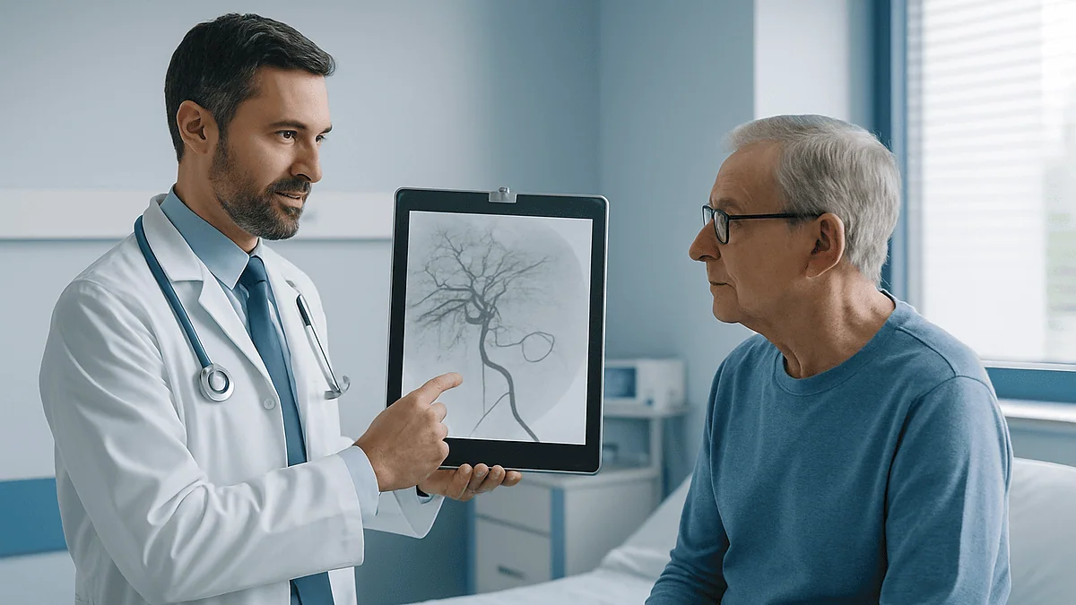
TARE radioembolization is performed in two visits by an interventional radiology team. First, a mapping angiogram threads a thin catheter from the wrist or groin into the liver arteries to chart blood flow, close off branches leading elsewhere, and measure lung shunting with a test tracer. Days to weeks later, tiny yttrium-90 microspheres are delivered through the same route into tumor-feeding arteries, where they lodge and irradiate the tumor from inside.
The folder on the kitchen table has a word in it nobody in the family can pronounce on the first try: radioembolization. Underneath, a nurse has written two dates in pencil, a week and a half apart, and the phrase “mapping first, then treatment.” It is a lot to hold at once. Liver tumor. Radiation, but from the inside. A catheter, but no incision to speak of.
People who ask how is TARE radioembolization performed are usually not looking for physics. They want to know why there are two appointments instead of one, whether they will be awake, what “microspheres” are, and why the doctor keeps mentioning the lungs when the problem is in the liver.
This explainer walks through the procedure in the order it actually happens, from the first scan to the weeks afterward, using what mainstream medical sources describe. It does not replace the conversation with your treating team, which is where every decision about your care belongs.
What is TARE radioembolization, in one plain paragraph?
Transarterial radioembolization, usually shortened to TARE, is a way of delivering radiation to liver tumors through the bloodstream rather than through the skin. The National Cancer Institute defines it as a procedure in which tiny radioactive beads are injected into the artery that supplies a tumor, so that the beads lodge in the tumor’s small vessels and release radiation directly into it. You may also hear it called Y90 therapy, after yttrium-90, the radioactive element inside the beads, or SIRT, short for selective internal radiation therapy.
Three words in that name carry the whole idea. Transarterial means the route is an artery: a catheter, a thin flexible tube, travels through the arterial system to the liver. Radio refers to the radiation source. Embolization means partially blocking small vessels; the beads are physically small enough to wedge in the tiny arteries within a tumor and stay there.
Why the liver, specifically? Healthy liver tissue draws most of its blood from the portal vein, while liver tumors draw most of theirs from the hepatic artery. That asymmetry is the opening. Anything sent down the hepatic artery reaches tumor far more than it reaches normal liver, which is the same logic that underlies chemoembolization, described later.
One more distinction matters. TARE is not external beam radiotherapy, where a machine aims beams through the skin over many sessions. Here the radiation source travels in and stays in, and the treatment is usually delivered in a single session per liver region. Cleveland Clinic and Mayo Clinic both describe it as a locoregional treatment, meaning it targets the liver rather than the whole body, and it is generally offered when surgery is not an option at that point in care.
How is TARE radioembolization performed? The two-visit shape of the treatment
The single most confusing thing for patients is that TARE is not one procedure but two, built on the same technique. Cleveland Clinic describes the pattern plainly: a planning angiogram first, then the treatment angiogram at a separate visit, typically one to a few weeks later.

Visit one is reconnaissance. An interventional radiologist, a physician who performs image-guided procedures through blood vessels, maps the liver’s arterial tree, temporarily closes off branches that lead to the stomach or bowel, and injects a harmless test tracer to see where particles would travel. A nuclear medicine scan afterward shows how much tracer escaped to the lungs.
Visit two is delivery. The team returns to the same arterial route, positions the catheter exactly where the mapping showed it should go, and releases the yttrium-90 microspheres. The word microsphere just means a very small bead; these are made of glass or resin and are on the order of a few dozen micrometers across, roughly a third the width of a human hair, according to physics reviews indexed on PubMed.
Between the visits, planning happens out of sight. A medical physicist, a specialist who calculates radiation delivery, uses the mapping scans to work out how much activity to prepare and how to split it if more than one liver region is treated. Patients do not see this step, but it is the reason the first visit cannot be skipped.
Each visit is usually a same-day or overnight event. Both are done through a puncture in the skin at the wrist or the top of the thigh rather than an incision, which is why recovery is measured in days, not weeks. The sections below take each visit in turn.
Before mapping: what the workup usually involves
Nobody arrives at a mapping angiogram cold. The National Cancer Institute’s treatment summary for adult liver cancer notes that liver-directed therapies are considered within a wider plan, and the workup reflects that.
Imaging comes first. A contrast-enhanced CT or MRI of the liver shows where the tumors are, how many, and how they sit relative to the main arteries and the portal vein. This is the picture the interventional radiologist studies before the first catheter goes in.
Blood tests come next. Liver function tests measure how well the non-tumor liver is coping; the whole strategy depends on sparing enough working tissue. Bilirubin, albumin, and clotting measures are commonly reviewed, alongside kidney function because contrast dye is filtered by the kidneys. Tumor markers may be checked as a baseline for later comparison.
Then comes the conversation that decides eligibility. A multidisciplinary tumor board, a meeting of surgeons, oncologists, radiologists, and hepatologists, usually weighs the options together. Mayo Clinic describes this team approach as standard for liver cancer, because the same scan can point toward surgery, ablation, embolization, systemic therapy, or a combination depending on liver reserve and overall health.
Practical instructions follow. You will typically be told when to stop eating before each procedure, which of your regular medicines to hold and which to take, and whether a blood thinner needs adjusting. Those instructions come from your prescribing clinician and are specific to you; nothing in this article should override them.
Finally, expect a consent discussion that covers risks, alternatives, and what happens if mapping shows the procedure cannot safely proceed. That outcome is not a failure of care. It is exactly what the mapping visit exists to catch.
Mapping angiogram before Y90: what actually happens on the table
You are awake, or lightly sedated, lying flat in a room that looks like a cross between an operating theater and an imaging suite. A large X-ray camera hangs over the table. The mapping angiogram, an X-ray study of blood vessels using contrast dye, is about to begin.
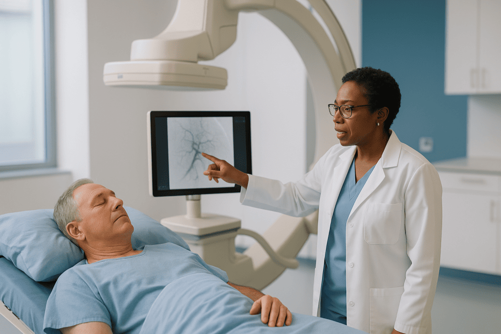
The skin at the wrist or upper thigh is cleaned and numbed. Through a needle puncture, the interventional radiologist places a short sheath into the artery, then threads a catheter up toward the liver under live X-ray guidance, called fluoroscopy. You will feel pressure, sometimes a warm flush when contrast is injected, but the inside of blood vessels has no pain nerves, so the catheter’s travel is not felt.
Once in the hepatic artery, the team injects contrast and watches it flow. This is the actual map: which branches feed the tumor, whether the anatomy is textbook or has variants, and, crucially, whether any small vessels run from the liver’s arteries toward the stomach, duodenum, or pancreas. Radioactive beads escaping to those organs can cause ulcers, so the radiologist may block such branches with tiny coils or plugs during this same session. Cleveland Clinic lists this protective step as part of standard planning.
Then a test dose of a tracer, a non-therapeutic radioactive particle of similar size to the treatment beads, is injected where the real microspheres would go. Afterward you move to a nuclear medicine camera, which photographs where the tracer landed.
The catheter comes out, pressure or a small closure device seals the puncture, and you rest for a few hours. Most people go home the same day. Total time on the table is commonly one to two hours, according to patient-education descriptions from Cleveland Clinic, though anatomy that needs extra coiling can lengthen it.
Why the lungs matter: the shunt scan and dose planning explained
The most common question after mapping is why a liver treatment involved a lung scan. The answer is a plumbing problem called shunting.
Some liver tumors contain abnormal channels that connect arteries directly to veins, bypassing the tiny capillary bed where beads are supposed to lodge. Particles that slip through those channels ride the venous blood back to the heart and are pumped into the lungs, which act as the body’s next filter. A small amount is unavoidable and harmless. A large amount could deliver meaningful radiation to lung tissue, which is the one complication planners work hardest to avoid.
The tracer study from the mapping visit measures this directly. The nuclear medicine camera counts how much activity ended up in the lungs versus the liver and reports it as a lung shunt fraction, a percentage. Published reviews indexed on PubMed describe thresholds above which teams either reduce the planned activity, modify the approach, or advise against TARE altogether. The exact cutoffs are a clinical judgment for your team and are not something this article will quote.
The same scan also confirms that no tracer reached the stomach or bowel. If it did, the radiologist may need to block another vessel before treatment day.
Dosimetry, the calculation of how much radiation to deliver, then happens off the table. The physicist combines the shunt result, the volume of liver being treated, tumor size, and the relative uptake between tumor and normal liver into a plan. Newer methods use the tracer scan itself as a preview of where the real beads will concentrate. None of this is visible to the patient, but it is the reason the treatment visit is usually scheduled a week or more later: the plan must be finalized and the radioactive beads ordered and calibrated for a specific date and time, since yttrium-90 decays steadily from the moment it is made.
How is TARE radioembolization performed on treatment day? From catheter to microspheres
Treatment day looks almost identical to mapping day, which many patients find reassuring. Same room, same puncture site, same route. The differences are the shielded container that arrives from the radiopharmacy and the extra people in the room: a nuclear medicine physician or physicist, and staff wearing dosimeters, small badges that record radiation exposure.
After numbing and sheath placement, the interventional radiologist navigates the catheter to the exact position chosen during mapping, often using a microcatheter, a very fine inner tube, to reach a specific tumor-feeding branch. A quick contrast check confirms the position matches the plan and that any previously coiled vessels remain closed.
Then the delivery. The yttrium-90 microspheres sit in a vial inside an acrylic shield. Using a sterile tubing system, the team slowly pushes the beads through the catheter with intermittent flushes, pausing to check flow under fluoroscopy. If flow slows because vessels are filling, the injection is slowed too. The goal is for the beads to travel with arterial blood into the tumor’s smallest vessels and stop there. Cleveland Clinic describes the infusion itself as taking only minutes, with the whole procedure commonly lasting around an hour.
You will not feel the radiation. Some people notice a dull ache in the right upper abdomen during or shortly after the infusion, which staff can treat.
Once delivery is complete, the catheter and sheath are withdrawn and the puncture sealed. Many centers then take a confirmatory nuclear medicine scan, called a bremsstrahlung or PET scan, that shows where the beads actually landed. Afterward you recover for several hours while the puncture site is monitored. Depending on the center, you go home that evening or stay one night. The radiation is now working from inside the tumor, and it will keep doing so for days.
What the yttrium-90 microspheres actually do inside the tumor
Once lodged, the beads become tiny, stationary radiation sources. Understanding a few numbers about the physics explains why the treatment is delivered once and why the safety rules afterward are short.
Yttrium-90 is a beta emitter. Beta particles are fast electrons that travel only a short distance in tissue before stopping. Physics reviews indexed on PubMed put the average travel at a few millimeters and the maximum at roughly a centimeter. That short reach is the point: radiation concentrates within and immediately around each bead, saturating tumor that has drawn the beads in, while normal liver a short distance away receives far less.
Because tumors are richly supplied by arteries and normal liver is not, beads cluster preferentially in tumor. Imaging after treatment routinely shows a much higher concentration in tumor than in surrounding tissue, which is what dosimetry relies on.
The half-life of yttrium-90, the time for half its radioactivity to decay, is about 64 hours, according to the same published sources. That means the beads deliver most of their radiation within the first week or two after treatment, then fade to negligible activity. The beads themselves are inert glass or resin and remain in the liver permanently without causing harm; they are not removed.
Radiation damages the DNA of cells it passes through. Tumor cells, which divide often, cope poorly with that damage and die over the following weeks. This is why imaging is not repeated immediately: the visible response develops over about two to three months, per Mayo Clinic’s description of follow-up after liver-directed therapy, and early scans can look worse before they look better because of swelling and inflammation.
Yttrium-90 emits almost no gamma radiation, the penetrating type that escapes the body. That is why the precautions described later are modest compared with some other radioactive treatments.
Who is TARE radioembolization usually for, and who is usually asked to wait?
TARE sits in a specific place on the treatment map. The National Cancer Institute’s liver cancer treatment summary lists embolization approaches among options for tumors that cannot be removed surgically, and mainstream sources describe several situations where teams consider it.
The first is hepatocellular carcinoma, the most common primary liver cancer, when the tumor is confined to the liver but too large, too numerous, or too awkwardly placed for resection or ablation. Cleveland Clinic and Mayo Clinic both list radioembolization among treatments for this scenario.
The second is liver metastases, cancer that has spread to the liver from elsewhere, most often colorectal cancer or neuroendocrine tumors, when the liver is the dominant site of disease and systemic therapy alone is not controlling it.
A third role is as a bridge or downstaging tool: shrinking tumors so that a person may become eligible for surgery or transplant, or holding disease steady while waiting. Some teams also use a focused version, sometimes called radiation segmentectomy, to treat a single liver segment intensively when ablation is not feasible.
Who is usually asked to wait, or steered elsewhere? Several situations, described in guideline-level sources, make teams cautious. Poor liver function is the main one: if the non-tumor liver is already struggling, adding radiation risks liver failure. A high lung shunt fraction on mapping, discussed earlier, is another. Blood flow toward the gut that cannot be blocked, a blocked portal vein in some contexts, extensive disease outside the liver, and prior radiation to the liver all weigh against proceeding. Pregnancy is a contraindication.
None of these are absolute rules applied by a checklist. The tumor board weighs them against alternatives, and the decision belongs to that team with you.
Radioembolization vs chemoembolization: how the liver-directed options compare
Patients often meet TARE alongside a sibling procedure, transarterial chemoembolization, or TACE, and struggle to tell them apart. Both use the same arterial route and the same reasoning about tumor blood supply. The payload differs.
In TACE, the catheter delivers chemotherapy, sometimes loaded onto beads, together with material that blocks the artery. The blockage starves the tumor of oxygen while the drug concentrates locally. In TARE, the beads are so small that blood flow is only partly reduced; the treatment effect comes from radiation, not from cutting off supply. Mayo Clinic and the National Cancer Institute describe both as liver-directed treatments for unresectable tumors.
| Feature | TARE (Y90) | TACE | Thermal ablation | External beam radiation |
|---|---|---|---|---|
| Route | Hepatic artery catheter | Hepatic artery catheter | Needle through skin into tumor | Beams from outside the body |
| Active agent | Radiation from beads | Chemotherapy plus vessel blockage | Heat or cold | Radiation |
| Sessions | Mapping visit, then usually one treatment per liver region | Often repeated over months | Usually one session per tumor | Several sessions over days to weeks |
| Typical short-term effects | Fatigue, mild nausea, abdominal ache; usually milder than TACE per Cleveland Clinic | Post-embolization syndrome with fever and pain, often more pronounced | Local pain, low-grade fever | Fatigue, skin changes, nausea |
| Usual hospital stay | Same day or one night | Often one or more nights | Same day or one night | Outpatient |
Which is chosen depends on tumor number and size, liver function, portal vein status, prior treatments, and what the team is trying to achieve. Published comparisons indexed on PubMed exist, but they do not crown one approach for every patient, and this article makes no such claim. Your team’s recommendation is the one that counts.
Y90 radioembolization side effects and risks in plain language
Every honest explainer has to say what can go wrong, without dramatizing it. Mainstream sources sort the effects of TARE into common and expected, uncommon and treatable, and rare but serious.
Common and expected comes first. Cleveland Clinic describes a cluster called post-embolization syndrome: fatigue, low-grade fever, mild nausea, loss of appetite, and a dull ache under the right ribs. It typically begins within a day or two and eases over one to two weeks. Mild, temporary changes in liver blood tests are also usual as the treated tissue reacts. Bruising or tenderness at the wrist or groin puncture site is routine.
Uncommon and treatable includes ulcers in the stomach or duodenum, which happen if beads reach the gut despite mapping; this is exactly why vessels are coiled beforehand. Inflammation of the gallbladder or pancreas can occur for the same reason. Bleeding or a small clot at the puncture site, or an allergic reaction to contrast dye, are risks shared by every angiogram.
Rare but serious effects deserve naming. Radiation-induced liver disease, sometimes called radioembolization-induced liver disease, is a delayed injury to the non-tumor liver that can appear weeks to months later with jaundice and fluid buildup; the risk rises when liver reserve was already limited, which is why eligibility is scrutinized. Radiation pneumonitis, inflammation of the lungs, is the reason for the shunt study and is uncommon when planning thresholds are respected. Infection and, very rarely, liver abscess have been reported.
Published reviews indexed on PubMed provide rates for each of these, and your consent discussion should include the figures your center uses. What matters for a patient is knowing which symptoms are ordinary and which are not, covered in the red-flag section near the end.
Radioembolization recovery time: what the following days and weeks usually look like
The pattern most people describe is a slow week, then a gradual climb back. Here is the typical arc, drawn from patient-education material at Cleveland Clinic and Mayo Clinic, with the caveat that individual experience varies and your team’s instructions come first.
Hours after treatment. You lie flat for a period so the artery puncture seals, longer for a groin site than a wrist site. Staff check the site, your pulse, and your blood pressure. Some abdominal ache and nausea are common and are treated as they arise. Most people are discharged that evening or the next morning.
Days one through three. Fatigue is the headline. Appetite may be poor and a low-grade fever can come and go. Keep the puncture site clean and dry as instructed, avoid heavy lifting, and drink fluids unless you have been told to restrict them. Radiation precautions, covered next, apply during this window.
Week one to two. Energy usually starts returning. Many people are back to desk work or light routines within one to two weeks, per Cleveland Clinic’s description. A blood test around this time is common to check liver function.
Weeks three to eight. This is the quiet stretch. The beads have largely finished delivering their radiation, but the tumor’s response is still unfolding. Feeling well in this period says little either way about how the scan will read.
Around two to three months. Follow-up imaging, usually CT or MRI, and often a tumor marker check. Mayo Clinic describes this interval as typical for assessing response to liver-directed therapy. The team then decides whether to treat another liver region, watch, or move to a different approach.
Systemic therapies, if you were on them, are paused and restarted according to your oncologist’s plan; do not change them yourself.
Radiation safety at home after Y90: what the precautions really are
The word radiation makes families picture lead aprons and sealed rooms. The reality after Y90 is far more ordinary, and understanding why helps everyone relax.
As described earlier, yttrium-90 emits beta particles that travel only millimeters in tissue and almost no gamma rays. The beads are locked inside the liver. Very little radiation reaches the body surface, and almost none reaches another person standing beside you. This is different from treatments such as radioactive iodine, where the isotope circulates and is excreted.
Still, centers give a short list of common-sense measures for the first several days, typically up to about a week, according to Cleveland Clinic’s patient guidance. These usually include limiting prolonged close contact with young children and pregnant people, such as not holding a child on your lap for extended periods, and sleeping apart from a partner if advised. Bodily fluids carry negligible activity, but ordinary hygiene, flushing twice and washing hands, is often suggested for the first few days as a belt-and-braces measure.
You may be given a card or letter stating that you have received a radioactive treatment. Airport security scanners can occasionally detect low-level radiation for a few weeks, and the card explains why. Keep it with you if you travel during that period.
Within a couple of weeks, the activity has decayed to a level that requires no special measures. The beads remain, permanently and harmlessly, as inert glass or resin.
Your own center’s written instructions are the ones to follow, since protocols differ slightly by bead type and regulatory setting. If a family member is anxious, ask the nuclear medicine team to talk them through it; they field these questions daily and can explain the numbers behind the reassurance.
What people often get wrong about radioembolization
Some misunderstandings surface again and again in clinic. Correcting them early saves worry.
“It is a kind of chemotherapy.” No. TARE delivers radiation, not drugs. The confusion comes from its cousin TACE, which does deliver chemotherapy through the same route. TARE does not cause hair loss and does not suppress bone marrow the way systemic chemotherapy can.
“The mapping visit is just a formality.” The opposite. Mapping is where the team discovers vessels that could carry beads to the stomach, measures lung shunting, and gathers the data for dosimetry. Sometimes mapping shows that treatment cannot safely proceed, and finding that out beforehand is the whole point.
“I will be radioactive and dangerous to my family.” The beta radiation from yttrium-90 barely leaves the liver, let alone the body. Precautions are brief and modest, as the previous section explains.
“The beads dissolve or are removed later.” They stay permanently. They are inert once the radiation decays and cause no harm.
“If the first scan looks bigger, the treatment failed.” Early imaging can show swelling and inflammation in the treated area. Mayo Clinic notes that response is usually assessed at around two to three months, and radiologists read these scans with that timeline in mind.
“It replaces surgery or transplant.” For some people it is offered because surgery is not an option; for others it is used to shrink tumors so surgery or transplant becomes possible. It is one tool within a plan, not a substitute for the plan.
“Newer or more expensive always means better.” Published reviews indexed on PubMed compare liver-directed options carefully and do not find a single winner for every situation. Fit to the individual patient matters more than novelty.
Questions to ask your care team before TARE radioembolization
A good consultation leaves room for questions, and having them written down helps when the room feels overwhelming. These are the ones that tend to matter most, drawn from what patients commonly raise and what mainstream sources flag as decision points.
- Why are you recommending TARE for me rather than TACE, ablation, surgery, or systemic therapy alone, and what would change that recommendation?
- What did my liver function tests show, and how much healthy liver reserve do I have?
- Which part of my liver will be treated: one segment, one lobe, or both, and will that mean one treatment session or two?
- What will you be looking for on the mapping angiogram, and what findings would lead you to advise against proceeding?
- If vessels need to be coiled, what does that involve and does it change anything for me afterward?
- What is my lung shunt result, and how does it affect the plan?
- Which of my regular medicines should I hold or continue before each procedure, and who confirms that?
- Will I go home the same day or stay overnight, and who should drive me?
- What side effects should I expect in the first two weeks, and which ones should prompt a call?
- What radiation precautions does your center advise at home, and for how long?
- When will follow-up imaging happen, and what will you be looking for?
- What happens next if the scan shows a good response, and what happens if it does not?
- Are there clinical trials relevant to my situation that I should know about?
Ask the team to write answers in the folder, next to the pencil dates. Bring someone with you if you can; a second set of ears catches what the first misses. And ask who to call after hours, because the next section is about exactly that.
When to call your doctor after radioembolization
Most of the first two weeks after TARE is fatigue and mild discomfort that fades. A small number of symptoms are different in kind and should not wait for the scheduled follow-up. Mainstream sources including Cleveland Clinic and Mayo Clinic list the following as reasons to contact your team promptly, and to seek emergency care if severe.
Call the same day if you notice:
- Fever that is higher than the low-grade fever you were told to expect, or any fever with shaking chills, which can signal infection or abscess.
- Abdominal pain that is severe, worsening, or not controlled by the measures your team recommended.
- Vomiting that prevents you from keeping down fluids, or vomit that is dark or contains blood, which can indicate a stomach or duodenal ulcer.
- Black, tarry, or bloody stools, another sign of gut bleeding.
- Yellowing of the skin or the whites of the eyes, dark urine, or a swelling belly, which can indicate the liver is struggling and can appear weeks after treatment.
- New confusion, drowsiness, or unusual sleepiness, which can accompany liver dysfunction.
- A puncture site that swells rapidly, bleeds through a dressing, becomes hot and red, or a limb below it that turns cold, pale, or numb.
- New or worsening shortness of breath or a persistent dry cough, which the team will want to assess given the lungs’ role in this treatment.
Seek emergency care immediately for heavy bleeding from the puncture site that pressure does not control, chest pain, sudden severe breathlessness, or fainting.
Keep the after-hours number your team gave you somewhere visible. When in doubt, call; interventional radiology and oncology teams expect these calls and would rather hear from you early. The final decision about what any symptom means, and what to do about it, rests with the clinicians who know your case.
Frequently asked questions
How is TARE radioembolization performed and is it surgery?
It is not surgery in the conventional sense. An interventional radiologist passes a thin catheter through a needle puncture at the wrist or groin into the liver’s arteries under X-ray guidance, first to map the vessels and later to deliver yttrium-90 microspheres. There is no incision, you are awake or lightly sedated, and most people go home the same day or after one night.
Why do I need a mapping angiogram before Y90 treatment?
Mapping identifies exactly which arteries feed the tumor, blocks any small branches that could carry beads to the stomach or bowel, and uses a test tracer to measure how much would escape to the lungs. Those results determine whether treatment is safe and how the radiation is planned. Occasionally mapping shows TARE should not proceed, which is precisely why the step exists.
How long is the gap between mapping and treatment?
Typically one to a few weeks, according to patient-education material from Cleveland Clinic. The interval allows the physicist to finalize dosimetry and lets the radiopharmacy prepare beads calibrated for a specific date and time, since yttrium-90 begins decaying as soon as it is produced. Your center will give you the exact schedule.
What are the most common Y90 radioembolization side effects?
Fatigue, low-grade fever, mild nausea, poor appetite, and a dull ache under the right ribs, a cluster called post-embolization syndrome, are the most common and usually ease within one to two weeks per Cleveland Clinic. Temporary changes in liver blood tests and bruising at the puncture site are also routine. Less common risks include gut ulcers, gallbladder inflammation, and delayed liver injury.
What is the difference between radioembolization vs chemoembolization?
Both use a catheter in the hepatic artery, but the payload differs. Chemoembolization delivers chemotherapy along with material that blocks the artery; radioembolization delivers radiation from tiny beads and only partly reduces blood flow. Chemoembolization is often repeated and tends to cause more pronounced post-procedure symptoms, while radioembolization is usually a single treatment per liver region. Your team chooses based on liver function, tumor pattern, and goals.
What is the typical radioembolization recovery time?
Most people feel tired for the first week, with appetite and energy gradually returning so that light routines and desk work resume within one to two weeks, according to Cleveland Clinic. Follow-up imaging to assess the tumor’s response is usually scheduled around two to three months after treatment. Individual recovery varies with liver function and overall health.
Will I be radioactive and need to stay away from my family?
Only modestly and briefly. Yttrium-90 emits beta particles that travel millimeters and almost no penetrating gamma rays, so very little radiation leaves your body. Centers typically advise limiting prolonged close contact with young children and pregnant people for several days to about a week. Follow your own center’s written instructions, which may vary slightly.
Do the microspheres ever come out of the liver?
No. The glass or resin beads lodge permanently in the tumor’s small vessels and remain there after the radiation has decayed. They are inert and cause no known harm in place. They are not dissolved, excreted, or surgically removed, and imaging years later may still show them.
Who is usually not a candidate for TARE radioembolization?
People with poor liver function, a high lung shunt fraction on mapping, blood flow to the gut that cannot be blocked, extensive cancer outside the liver, or prior liver radiation are often steered toward other options, and pregnancy is a contraindication. These are judgments a multidisciplinary tumor board weighs against alternatives, not rules applied by checklist.
Does a bigger-looking tumor on the first scan mean the treatment failed?
Not necessarily. Radiation causes swelling and inflammation in the treated area, so early imaging can appear larger or unchanged before shrinkage becomes visible. Mayo Clinic describes response assessment at around two to three months for this reason. Radiologists interpret post-TARE scans with that timeline in mind, and your team will explain what your images show.
References
- Cleveland Clinic – Liver Cancer
- NHS – Liver cancer: Treatment
- PubMed – Yttrium-90 radioembolization for liver tumors (indexed literature)
This article is for general information only and is not a substitute for professional medical advice. Please consult a qualified doctor about your individual situation.

More from the Blog

Does Acute Myeloid Leukemia Treatment Include a Transplant? How Genetic Findings Decide
Acute myeloid leukemia treatment includes a stem cell transplant for some people but not all. After chemotherapy brings the disease into remission, the care…
Surgery or Chemoradiation for Cervical Cancer? What Makes Each Path the Better Fit
Surgery and chemoradiation treat cervical cancer about equally well when each is matched to the right stage. Surgery is usually chosen for small tumors…

How Immunotherapy for Melanoma Skin Cancer Is Given, Cycle by Cycle
Immunotherapy for melanoma is usually given as an intravenous infusion in repeating cycles, typically every few weeks, with blood tests and a symptom review…

How Long After Radioiodine Treatment Does the Thyroid Respond? A Realistic Timeline
Radioactive iodine does not work immediately. For an overactive thyroid, hormone levels usually begin falling within a few weeks, and most people notice the…

Questions to Ask Your Dermatologist or Oncologist Before Skin Cancer Treatment
Before skin cancer treatment, ask your dermatologist or oncologist what type and stage of skin cancer you have, what the pathology report says, which…
Esophagectomy for Esophageal Cancer: What Happens in the Operating Room and After
An esophagectomy removes part or most of the esophagus, usually along with nearby lymph nodes, and rebuilds the swallowing tube by pulling the stomach…