

Hysteroscopic Fibroid Removal: Treating Fibroids Inside the Uterine Cavity Without Incisions

Key Takeaways
- Hysteroscopic fibroid removal reaches only fibroids that touch the uterine cavity; fibroids buried in the wall or on the outer surface need a different route.
- Because no cut is made through the uterine muscle, the procedure leaves no skin scar and does not on its own usually change future delivery plans, unlike abdominal myomectomy.
- Fluid absorbed during the operation is counted continuously, and stopping early when a limit is reached is a built-in safety step, which is why large fibroids may be planned as two sessions.
- Uterine perforation and scar tissue inside the cavity are the procedure-specific risks that matter most, particularly for anyone planning pregnancy.
- Removed fibroids do not regrow, but new fibroids can form; fibroids typically shrink after menopause as hormone levels fall.
- No diet, herb or supplement has been shown in controlled trials to eliminate existing fibroids, despite widespread online claims.
Hysteroscopic fibroid removal, also called hysteroscopic myomectomy, treats fibroids that grow into the uterine cavity by passing a thin camera and cutting instrument through the vagina and cervix, so no abdominal incision is needed. It is usually a same-day procedure for submucosal fibroids causing heavy bleeding or fertility concerns. Recovery is typically measured in days, though suitability, risks and alternatives depend on fibroid size, position and personal goals, decided with the treating gynecologist.
The ultrasound report lands in the patient portal before the follow-up call does, and one word in it keeps snagging the eye: submucosal. A fibroid, the sonographer had said, sitting right where a period begins. That explains the eleven-day cycles, the iron tablets, the meetings rearranged around the heaviest mornings. What it does not explain is what happens next.
For many people in that position, the option raised first is hysteroscopic fibroid removal, a way of reaching a fibroid from inside the uterus rather than cutting through the abdomen to get to it. The idea sounds almost too tidy: no scar, home the same day, a camera doing the looking and a slim instrument doing the work.
Tidy is not the same as simple. Which fibroids this approach can reach, what the procedure actually involves, where the real risks sit and what the weeks afterward tend to look like deserve a clear, unhurried explanation. That is what follows.
What is hysteroscopic fibroid removal, and why does inside the cavity matter?
Fibroids are noncancerous growths made of muscle and fibrous tissue that develop in or around the uterus. They are common: the NHS estimates that around one in three women develops them at some point, most often between the ages of 30 and 50. Many cause no symptoms at all and are found by chance.
Where a fibroid sits changes almost everything about how it behaves. Doctors describe three broad positions. Subserosal fibroids bulge from the outer surface of the uterus. Intramural fibroids grow within the thick muscular wall. Submucosal fibroids push inward, distorting the endometrium, the lining that builds up and sheds each month.
That last group is the focus here. The uterine cavity is the small space inside the uterus where menstrual blood collects and where an embryo implants. A submucosal fibroid crowds that space, enlarges the bleeding surface and disturbs the way the lining contracts to stop a period. Clinically, this is why a fibroid the size of a grape sitting in the cavity can cause heavier bleeding than a far larger one on the outside wall.
Hysteroscopic fibroid removal takes advantage of that same geography. A hysteroscope, a thin telescope with a light and camera, is passed through the vagina and cervix into the cavity, and the fibroid is removed from within. The medical term is hysteroscopic myomectomy: myomectomy simply means removing a fibroid while leaving the uterus in place, which distinguishes it from hysterectomy, the removal of the uterus itself.
Because the surgeon works through a natural passage, there is no cut in the skin and no cut through the uterine muscle. That single fact drives most of the advantages, and a few of the limitations, that the rest of this article explains.
Submucosal fibroid removal: which fibroids actually qualify?
Not every fibroid labelled submucosal is equally reachable from inside. Gynecologists grade how deeply a fibroid is anchored in the muscle wall, and that depth largely decides whether a hysteroscopic approach is sensible.
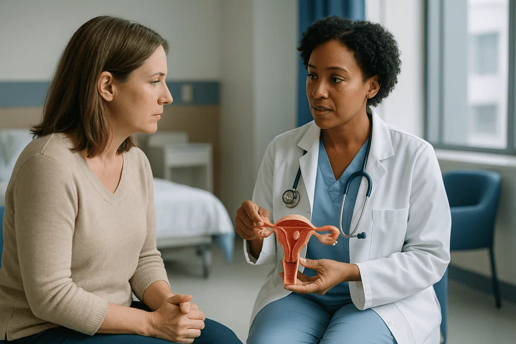
At one end are fibroids that hang entirely within the cavity on a stalk, like a mushroom on a stem. These are the most straightforward to remove through the hysteroscope. Next come fibroids that sit mostly in the cavity with a base embedded in the wall. Then there are fibroids buried mainly within the muscle with only a portion breaking the surface of the lining. The deeper the fibroid extends into the wall, the harder it is to remove completely in one session and the greater the chance that a second procedure, or a different approach altogether, will be recommended.
Size matters too. Hysteroscopic removal is generally reserved for smaller fibroids, because every piece of tissue has to be shaved into strips or fragments and drawn out through the cervix. The Mayo Clinic describes the technique as suited to fibroids contained inside the uterus, and surgeons often plan a staged procedure when a fibroid is larger than they can safely remove within one operating time.
Number counts as well. One or two cavity fibroids are a different proposition from a uterus dotted with many fibroids of mixed positions, where a hysteroscopic procedure might treat the bleeding source but leave the bulk symptoms untouched.
What the approach cannot do is reach intramural or subserosal fibroids that do not touch the cavity. For those, the options run through laparoscopic or open myomectomy, uterine artery embolization, medicines or hysterectomy, discussed later. The honest question to ask is not whether a fibroid can be removed, but whether removing this particular fibroid through this particular route is likely to address the symptom that prompted the visit.
How hysteroscopic fibroid removal actually works, step by step
Picture a room-sized balloon that has to be inflated before anyone can see the walls. The uterine cavity is normally collapsed, its front and back walls touching. The first job of the procedure is to open it.
After anesthesia takes effect, the surgeon gently widens the cervix, the neck of the uterus, using slim dilators. The hysteroscope is then passed through. Sterile fluid, usually saline, flows through the scope to distend the cavity, a term that simply means holding it open with gentle pressure so the camera has a view. The image appears on a screen, and the fibroid is identified along with its base.
Removal happens in one of two main ways. A resectoscope carries a fine wire loop that uses electrical energy to shave the fibroid into strips, a little like planing wood. A newer generation of mechanical tissue removal devices instead uses a rapidly moving blade inside a hollow tube that cuts and suctions tissue simultaneously, without electrical current in the cavity. Both approaches are established; the choice depends on the fibroid, the surgeon’s training and equipment available. Neither is inherently superior for every case.
Throughout, a nurse or technician tracks how much fluid goes in and how much comes out. The difference is fluid the body has absorbed through open blood vessels in the uterine wall, and keeping that number within safe limits is one of the central safety tasks of the operation.
Once the fibroid is removed down to the level of the surrounding lining, the fragments are sent to a pathology laboratory for routine examination, the scope is withdrawn and nothing is stitched. Cleveland Clinic notes that hysteroscopic procedures can range from a few minutes for a diagnostic look to more than an hour when tissue is being removed, so the exact time varies widely with what is being done.
Who is hysteroscopic myomectomy usually for, and who is usually asked to wait?
The typical candidate has a fibroid inside the cavity and a problem that fibroid is plausibly causing. Heavy menstrual bleeding is the most common trigger, particularly when it has led to iron-deficiency anemia, the shortage of red blood cells that leaves people breathless on stairs and tired by mid-afternoon. Bleeding between periods, difficulty conceiving or repeated early pregnancy loss with a cavity fibroid on imaging are other reasons gynecologists raise the option.
Equally important is who is usually steered elsewhere or asked to wait.
- People whose fibroids sit mainly in the wall or on the outer surface, where a hysteroscope cannot reach.
- Those with a very large or deeply embedded cavity fibroid, where a staged or abdominal approach may be safer.
- Anyone with an active pelvic infection, which is treated first.
- Those who are pregnant; the procedure is not performed during pregnancy.
- People with severe anemia, who may be asked to build up their blood count with medical support before surgery so they can tolerate any blood loss.
There is a third group worth naming: people with fibroids who have no troublesome symptoms. Both the Mayo Clinic and NHS describe watchful waiting as a legitimate plan in that situation, because fibroids are noncancerous and tend to shrink after menopause when estrogen levels fall. Someone in their late forties with mild symptoms may reasonably choose to monitor rather than operate, and a good clinician will present that option rather than default to a procedure.
Timing within the menstrual cycle also matters for those who do proceed. Surgeons often schedule the operation for the days just after a period, when the lining is thinnest and the view is clearest. None of these decisions belong to a checklist; they emerge from the conversation between the patient and the treating team, weighing symptoms, imaging, future pregnancy plans and personal priorities.
How do doctors confirm a fibroid is inside the cavity before operating?
A pelvic exam can suggest an enlarged or irregular uterus, but it cannot say where a fibroid sits. Imaging does that, and the sequence usually builds from simple to detailed.
Transvaginal ultrasound is the standard first step. A slim probe placed in the vagina uses sound waves to build a picture of the uterus, showing the size and rough position of fibroids. Because the cavity is collapsed, ultrasound alone can struggle to tell a fibroid that bulges into the cavity from one that merely sits close to it.
Saline infusion sonography solves that problem. A small amount of sterile fluid is introduced through the cervix during the ultrasound, opening the cavity so anything protruding into it stands out in sharp outline. The Mayo Clinic lists this among the tests used to clarify fibroid position when cavity involvement is suspected.
Magnetic resonance imaging, or MRI, gives the most detailed map of fibroid number, size and depth within the wall. It is not needed for everyone, but it becomes valuable when a uterus holds several fibroids, when surgery is being planned and the surgeon wants to know how deep a cavity fibroid is anchored, or when other uterine conditions need to be told apart.
Diagnostic hysteroscopy, a look inside with the camera but no cutting, may be done as a separate short procedure or at the start of the operation itself. It confirms what imaging suggested and lets the surgeon see the fibroid’s base directly.
Alongside imaging, a blood count checks for anemia, since heavy bleeding over months quietly drains iron stores. Correcting that before surgery is part of preparing the whole person, not just the uterus. Each result feeds the decision about whether hysteroscopic removal is the right route, and the treating team interprets them together rather than acting on any single scan.
What happens before the procedure: preparation, anesthesia and medicines
Preparation is mostly about making the surgeon’s view clearer and the patient’s recovery smoother, and some of it starts weeks ahead.
A thin uterine lining helps. Beyond scheduling surgery just after a period, some clinicians prescribe a course of hormonal medicine to thin the endometrium and, in some cases, shrink the fibroid beforehand. One class used for this purpose, gonadotropin-releasing hormone agonists, works by temporarily switching off the ovaries’ estrogen production, creating a short-term menopause-like state; the Mayo Clinic describes their preoperative use to reduce fibroid size and improve anemia. These medicines can bring hot flashes and other side effects, they are used for defined periods, and the decision to prescribe them, at what strength and for how long, sits with the prescribing clinician. Nothing about them should be started or stopped on the basis of an article.
Iron replacement, by tablet or infusion, may be arranged if anemia is present. Some surgeons also use a cervical softening medicine shortly before the procedure to make dilation gentler; again, that is a clinical judgment.
Anesthesia options range from general anesthesia, where the patient is fully asleep, to regional anesthesia numbing the lower body, to local anesthetic with sedation. Small cavity fibroids are sometimes removed in an office setting with local anesthesia; larger ones are done in an operating room. Johns Hopkins and Cleveland Clinic both describe this spectrum and note that the choice depends on the extent of the procedure and the patient’s health.
Practical instructions are ordinary but matter: when to stop eating and drinking, which regular medicines to take that morning, and arranging someone to drive home, since sedation lingers for hours. Patients are typically asked to report any chance of pregnancy, and a pregnancy test is often done on the day. Writing down the questions that surfaced at 2 a.m. is as much preparation as fasting is.
Hysteroscopic myomectomy risks: what the evidence actually shows
No incision does not mean no risk. The complications of hysteroscopic myomectomy are different from those of abdominal surgery, and patient-facing summaries from Johns Hopkins, Cleveland Clinic and the Mayo Clinic describe serious problems as uncommon, but understanding them is part of informed consent.
Uterine perforation is the one surgeons watch for most closely. It means an instrument passes through the wall of the uterus. Often this is recognized immediately and heals without further treatment, but if it occurs while an energy device is active, nearby bowel or bladder can be injured, which may require a laparoscopy to inspect and repair. Deeply embedded fibroids and a thin uterine wall raise the chance.
Fluid overload is specific to this operation. Distension fluid absorbed through open vessels can dilute the body’s salts and strain the heart and lungs if volumes climb, which is why fluid in and fluid out are counted continuously and the procedure is stopped if a preset limit is reached. Stopping early is a safety decision, not a failure, and is one reason large fibroids are sometimes planned as two sessions.
Bleeding can occur from the fibroid bed. Most settles with time or a temporary balloon placed in the cavity; transfusion is rare. Infection of the uterus or pelvis is possible, as after any procedure through the cervix.
Intrauterine adhesions, bands of scar tissue that form when raw surfaces inside the cavity heal against each other, deserve particular mention for anyone planning pregnancy. They can reduce menstrual flow and interfere with implantation. Removing fibroids on opposing walls in the same session increases the risk, and some surgeons plan around that.
Finally, incomplete removal, anesthesia reactions and the rare finding on pathology of an unexpected tumor are all discussed at consent. Ask how often each has arisen in your surgeon’s own practice; a candid answer is a good sign.
How does hysteroscopic fibroid removal compare with other fibroid treatments?
Fibroid care is a menu rather than a ladder, and the right choice depends on where the fibroids are, how big they are, what symptoms dominate and whether pregnancy is planned. The table summarizes the main options as described by the Mayo Clinic and NHS; recovery figures are typical ranges from those sources, not promises.
| Option | Fibroids it suits | Incision? | Uterus kept? | Typical recovery |
|---|---|---|---|---|
| Hysteroscopic myomectomy | Smaller fibroids inside the cavity | None | Yes | Days; usually home same day |
| Laparoscopic or robotic myomectomy | Fibroids in the wall or on the outer surface, limited number | Several small abdominal cuts | Yes | Around one to two weeks or more |
| Open (abdominal) myomectomy | Large or numerous fibroids | One larger abdominal cut | Yes | Several weeks |
| Uterine artery embolization | Symptomatic fibroids in the wall when pregnancy is not planned | Needle puncture in groin or wrist | Yes | About one to two weeks |
| Hormonal medicines (various classes) | Bleeding control; temporary shrinkage | None | Yes | Ongoing while taken |
| Hysterectomy | Any fibroids when family is complete and other options failed or declined | Vaginal, laparoscopic or abdominal | No | Several weeks; NHS cites roughly six to eight |
| Watchful waiting | Fibroids with mild or no symptoms | None | Yes | Not applicable |
Two patterns stand out. First, hysteroscopic removal is the only surgical option with no incision at all, but it is also the most restricted in which fibroids it can reach. Second, hysterectomy is the only approach after which fibroids cannot return, at the cost of future pregnancy and a longer recovery. Everything else manages fibroids rather than eliminating the tendency to form them.
Medicines sit in the table for a reason. For some people, hormonal treatments control bleeding well enough that surgery is deferred or avoided. For others they buy time before an operation. Which, if any, is appropriate is a prescribing decision made with the clinician who knows the full history.
Hysteroscopic myomectomy recovery time: the first days and weeks
Most people leave the same day, once they are awake, passing urine and comfortable. Johns Hopkins describes recovery from hysteroscopic procedures as generally quick, with mild cramping and light vaginal bleeding or watery discharge for a day or two being expected. Cleveland Clinic notes that many people return to routine activities within a day or two, though anyone who had general anesthesia is advised to rest and avoid driving for the first 24 hours.
The days that follow have a recognizable shape.
In the first two or three days, cramping resembling a period is common and typically eases with rest, warmth and over-the-counter pain relief as advised by the care team. Discharge may be pink or brownish and can continue intermittently for a week or two as the fibroid bed heals. A small amount of spotting is expected; soaking through pads hourly is not, and the red-flag section later covers that.
Through the first couple of weeks, surgeons usually ask patients to avoid anything placed in the vagina, including tampons, douching and intercourse, and to skip baths and swimming in favor of showers, to lower infection risk while the cervix and cavity heal. The exact interval varies; follow the specific instructions given rather than a general figure.
The first period afterward may arrive on schedule or be a little early or late, and it can be lighter or briefly heavier than usual. Patterns tend to settle over the following cycles. This is also when the real test of the procedure begins: whether bleeding is more manageable than before.
A follow-up visit is usually arranged to review the pathology result, check healing and talk through next steps, including any plan for pregnancy or ongoing symptom monitoring. If anemia was present, restoring iron stores continues for weeks to months and is monitored by blood tests, with treatment guided by the clinician. Energy returns as the blood count does, not the morning after the operation.
Hysteroscopic myomectomy and pregnancy: what is known about fertility
The uterine cavity is where a pregnancy has to start, so a fibroid occupying it is a natural suspect when conception is difficult or early losses recur. The Mayo Clinic notes that fibroids usually do not interfere with getting pregnant, but that submucosal fibroids are the type most likely to affect fertility or contribute to pregnancy loss.
Removing a cavity fibroid is therefore often recommended as part of a fertility work-up, and the logic is sound: restore a normal cavity shape and lining surface, and implantation has a fairer chance. What the evidence can and cannot say deserves stating plainly. Observational studies suggest removing submucosal fibroids improves the chance of conception and reduces miscarriage compared with leaving them in place, but the number of high-quality randomized trials is small, and results vary with fibroid size, depth and the presence of other fertility factors. No clinician can promise that removing a fibroid will lead to a pregnancy.
Two practical points follow. First, because the procedure removes raw tissue from the lining, surgeons want the cavity healed before conception is attempted; the recommended waiting interval is individual and set by the treating team, often after a check of the cavity by ultrasound or a second look with the hysteroscope to confirm no adhesions have formed.
Second, the absence of a cut through the uterine muscle matters for later delivery. After abdominal or laparoscopic myomectomy, obstetricians often advise cesarean birth because the scar in the muscle wall could weaken under labor. Hysteroscopic removal does not make an incision in the muscle, so on its own it does not usually change delivery planning, though any pregnancy after fibroid surgery should be discussed with the obstetric team, who will weigh the full history including how deeply the fibroid extended into the wall.
Do fibroids come back after hysteroscopic removal?
A fibroid that has been completely removed does not regrow from nothing. What can happen is that a portion left behind in the wall enlarges again, or entirely new fibroids form elsewhere in the uterus. The Mayo Clinic is candid that after any myomectomy, new fibroids may develop and may or may not need treatment.
Several factors shape that likelihood. A uterus that has already produced several fibroids has demonstrated a tendency to make more. Younger age at surgery means more years of ovarian hormone exposure ahead, since fibroids depend on estrogen and progesterone to grow. Conversely, someone within a few years of menopause may see any remaining or new fibroids shrink naturally as hormone levels fall, which the NHS describes as the usual course.
For the cavity specifically, the fibroid that caused heavy bleeding is gone, and that is the symptom most likely to improve. If bleeding creeps back over subsequent years, it may signal a new cavity fibroid, regrowth of a deep base, or an unrelated cause such as a polyp or a lining disorder, and it warrants fresh assessment rather than assumption.
There is no proven way to prevent new fibroids from forming. Diet, exercise and weight are associated with fibroid risk in population studies, and general cardiovascular health is worth pursuing for its own sake, but no lifestyle program has been shown to eliminate fibroids or stop them recurring. Claims otherwise, which appear frequently online, are not supported by the evidence.
What does help is a simple monitoring habit: noting cycle length, heavy days and any bleeding between periods, so that a change is noticed early rather than normalized. A follow-up plan agreed with the gynecologist, whether that is a symptom review at intervals or an ultrasound if anything shifts, keeps the door open without turning every cycle into a worry.
What people often get wrong about hysteroscopic fibroid removal
Misunderstandings cluster around this procedure precisely because it sounds so minor. A few deserve correcting.
It is the same as a D and C. Dilation and curettage scrapes the lining blindly. Hysteroscopic myomectomy removes a specific growth under direct camera vision. The dilation step is shared; everything else differs.
No incision means no real risk. The risks are simply different: perforation, fluid absorption and cavity scarring rather than wound infection or hernia. The safety monitoring inside the operating room is intensive for exactly that reason.
It removes all the fibroids. It removes those reachable from the cavity. Fibroids in the wall or on the outer surface remain, and if bulk or pressure was the main problem, this procedure will not address it.
Fibroids are cancer, or turn into cancer. Fibroids are benign. The Mayo Clinic notes that a cancerous form, leiomyosarcoma, is rare and is not thought to arise from an existing fibroid. Tissue removed is examined by pathology as a routine safeguard, not because cancer is expected.
A special diet or supplement can eliminate fibroids. Some of the highest-ranking pages on this topic promise exactly that. Population studies link certain dietary patterns with fibroid risk, but no diet, herb or supplement has been shown in controlled trials to shrink or remove existing fibroids. Treating fibroids with unproven regimens while bleeding continues can deepen anemia.
Everyone with fibroids ends up with a hysterectomy. Hysterectomy remains one option among many. Plenty of people manage fibroids with medicines, a single myomectomy or simply time and menopause.
Heavy periods are just something to live with. Bleeding that dictates the calendar or drains iron stores is a medical problem with several evidence-based treatments. Deciding not to treat is a valid choice; not knowing that treatment exists is not.
Questions to ask your care team before hysteroscopic myomectomy
A consultation goes better with the right questions written down. These are the ones that tend to matter most, drawn from what patients commonly wish they had asked.
- Which of my fibroids are inside the cavity, and how deeply does each one extend into the wall?
- Is the fibroid you plan to remove the likely cause of my main symptom, and which symptoms would this procedure not help?
- Do you expect to remove it completely in one session, or is a staged plan likely?
- Which technique will you use, a resectoscope or a mechanical tissue removal device, and why for my case?
- What kind of anesthesia do you recommend, and will this be in an office or an operating room?
- Do you advise any medicine beforehand to thin the lining or shrink the fibroid, and what should I expect from it?
- How do you monitor and limit fluid absorption during the procedure?
- What is your plan if the uterus is perforated or the fibroid cannot be fully removed?
- I am hoping to become pregnant. How does that change your approach, and how will you check for scar tissue afterward?
- What activity, bathing and intercourse restrictions should I follow, and for how long?
- When and how will I get the pathology result?
- What are the alternatives for me specifically, including doing nothing for now?
- How will we follow up, and what would prompt another look?
Consider bringing someone along to take notes, and ask whether the consultation can be summarized in writing. It is entirely reasonable to request time to think before scheduling, and to ask for a second opinion within the same health system or elsewhere. A surgeon confident in the plan will not be troubled by that request. The goal of these questions is not to test the clinician but to leave the room understanding what will happen to your body, why this route was chosen and what the plan is if things do not go to script.
When to call your doctor after hysteroscopic fibroid removal
Light spotting, watery or brownish discharge and period-like cramping for a few days are expected. Symptoms that break that pattern are not, and the care team will want to hear about them promptly rather than at the scheduled follow-up. Discharge instructions will include a number to call, including out of hours; keep it somewhere visible.
Contact your doctor or the surgical team the same day for any of the following:
- Heavy bleeding that soaks through a pad in an hour or less, or passing large clots.
- A fever, chills or feeling generally unwell.
- Pelvic pain that is severe, worsening or not eased by the pain relief you were advised to use.
- Vaginal discharge with a foul smell.
- Difficulty passing urine, or pain when doing so.
- Nausea and vomiting that prevents keeping fluids down.
- Dizziness, faintness or a racing heartbeat, which can signal blood loss or infection.
Seek emergency care immediately, without waiting for a call back, for sudden severe abdominal pain, especially with a rigid or swollen abdomen; fainting; shortness of breath, chest pain or coughing up blood; or a swollen, painful calf. The first group can indicate a perforation or internal bleeding; the second can indicate a blood clot in the lung or leg, uncommon after this procedure but serious whenever it occurs.
Beyond the immediate recovery period, arrange a review if heavy bleeding returns over the following months, if periods stop or become very scant, which can suggest scarring inside the cavity, or if you are trying to conceive and want the cavity checked. None of these signs diagnose a specific problem on their own. They are reasons for assessment, and the treating team decides what, if anything, needs to be done.
Frequently asked questions
Is hysteroscopic fibroid removal painful?
The procedure itself is done under anesthesia, so it is not felt at the time. Afterward, most people describe cramping similar to a period for a day or two, along with light bleeding or watery discharge, according to Johns Hopkins and Cleveland Clinic patient guides. Pain that is severe, escalating or not controlled by the relief your team advised is not typical and should be reported the same day.
How long is hysteroscopic myomectomy recovery time?
Recovery is usually measured in days rather than weeks. Most people go home the same day and return to ordinary activities within a day or two, per Cleveland Clinic and Johns Hopkins. Restrictions on tampons, intercourse and bathing typically last a couple of weeks while the cavity heals, and your surgeon will give the exact interval. Restoring iron stores after prolonged heavy bleeding takes longer and is monitored separately.
What is submucosal fibroid removal and why is it different?
Submucosal fibroid removal targets fibroids that bulge into the uterine cavity, the space where menstrual bleeding begins and pregnancies implant. These fibroids cause heavy bleeding out of proportion to their size because they enlarge and distort the lining. Their position makes them reachable through the cervix with a hysteroscope, so they can be removed without any abdominal incision, unlike fibroids in the wall or on the outer surface.
What are the main hysteroscopic myomectomy risks?
The main risks are uterine perforation, absorption of too much distension fluid, bleeding, infection and scar tissue forming inside the cavity. Incomplete removal requiring a second procedure is also possible with larger or deeply embedded fibroids. Patient information from Johns Hopkins, Cleveland Clinic and the Mayo Clinic describes serious complications as uncommon, but each is discussed at consent and monitored for during and after the operation.
Can I get pregnant after hysteroscopic myomectomy, and how soon?
Pregnancy is possible after the procedure, and removing a cavity fibroid is often part of fertility care because the Mayo Clinic notes submucosal fibroids are the type most likely to affect fertility. The waiting interval before trying to conceive is set individually by the treating team, usually after the cavity has healed and sometimes after a check for scar tissue. No one can promise that removal will result in pregnancy.
Will I need a cesarean after hysteroscopic fibroid removal?
Hysteroscopic removal does not cut through the uterine muscle wall, so on its own it does not usually lead to a recommendation for cesarean birth, unlike abdominal or laparoscopic myomectomy, where the muscle scar is a concern during labor. That said, delivery planning depends on the whole history, including how deep the fibroid extended, and should be discussed with the obstetric team during pregnancy.
Do fibroids come back after hysteroscopic removal?
A completely removed fibroid does not regrow, but new fibroids can develop elsewhere in the uterus, and a deep base left in the wall can enlarge again. The Mayo Clinic notes that new fibroids may appear after any myomectomy. Younger age and a history of multiple fibroids raise the likelihood; approaching menopause lowers it, since fibroids tend to shrink as hormone levels fall.
Does hysteroscopic fibroid removal work for large fibroids?
It is generally reserved for smaller fibroids inside the cavity, because every fragment must be shaved and removed through the cervix, and fluid absorption limits how long the surgeon can safely operate. Larger cavity fibroids are sometimes tackled in two planned sessions or shrunk beforehand with hormonal medicine prescribed by the clinician. Fibroids in the wall or on the outer surface need a laparoscopic, abdominal or other approach.
Will medicine or diet get rid of fibroids without surgery?
Hormonal medicines can reduce bleeding and temporarily shrink fibroids while they are taken, but fibroids commonly regrow when treatment stops, according to the Mayo Clinic. No diet, herb or supplement has been shown in controlled trials to eliminate existing fibroids. Whether medicine is a suitable alternative or a step before surgery is a decision for the prescribing clinician based on symptoms, fibroid position and pregnancy plans.
Is hysteroscopic myomectomy the same as a D and C?
No. Dilation and curettage scrapes the uterine lining without direct vision, whereas hysteroscopic myomectomy uses a camera to see the fibroid and a resectoscope or tissue removal device to remove that specific growth. Both begin with gently widening the cervix, which is where the confusion arises. The tissue removed during myomectomy is sent for routine pathology examination as a standard safeguard.
References
- NHS – Fibroids: Treatment
- Cleveland Clinic – Hysteroscopy
- MedlinePlus – Uterine Fibroids
- NIH NICHD – What are the treatments for uterine fibroids?
This article is for general information only and is not a substitute for professional medical advice. Please consult a qualified doctor about your individual situation.

More from the Blog

Ovarian Cysts and Back Pain: Why the Lower Back Hurts
Yes, ovarian cysts can cause back ache. A cyst on an ovary can press on nearby tissue or send pain along shared nerve pathways,…

Period Pain Relief: Evidence-Ranked Options Beyond Just Rest
The best-supported options for period pain relief are over-the-counter anti-inflammatory pain relievers started at the first sign of cramps, continuous low-level heat on the…

Fibroids After 40: How Age and Approaching Menopause Change the Treatment Discussion
Fibroids after 40 are usually treated according to symptoms, not age alone. Because fibroids depend on estrogen and progesterone, many shrink after menopause, so…

Preparing for Ovarian Cyst Surgery: Pelvic Exam, Fasting and the Medicines to Pause
Preparing for ovarian cyst surgery usually means a pre-operative assessment with a pelvic exam and ultrasound, blood tests, and a review of every medicine…

Prolapse Treatment in Older Women: How Health, Activity and Goals Guide the Options
Prolapse treatment in older women is chosen by weighing three things together: how much the prolapse bothers daily life, the woman's overall health and…

Who Is a Candidate for Robotic Hysterectomy? Fibroids, Endometriosis and Uterus Size
Robotic hysterectomy candidates are usually people with a benign condition such as fibroids, endometriosis, adenomyosis or heavy bleeding that has not settled with other…