

Imaging in Implantology: How Panoramic X-Rays and 3D Scans Guide Implant Position and Depth

Key Takeaways
- A panoramic X-ray shows bone height and the general path of the nerve canal, but it magnifies unevenly and has no cheek-to-tongue dimension, so it cannot measure implant width or exact depth on its own.
- A cone beam CT (CBCT) produces a cross-section of the jaw at the planned site, letting the clinician draw a virtual implant of a chosen length, diameter, angle and depth directly onto your anatomy.
- In the lower jaw, implant depth is set by the distance to the mandibular canal; in the upper back jaw, by the distance to the sinus floor, and the scan is what makes grafting or sinus-lift conversations concrete.
- Radiation from CBCT varies by more than an order of magnitude between machines and settings, and choosing a small field of view is the single biggest lever for keeping dose low.
- Titanium implants are not ferromagnetic, so having them does not prevent an MRI; you declare them to the radiographer, and any magnet-retained denture is removed beforehand.
- Imaging returns after surgery at predictable points: a confirmation X-ray on the day, a possible periapical film during the months of bone healing, and routine films at check-ups over the years.
Implantology imaging combines two-dimensional dental X-rays, most often a panoramic film, with a three-dimensional cone beam CT (CBCT) scan when the clinician needs to see bone width, bone height and nearby structures such as the nerve canal or sinus floor. These images help the surgical team plan where an implant can sit and how deep it can safely go. The choice of scan is individual and rests with the treating team.
The chair tilts back, a plastic bite stick goes between your front teeth, and a machine the size of a coat rack swings slowly around your head. Twenty seconds later there is a grayscale picture of every tooth you own on the screen, and the dentist is pointing at a gap that used to hold a molar. Then comes the sentence that sends many people to a search engine: “Before we plan the implant, I’d like a 3D scan.”
That moment is where implantology imaging earns its keep. A dental implant is a titanium post placed into the jawbone to hold a replacement tooth, and the bone around it is not a uniform block. It thins, curves, hides a nerve canal in the lower jaw and shares a wall with the sinus in the upper jaw. None of that is visible to the naked eye.
This explainer walks through what each image shows, what it cannot show, how the pictures translate into a plan for position and depth, and which questions are worth asking before anyone drills.
What implantology imaging actually does
Strip away the technology and the job is simple: measure the bone you cannot see, and map anything you must not hit. Everything else follows from those two tasks.
A dental implant needs a bed of bone around its entire length, with enough left over on the cheek side and the tongue side to stay stable while it heals. Osseointegration, the process by which living bone grows onto the implant surface and locks it in place, only happens when that bone is present and healthy. Mayo Clinic describes this bone-healing phase as taking several months, which is one reason surgeons want a precise picture before they start rather than a rough one.
Imaging answers a set of concrete questions. How tall is the bone from the gum crest down to the nerve canal or up to the sinus floor? How wide is it at the level where the implant will sit? Is the bone dense or soft, according to how it appears on the scan? Are there leftover root fragments, cysts or an infection that would need treatment first? Where exactly does the adjacent tooth’s root curve, and will the implant clear it?
Two families of images answer these questions. Flat, two-dimensional X-rays such as the panoramic film give the overview: how many teeth, how much bone in general, anything obviously wrong. Three-dimensional scans, principally cone beam computed tomography (CBCT), give the cross-section: a slice through the jaw at the planned site, with measurable width, height and angle.
The distinction matters because a flat picture squashes depth into nothing. On a panoramic film the nerve canal and the future implant can look comfortably apart while, in three dimensions, one sits directly above the other. That is the gap implantology imaging exists to close.
What imaging is often used before implant surgery?
Most implant journeys begin with the images the dentist already has, and add more only when a specific question cannot be answered. The sequence usually looks like this.
Periapical X-rays. A periapical film is a small intraoral X-ray showing one or a few teeth from crown to root tip. MedlinePlus lists it among the standard dental X-rays. It gives fine detail on a single site but no sense of bone width, and it can distort if the sensor is not perfectly aligned.
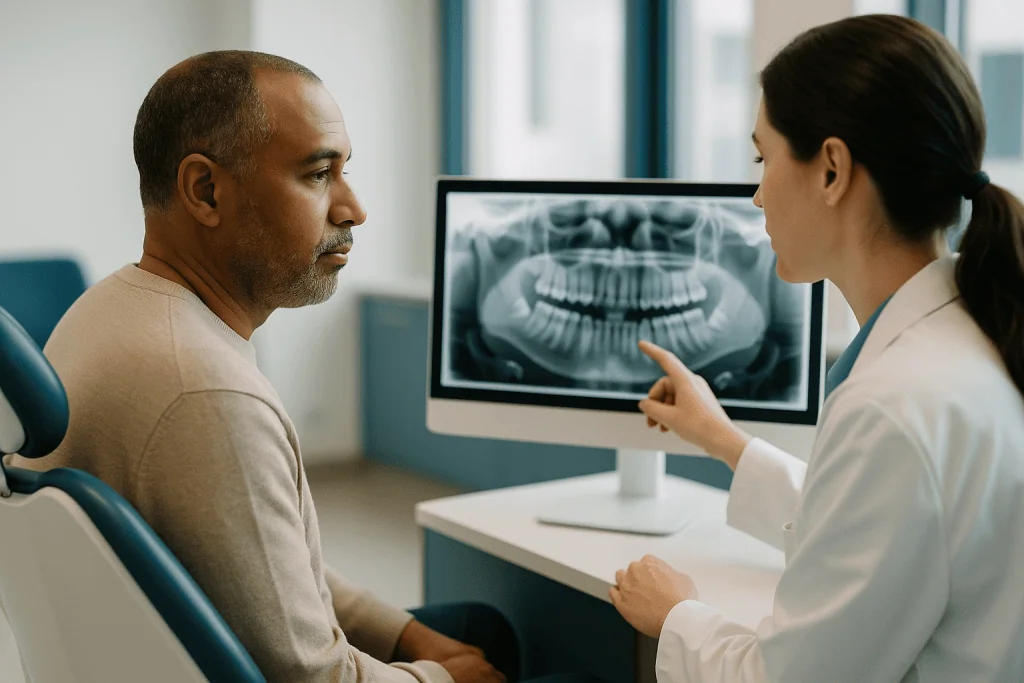
Panoramic X-ray. Also called an OPG, this is the sweeping image taken by a machine that orbits the head. It shows both jaws, all teeth, the sinuses and the outline of the nerve canals on one film. It is the workhorse screening image and often the first thing an implant dentist studies.
Cone beam CT (CBCT). A CBCT scanner rotates once around the head and reconstructs a three-dimensional volume of the jaws. Unlike a hospital CT, which the NHS describes as a series of X-ray images taken from different angles and combined by computer, a dental CBCT uses a cone-shaped beam and a smaller field, and is designed for hard tissue such as bone and teeth. It is the image that shows bone width, exact nerve position and the shape of the sinus floor.
Intraoral scans or impressions. These are not X-rays at all. A digital scanner or a tray of impression material captures the surface of the teeth and gums. Merged with the CBCT data, they let the team see where the crown will sit in relation to the bone underneath.
Not every patient needs every step. A single-tooth gap with generous bone and clear landmarks may be planned on far less imaging than a full arch, a site next to the nerve, or a jaw that has lost bone after years without teeth.
Panoramic X-ray for implants: what it shows and what it misses
The panoramic film is loved for good reasons. It is quick, comfortable, widely available, and it delivers the whole mouth in one glance. For an implant dentist it acts as a map before the detailed survey.
On a good panoramic image the clinician can see the general height of bone in the edentulous area, the term for a region with no teeth. The mandibular canal, the tunnel in the lower jaw carrying the inferior alveolar nerve that supplies feeling to the lower lip and chin, appears as a faint dark band running along the lower jaw. The mental foramen, the exit hole for that nerve near the premolars, shows as a small round shadow. The floor of the maxillary sinus, the air-filled space above the upper back teeth, traces a curve above the upper molars. Retained roots, impacted teeth and large areas of infection are usually obvious.
What the film cannot do is measure reliably. Panoramic machines magnify the image, and the magnification varies across the film and between machines, which is why the review of CBCT in implant dentistry published on PubMed lists distortion and lack of cross-sectional information among the limitations of two-dimensional imaging. A bone height that looks generous may be exaggerated by a small percentage, and in implant planning a few millimeters is the whole game.
The larger gap is width. A panoramic film has no cheek-to-tongue dimension. A ridge that looks tall may be knife-edge thin. Bone can also be concave on the tongue side of the lower jaw, a hollow known as the submandibular fossa, into which a badly angled implant could perforate. None of this appears on a flat image.
So the panoramic film sets the agenda. It tells the surgeon which questions still need answering, and for many sites those answers come from the 3D scan.
CBCT scan for dental implants: what actually happens
People often expect a CBCT scan to feel like a hospital CT, complete with a tunnel and a moving table. It is far less dramatic.
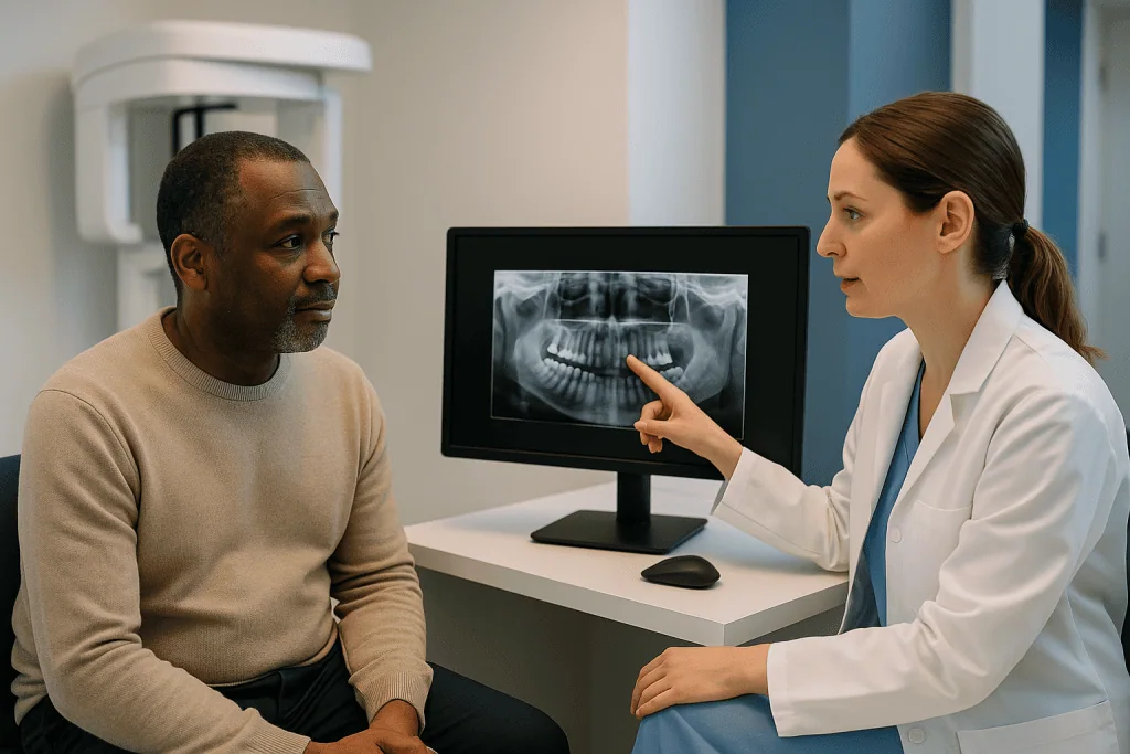
You either sit or stand, your chin rests on a small support, and you may bite lightly on a marker to keep still. Jewelry, glasses, hearing aids and removable dentures come out, because metal scatters the beam and creates streaks in the image. The staff step behind a screen, the arm of the scanner makes a single slow circuit around your head, and the acquisition is over in well under a minute. Holding still matters more than anything else you do; even a small movement blurs the reconstruction.
What the machine has captured is a cylinder of data called the field of view. A small field might cover a few teeth in one jaw; a large one might take in both jaws and the sinuses. The clinician chooses the smallest field that answers the question, both to keep radiation down and to keep the image sharp.
Back at the screen, software slices that cylinder in any direction. The view that matters most for implants is the cross-section, a slice cut across the ridge from cheek to tongue. On it the bone looks like a rounded triangle or a tall rectangle, the nerve canal appears as a small dark circle, and the clinician can draw a virtual implant of the intended length and diameter directly onto the picture. The measuring tools are accurate to a fraction of a millimeter when the patient has stayed still.
There is no contrast dye, no injection and no preparation beyond removing metal. Pregnant patients should tell the team before any X-ray, as the NHS advises for all imaging; the decision to postpone or proceed with shielding sits with the clinician.
Do I need a CT scan for a tooth implant? Who is usually scanned and who is asked to wait
The honest answer is: often, but not always, and the decision belongs to the person placing the implant.
The systematic review of CBCT in implant dentistry available on PubMed notes that professional guidelines increasingly favor cross-sectional imaging for implant planning, while stressing that it should be justified case by case rather than ordered as a routine for everyone. In practice, a 3D scan is usually requested when the two-dimensional images leave a clinically important question unanswered.
Situations where a CBCT is commonly requested include a site in the lower jaw close to the mandibular canal or the mental foramen; upper back teeth where the sinus floor dips low and a sinus lift, a procedure that adds bone beneath the sinus membrane, may be needed; a ridge that appears narrow or has been without teeth for many years; multiple implants or a full arch, where angulation between implants matters; a planned surgical guide, which cannot be made without 3D data; and any suspicion of pathology such as a cyst or a retained root.
A scan may be deferred or judged unnecessary when the panoramic and periapical films show tall, wide bone well clear of any landmark, and the surgeon is confident the extra information would not change the plan.
Some patients are asked to wait rather than scan. If a tooth has just been removed, the socket is still filling with new bone, and a scan taken too early shows a hollow that will look very different after healing. Active gum infection, an untreated sinus problem visible on the panoramic film, or uncontrolled medical conditions are typically addressed first. Cleveland Clinic lists healthy gums and adequate bone among the general requirements before implant surgery is considered.
Asking “what would this scan change about my plan?” is a fair question, and a good team will have a clear answer.
How 3D dental scans before an implant guide position and depth
Position and depth are decided on the screen long before the drill is picked up. Here is how the cross-sectional image turns into a plan.
Depth is set by the landmark below or above. In the lower jaw, the clinician measures from the crest of the bone down to the roof of the mandibular canal and plans an implant short enough to leave a margin of intact bone above the nerve. In the upper back jaw, the same measurement runs up to the sinus floor. If the available height is too short for a stable implant, the options are a shorter implant, bone grafting to add height, or a sinus lift, and the scan is what makes that conversation concrete.
Width sets the diameter and the angle. The implant needs bone on both sides along its full length. If the ridge is thin at the top but widens below, the surgeon may plan a narrower implant or tilt it to follow the bone. If the tongue-side wall undercuts, the angle is adjusted so the tip does not exit the bone.
The future crown decides the ideal spot. When the intraoral scan is merged with the CBCT, the software shows the shape of the tooth that will eventually sit on top. An implant placed perfectly in bone but pointing toward the cheek would produce an awkward crown, so the planned position is a negotiation between where the bone is and where the tooth needs to be.
Neighbors are checked. Adjacent roots are traced on the slices so the implant clears them, and the distance to the next tooth is measured so the gum between them has room.
The output is a virtual implant with a set length, diameter, angle and depth, drawn on your own anatomy. Everything in surgery then aims to reproduce that drawing.
Dental implant imaging types compared
Each image answers a different question, and the table below summarizes what each one is usually good for and where it falls short.
| Imaging type | What it shows well | What it cannot show | Typical role in implant planning |
|---|---|---|---|
| Periapical X-ray | Fine detail of one or two teeth and the bone immediately around them; root shape; small infections | Bone width; distant landmarks; the whole jaw at once | Checking a single site or an adjacent root; follow-up after placement |
| Panoramic X-ray | Overview of both jaws, all teeth, sinus outline, approximate nerve canal path, gross pathology | Accurate measurement; cheek-to-tongue width; undercuts; exact nerve depth | Initial screening and deciding whether a 3D scan is needed |
| Cone beam CT (CBCT) | Cross-sectional bone width and height; exact position of nerve canal and sinus floor; bone contour; data for surgical guides | Soft tissue detail; artifacts near existing metal; slight blur if patient moves | Detailed planning of implant position, depth, angle and diameter |
| Intraoral scan or impression | Surface of teeth and gums; bite relationship; shape of the future crown | Anything below the gum line | Merged with CBCT to align the implant with the final tooth |
The pattern is that no single image does the whole job. A panoramic film without a CBCT leaves width and exact depth uncertain; a CBCT without a surface scan shows bone but not how the tooth will look and function; a periapical film alone is too narrow for planning yet ideal for checking a healed implant later.
The systematic review on PubMed also notes that CBCT image quality and radiation dose vary widely between machines and settings, which is why the same scan is not identical everywhere. Teams choose the combination that answers their specific questions for your specific jaw, and that judgment cannot be replaced by a table.
How much radiation does implantology imaging involve?
Radiation is the question people are often slightly embarrassed to ask, and it deserves a plain answer.
All dental X-rays use ionizing radiation, the kind that can in principle damage cells. MedlinePlus states that the amount from dental X-rays is very low, and that the exam is considered safe, while advising anyone who is pregnant to tell the dentist beforehand. The NHS makes the same general point about X-rays: the dose is small, the benefit in most cases outweighs the risk, and staff take steps to keep exposure as low as reasonably possible.
Within dentistry there is a hierarchy. A periapical film delivers the least. A panoramic film delivers more, though still a small amount. A CBCT scan delivers more again, and here the range is wide. The systematic review of CBCT in implant dentistry indexed on PubMed reports that effective doses from dental CBCT vary by more than an order of magnitude depending on the machine, the size of the field of view and the exposure settings, and that small-field, low-dose protocols can sit close to conventional dental imaging while large-field, high-resolution scans sit considerably higher, though still well below a typical medical CT of the head.
Three practical points follow. First, field of view is the main lever: a scan of a few teeth exposes far less tissue than a scan of both jaws, so asking whether a limited field would suffice is reasonable. Second, a scan that changes the surgical plan is worth its dose; one taken “just in case” with no question to answer is harder to justify, which is the position most professional bodies take. Third, repeat scans add up, so bringing previous images to a new dentist can avoid duplication.
A thyroid collar or lead apron is sometimes used for two-dimensional films; with CBCT, positioning and field selection matter more. Your team can tell you what protocol they use and why.
From scan to surgical guide: how digital planning translates to the drill
A virtual implant on a screen is only useful if the surgeon can reproduce it in a real mouth. Two approaches make that transfer.
Freehand placement with the plan in view. The surgeon studies the cross-sections, notes the planned depth and angle, and places the implant by eye and by feel, checking depth with marked drills and sometimes a periapical X-ray mid-procedure. Experienced clinicians do this routinely for straightforward sites.
Guided surgery. Here the plan is exported to a surgical guide, a custom plastic template with metal sleeves that seats over the teeth or gums and directs the drill along the planned path at the planned depth. Making the guide requires both the CBCT, for the bone, and an intraoral scan or impression, for the surfaces the guide will rest on. The two data sets are superimposed by matching the teeth visible in both.
This is where the common question about impressions comes in. A conventional impression uses a tray of soft material that sets in the mouth; a digital impression uses a handheld scanner that photographs the teeth thousands of times a second. Either is usually completed within a single short appointment, and neither involves radiation. The scanner is slightly larger than an electric toothbrush and is moved slowly over the teeth while a model builds on a nearby screen.
Guides are not magic. Their accuracy depends on how well the guide seats, how still the patient held during the CBCT, and how faithfully the two scans were merged. In a jaw with few or no teeth to anchor on, the guide rests on gum or bone and is less predictable. Surgeons still verify visually and with intraoperative X-rays when needed.
Whether to use a guide, and which kind, is a clinical decision that weighs the complexity of the site against the added steps. Either way, the imaging does the planning; the surgeon does the surgery.
What the days and weeks after implant surgery usually look like, and when imaging returns
Imaging does not stop once the implant is in. It comes back at a few predictable points, though the exact schedule is set by your treating team.
Immediately after placement. A periapical or panoramic X-ray is often taken to confirm that the implant sits where it was planned, that it is not touching an adjacent root, and that it has not strayed toward the nerve canal or sinus. This is the moment the plan is checked against reality.
The first days. Mayo Clinic describes typical discomfort after implant surgery as swelling of the gums and face, bruising of the skin and gums, pain at the site and minor bleeding, all of which usually ease over the following days. No imaging is normally needed unless something unexpected happens.
The healing months. Bone grows onto the implant surface during osseointegration, which Mayo Clinic describes as taking several months. During this period some clinicians take a periapical film to check bone levels before the crown is made; others rely on clinical stability. If a bone graft or sinus lift was performed, healing may be staged and take longer, and an image may be taken to judge whether the grafted bone is ready before the implant is placed.
Before the crown. A further intraoral scan or impression captures the position of the implant so the laboratory can make the final tooth. This is a surface scan, not an X-ray.
Long term. Periodic periapical films at routine check-ups let the dentist watch the bone level around the implant over the years. Repeat CBCT is not routine and is generally reserved for a specific problem such as suspected infection, nerve symptoms or sinus involvement.
Everyone heals at a different pace, and the timings above are typical ranges from the cited sources rather than promises. Your team will tell you when each image is due and what it is for.
Can I have an MRI scan if I have dental implants?
This is one of the most searched questions about implants, and the reassurance is well grounded.
Magnetic resonance imaging (MRI) uses a strong magnet and radio waves, not X-rays, to build pictures of the body. The concern with any metal is that a ferromagnetic object, meaning one strongly attracted to magnets, could heat, move or be pulled by the field. Dental implants are made of titanium or titanium alloy, and increasingly of zirconia, a ceramic. Titanium is not ferromagnetic; it is not pulled by the MRI magnet, which is why it is also used for orthopedic joint replacements and surgical clips that are routinely imaged.
The NHS MRI guidance asks patients to tell staff about any metal in the body before the scan, and lists dental fillings and dental work among items that are generally fine but should be declared. Declaring is the important step. The radiographer decides, not the patient, and telling them lets them position and set up the scan appropriately.
Two practical points are worth knowing. First, metal near the area being imaged can create an artifact, a dark or distorted patch on the picture. An MRI of the brain or neck may show a small blur around the jaw from implants, crowns or braces; this rarely matters for the diagnostic question but the radiologist will note it. Second, some removable components are different from the implant itself. Magnetic denture attachments exist, and a removable denture retained by magnets should be taken out before an MRI. If you are unsure what holds your prosthesis in, ask the dentist who fitted it before your scan appointment.
Having implants also does not prevent future dental X-rays or CBCT. Metal does create streak artifacts on CBCT near the implant, which is one reason a scan taken before placement is more informative than one taken afterward.
What people often get wrong about implant imaging
Myths about dental scans travel fast, and several of them shape decisions for the worse.
“If I have a panoramic X-ray, I don’t need anything else.” The panoramic film shows height, not width, and its magnification is not uniform. For a site near the nerve canal or sinus, or a narrow ridge, it usually cannot answer the questions that decide implant depth and angle on its own.
“A 3D scan is the same as a hospital CT.” Dental CBCT uses a cone-shaped beam, a single rotation and a limited field, and is optimized for bone and teeth. The NHS describes hospital CT as a series of images from many angles combined by computer, usually with the patient lying in a ring-shaped scanner; the dose and set-up differ. The two are related but not interchangeable, and a CBCT does not show soft tissue the way a medical CT does.
“More imaging always means a safer implant.” Imaging reduces uncertainty, but only when it answers a question. A scan with no clinical question adds radiation without adding safety, which is why professional guidance asks for case-by-case justification.
“Guided surgery guarantees a perfect result.” A surgical guide reproduces a plan; it does not remove the need for judgment, and its accuracy depends on how well it seats and how good the underlying scans are. It is a tool, not an outcome.
“Implants make MRI impossible.” Titanium implants are not ferromagnetic and are declared, not removed, before an MRI, as covered above.
“The scan tells you whether the implant will succeed.” Imaging describes anatomy at one moment. Healing depends on gum health, general health, smoking, bite forces and oral hygiene over years. Cleveland Clinic lists several of these as factors that affect how implants fare. A good scan improves the odds of a well-placed implant; it cannot promise what happens afterward.
Questions to ask your care team about implantology imaging
A short, specific list of questions turns a scan from something done to you into something you understand. These are the ones that tend to produce useful answers.
- Which images do you already have of this area, and are they recent enough to plan from?
- What specific question will the 3D scan answer that the panoramic film cannot?
- Will you use a small field of view limited to this site, or a larger one, and why?
- How close is the planned implant to the nerve canal or the sinus floor on my scan? Can you show me the cross-section?
- Is there enough bone width and height, or are you considering a graft, a sinus lift or a shorter implant?
- Will you use a surgical guide, and if so, will I need a digital scan or an impression as well?
- What imaging will you take on the day of surgery to confirm the position?
- When will I be imaged again during healing, and what will you be looking for?
- If I already have a CBCT from another dentist, can you use it instead of repeating it?
- I am pregnant, or may be; how does that change the imaging plan?
- Do I have any magnetic denture attachments I should know about before a future MRI?
Two habits make these conversations easier. Ask to see the images on the screen; a cross-section with a virtual implant drawn on it is far clearer than any verbal description, and clinicians are usually glad to walk through it. And bring or request copies of prior imaging when you move between practices, so nothing is repeated unnecessarily.
None of these questions second-guess the team. They give you the vocabulary to follow the reasoning, and they make sure the decisions that matter, about position, depth and whether extra procedures are needed, are made with you in the room.
When to call your doctor or dentist
Imaging planning is calm, scheduled work, but the surgery it leads to has a short list of warning signs that should not wait for the next appointment. Mayo Clinic notes that complications are uncommon and usually minor, and that they can include infection, injury to nearby teeth or blood vessels, nerve damage causing pain, numbness or tingling in the teeth, gums, lips or chin, and sinus problems when upper-jaw implants protrude into the sinus. Contact your treating team promptly if you notice any of the following.
- Numbness, tingling or an electric-shock sensation in the lower lip, chin or tongue that persists after the local anesthetic should have worn off, or that appears later. This can indicate the implant is near or pressing on the nerve, and early assessment matters.
- Pain that increases after the first few days rather than easing, or that is not controlled by what you were advised to use.
- Swelling that keeps growing after the second or third day, spreads toward the eye or neck, or comes with difficulty swallowing or opening the mouth.
- Bleeding that does not slow with gentle pressure.
- Fever, chills or a bad taste with discharge from the surgical site.
- Nasal congestion, nosebleeds or a feeling of fluid moving between mouth and nose after an upper-jaw implant or sinus lift.
- A loose, mobile or clicking implant, or a healing cap that has come off.
- Any allergic-type reaction such as rash, facial swelling or wheeze after medicines you were given.
Difficulty breathing, rapidly spreading facial swelling that affects swallowing, or heavy bleeding that will not stop are emergencies; seek urgent medical care rather than waiting to reach the dental office.
For anything less dramatic but still worrying, call the team that placed the implant. They have your scans, know exactly where the implant sits in relation to the nerve and sinus, and can decide quickly whether a periapical film or a repeat CBCT is needed. Every decision about investigation and treatment rests with them.
Frequently asked questions
Do I need a CT scan for a tooth implant?
Often, but not always; the decision is your surgeon’s. A 3D cone beam CT is usually requested when two-dimensional X-rays cannot answer a specific question, such as how close the site is to the nerve canal or sinus, how wide the bone is, or when a surgical guide is planned. For a straightforward gap with generous bone well clear of landmarks, some clinicians plan on panoramic and periapical films alone.
What imaging is often used before implant surgery?
Most plans start with a panoramic X-ray for an overview of both jaws, sometimes with small periapical films of the site. If more detail is needed, a cone beam CT scan adds the cross-sectional view of bone width, height and nearby structures. A digital intraoral scan or conventional impression records the tooth surfaces so the implant can be aligned with the future crown.
How long does a dental implant impression or digital scan take?
Both are usually completed within a single short appointment and involve no radiation. A conventional impression uses a tray of soft material that sets in the mouth for a few minutes; a digital scan uses a handheld camera moved slowly over the teeth while a 3D model builds on screen. Your team can tell you which method they use and roughly how long to expect on the day.
Can I have an MRI scan if I have dental implants?
Yes, in general. Dental implants are made of titanium or zirconia, neither of which is ferromagnetic, so they are not pulled by the MRI magnet. The NHS asks patients to declare all metal and dental work before a scan so the radiographer can plan appropriately. Implants may cause a small artifact on images of the jaw area. Magnet-retained dentures are different and should be removed beforehand.
What is a CBCT scan for dental implants and does it hurt?
A cone beam CT is a three-dimensional X-ray of the jaws taken by a machine that rotates once around your head while you sit or stand still. It is painless, needs no injection or dye, and the exposure lasts well under a minute. The result is a volume of data that can be sliced in any direction to measure bone and locate the nerve canal and sinus.
Is a panoramic X-ray enough for implants?
Sometimes, but usually not on its own for a detailed plan. It shows bone height and gross anatomy across both jaws and is excellent for screening, but it does not show cheek-to-tongue width and its magnification varies, so measurements are approximate. For sites near the nerve, under the sinus, or in a thin ridge, most clinicians add a 3D scan before deciding position and depth.
How much radiation is in a 3D dental scan before an implant?
It varies widely with the machine, settings and field of view. A systematic review of CBCT in implant dentistry reports doses ranging from close to conventional dental X-rays for small-field scans to considerably higher for large-field, high-resolution scans, while remaining well below a medical head CT. Asking for the smallest field that answers the clinical question is the most effective way to limit exposure.
What are the main dental implant imaging types?
Four types cover most planning: periapical X-rays for fine detail of one or two teeth; panoramic X-rays for an overview of both jaws; cone beam CT for three-dimensional bone measurement and locating the nerve and sinus; and intraoral scans or impressions, which record tooth and gum surfaces without radiation. Each answers different questions, and teams combine them according to the complexity of the site.
Can a scan tell how deep my implant will go?
Yes, that is one of its main purposes. On the cross-sectional CBCT view the clinician measures from the bone crest to the roof of the nerve canal in the lower jaw, or to the sinus floor in the upper jaw, and plans an implant length that leaves a margin of intact bone. Width on the same slice guides the diameter and angle. The final depth is confirmed during surgery.
Will I need more scans after the implant is placed?
Some, but usually simple ones. An X-ray is often taken right after placement to confirm position, a periapical film may be taken during the months of bone healing, and routine films at check-ups monitor bone levels over the years. A repeat CBCT is not routine and is generally reserved for a specific concern such as suspected infection, nerve symptoms or sinus involvement.
References
This article is for general information only and is not a substitute for professional medical advice. Please consult a qualified doctor about your individual situation.

More from the Blog

Why Jaw Growth Decides When Implantology Can Place an Implant in a Younger Patient
Dental implants for teenagers are usually postponed until the jaw has stopped growing, because an implant fuses to bone and cannot move with the…

How Mini Implants Are Placed: Flapless Insertion, Same-Day Denture Fitting and What to Expect
A mini implant procedure places one or more narrow titanium posts into the jawbone, usually through a small pierce in the gum rather than…

All On 4 Healing Timeline: Soft Diet Weeks, Bone Integration and the Final Bridge Switch
All-on-4 healing usually unfolds in stages. Swelling and soreness peak in the first two to three days and settle over one to two weeks,…

Mini Implant Problems Explained: Loosening, Wear of Denture Attachments and When to Call
Mini implant problems most often fall into three groups: the implant itself loosening because bone has not bonded to it or has later receded,…

All On 6 Problems Explained Honestly: Screw Loosening, Bridge Fractures and Gum Inflammation
All on 6 problems usually fall into three groups: mechanical issues such as a loosened prosthetic screw or a chipped or fractured bridge, biological…

Is All On 4 Possible With Bone Loss? How Angled Implants Use the Bone You Still Have
Often, yes. All-on-4 with bone loss is frequently possible because the technique places four implants where bone usually remains, at the front of the…