

Is Doppler Ultrasound Needed for Suspected Torsion, and Why Must It Never Delay Surgery?

Key Takeaways
- Guidelines favor immediate surgical exploration over imaging when history and examination clearly point to testicular torsion, because every hour of lost blood flow reduces the chance of saving the testicle (Mayo Clinic).
- Testicular salvage is usual when surgery happens within roughly six hours of pain onset and becomes increasingly unlikely after about twelve, which is why Doppler ultrasound is reserved for uncertain cases (Mayo Clinic).
- Doppler ultrasound can show preserved or even increased blood flow in partial, early or intermittent torsion, so a normal scan does not exclude the diagnosis in a convincing clinical picture (NIH StatPearls).
- The whirlpool sign, a coiled appearance of the spermatic cord on ultrasound, is a direct image of the twist and is highly suggestive of torsion even when some testicular flow persists (NIH StatPearls).
- The loose testicular attachment that allows twisting is usually present on both sides, so surgeons routinely fix the unaffected testicle in the same operation (MedlinePlus).
- Recurrent episodes of sudden testicular pain that resolve on their own suggest intermittent torsion, a condition where elective fixation of both testicles is often discussed before a complete torsion occurs (NIH StatPearls).
Doppler ultrasound is useful when the diagnosis of testicular torsion is genuinely uncertain, but it is not required before surgery. When the history and examination point clearly to torsion, guidelines favor going straight to the operating room, because the testicle loses blood supply with every passing hour. Imaging is reserved for unclear cases, and it should never postpone an urgent urological opinion.
It is just after midnight, and a fifteen-year-old is sitting bolt upright in an emergency department bed, pale, one hand clamped over his groin, insisting the pain started an hour ago while he was asleep. His mother asks the question almost every parent asks in that moment: shouldn’t someone do a scan before they talk about an operation?
It is a reasonable question. The honest answer is that a doppler ultrasound for testicular torsion can be very helpful, and sometimes it is the wrong thing to wait for. A twisted testicle is one of the few true surgical emergencies in urology, and the clock began running before anyone reached the hospital.
This explainer walks through what the scan shows, where it can mislead, who is usually scanned first, who is usually not, and why the surgical team may politely decline to wait for the radiology department.
What actually happens when a testicle twists
Each testicle hangs inside the scrotum on a spermatic cord, a bundle that carries its artery, veins and the vas deferens. Testicular torsion means that cord has rotated on itself, kinking the vessels like a garden hose with a knot in it. Veins, which are thin-walled, collapse first; the testicle swells and congests. As the twist tightens, arterial blood stops too, and tissue starved of oxygen begins to die.
Why does it happen at all? Most people have a testicle anchored firmly to the scrotal wall. A minority are born with a looser attachment, often called the bell-clapper deformity because the testicle can swing freely like the clapper inside a bell. That anatomy is usually present on both sides, which is why surgeons routinely fix the unaffected testicle as well (MedlinePlus; NIH StatPearls).
Torsion is most common between the ages of 12 and 18, though it can occur in newborns and, less often, in adults (Mayo Clinic). The estimated incidence is about 1 in 4,000 males under 25 each year (NIH StatPearls). Triggers include sudden movement, minor injury, cold exposure or simply sleep; many patients wake with the pain.
The detail that shapes everything that follows is time. When surgery happens within roughly six hours of pain onset, the testicle can usually be saved; the chance of salvage falls steeply beyond that window, and after about 12 hours removal becomes increasingly likely (Mayo Clinic). That is the context in which every imaging decision is made: a test is only worth ordering if it will change management faster than it costs minutes.
Is Doppler ultrasound needed for every suspected testicular torsion?
No. This is the single most important point in the article, and it is where patients and families are most often surprised.
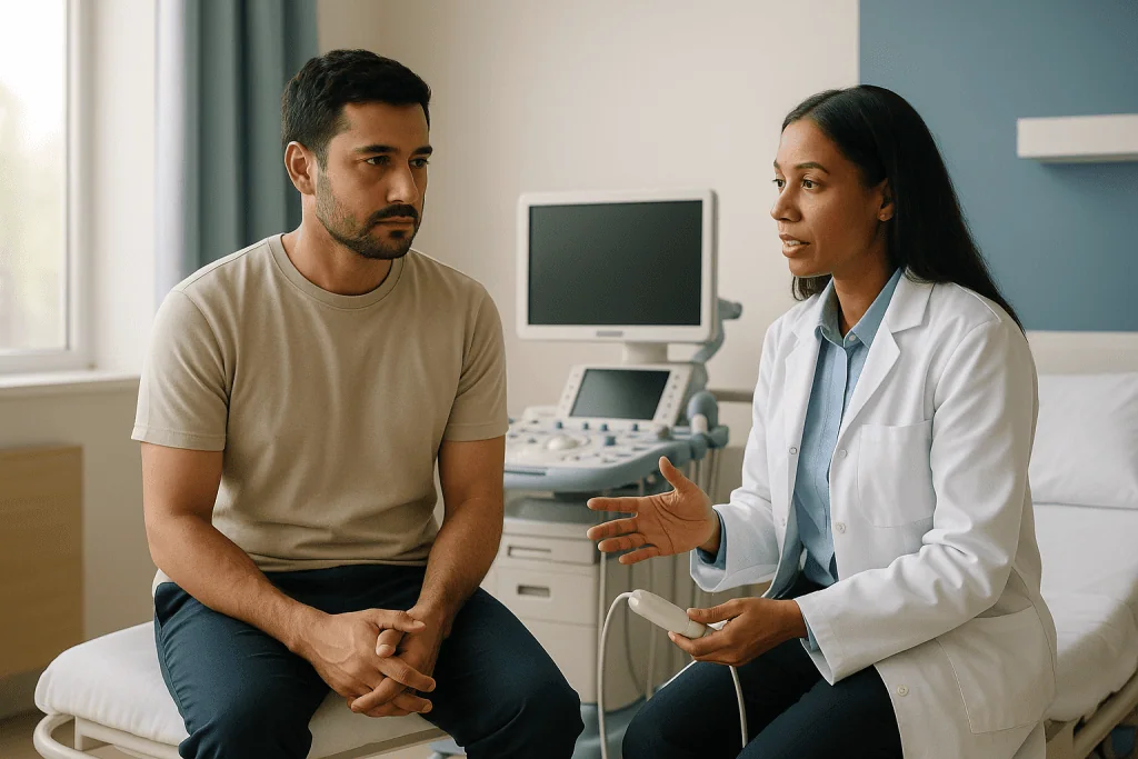
Doppler ultrasound is a standard ultrasound scan with an added mode that detects movement, in this case blood flowing through the testicle. The sonographer places a probe on the scrotum, compares both sides, and looks for flow in the testicle and the cord. In a straightforward case the scan takes minutes and is painless apart from the pressure on an already tender area.
Guideline-level practice across the UK, the US and Europe runs along the same line: if the clinical picture strongly suggests torsion, the patient should go to surgery without waiting for imaging. Scanning is for the uncertain middle ground, where the history is atypical, the examination is equivocal, or the pain has lasted long enough that a different diagnosis is plausible (Mayo Clinic; NIH StatPearls).
Why not scan everyone, just to be safe? Three reasons. First, an ultrasound can only be obtained as fast as a trained operator is available, and overnight that may mean a long delay. Second, a normal-looking scan does not rule torsion out, so a reassuring result in a textbook case can be actively dangerous. Third, surgical exploration is both the definitive test and the treatment: a surgeon can see the cord directly, untwist it and fix the testicle in a single procedure.
The phrase surgeons use is that ultrasound should be a bridge for the unsure, never a gate for the obvious. If the treating team recommends going straight to theatre, that recommendation reflects the evidence, not haste.
What does testicular torsion look like on ultrasound images?
Families who watch the screen usually see two things: a gray image of the testicle itself, and splashes of red and blue over it. The colors are the Doppler signal, each one representing blood moving toward or away from the probe.
In a healthy testicle, the color overlay is scattered through the tissue, and a waveform trace shows a pulsing arterial pattern with a gentler venous flow beneath it. In established torsion, the affected side is quiet. Color is absent or markedly reduced compared with the opposite testicle, and the waveform may be flat or show only high-resistance arterial spikes with no venous return, which is the earliest sign that outflow has been blocked (NIH StatPearls).
The gray-scale picture changes with time. Early on, the testicle may look normal or slightly enlarged. Over hours it becomes swollen and darker, and later patchy and heterogeneous, a sign that areas of tissue have already died. The epididymis, a coiled tube on the back of the testicle, often swells too.
One finding carries particular weight: the whirlpool sign. When the sonographer traces the cord upward, a twisted cord appears as a spiral or coiled mass, a direct image of the torsion rather than an inference from missing blood flow. Where it is seen, it is highly suggestive of torsion even when some flow persists in the testicle (NIH StatPearls).
Reactive fluid around the testicle, thickening of the scrotal skin and an abnormally high or horizontal position of the testicle round out the picture. None of these is required for the diagnosis, and absence of any one of them should not reassure anyone.
Why a Doppler ultrasound can miss testicular torsion
Ultrasound is a strong test in experienced hands, with reported sensitivity and specificity high enough that many departments rely on it for unclear cases (NIH StatPearls). Strong is not the same as perfect, and the ways it fails are worth understanding, because they are precisely the cases where waiting for a scan costs the most.

- Partial torsion. A cord twisted through a smaller angle can still carry some arterial blood. The screen shows color, the report says flow present, and the testicle continues to strangle slowly.
- Intermittent torsion. If the cord has untwisted by the time the probe touches the skin, flow may be normal or even increased, a reactive flush of blood after the blockage releases.
- Small testicles. In infants and prepubertal boys the vessels are tiny, and detecting flow is technically difficult even when it is present. A scan that shows little color on both sides is uninterpretable, not reassuring.
- Operator and equipment. Doppler settings, probe pressure and the sonographer’s experience all affect sensitivity. A scan performed by an on-call generalist at 3 a.m. may not match one by a specialist at midday.
- Late presentation. After many hours, inflammation around a dying testicle can produce flow in the surrounding tissue that is mistaken for flow inside it.
The practical consequence is a rule that experienced clinicians apply without apology: a negative scan in a patient whose story and examination fit torsion does not close the question. Surgical exploration, with its very low risk relative to a lost testicle, remains the right answer when suspicion is high (Mayo Clinic; NIH StatPearls).
How do doctors check for testicular torsion without a scan?
Before any imaging, the diagnosis is built from the story and a careful examination. The story matters most. Torsion classically begins abruptly, often within minutes, and the pain is severe, constant and frequently accompanied by nausea or vomiting. Pain that has crept up over days, burning on passing urine, or a fever at the start all push the thinking toward other diagnoses, though none of them excludes torsion (Mayo Clinic; MedlinePlus).
On examination the clinician looks at both sides and compares. A twisted testicle often sits higher than its partner and may lie horizontally rather than vertically, because the shortened, coiled cord pulls it upward. It is usually exquisitely tender throughout, firm, and swollen; the scrotal skin may be reddened. The epididymis may be felt in an unusual position, in front rather than behind.
One bedside test gets particular attention. Lightly stroking the inner thigh normally makes the testicle on that side rise, a reflex called the cremasteric reflex. In torsion this reflex is usually lost, because the twisted cord cannot transmit the pull. Its presence makes torsion less likely; its absence is suggestive but not definitive, especially in very young boys where the reflex is unreliable anyway (NIH StatPearls).
Some departments combine these findings into a structured score, grouping patients into low, intermediate and high risk. In published pediatric work, the highest-scoring patients were taken to surgery without imaging and the lowest-scoring were observed, with ultrasound reserved for the middle group (NIH StatPearls). Scores support judgment; they do not replace a senior clinician looking at the patient.
Lifting the scrotum sometimes relieves pain in epididymitis and not in torsion, a sign that has been quoted for decades. It is unreliable enough that it should never be used to decide against surgery.
Who goes straight to surgery, and who is usually scanned first?
Think of patients as falling into three groups, with the boundaries drawn by the treating team rather than by any fixed rule.
Straight to the operating room. A boy or young man with sudden severe pain of a few hours, a high-riding tender testicle and no cremasteric reflex belongs here. Imaging adds little and risks delay. The surgeon will usually ask the patient to stop eating and drinking, arrange anesthesia, and explore the scrotum as soon as a theatre is available (Mayo Clinic).
Scan first, then decide. This is the uncertain middle: a history that fits only partly, pain that has waxed and waned, a tender epididymis with a normally positioned testicle, or an adult whose age makes torsion statistically less likely. Here Doppler ultrasound can tip the balance either way, provided it can be obtained quickly and interpreted by someone comfortable with scrotal imaging.
Usually asked to wait and watch. Patients whose examination is clearly reassuring, whose pain has an obvious alternative explanation, or whose symptoms have resolved completely may be observed, scanned on a routine basis or referred for outpatient follow-up. Even then, clear instructions to return immediately if pain recurs are part of good care.
Two further groups deserve mention. Newborns with a hard, discolored scrotum are managed on their own pathway, because torsion in that age group has often occurred before birth and the testicle is rarely salvageable, though surgeons still frequently explore to protect the other side (NIH StatPearls). And patients with pain lasting more than a day are not written off; a late testicle may still be saved, and a dead one still needs removal to prevent infection and protect fertility.
Every one of these decisions belongs to the examining surgeon, who weighs the specific findings in front of them.
Scan first or surgery first: a side-by-side summary
The table below condenses the reasoning clinicians use. It is a summary of general practice, not a decision tool for patients or families; the treating team holds the decision.
| Question the team is asking | Points toward immediate surgery | Points toward Doppler ultrasound first |
|---|---|---|
| How did the pain start? | Sudden, severe, often waking the patient; nausea or vomiting | Gradual onset over a day or more; burning with urination |
| How long has it lasted? | Hours, within the window where salvage is likely (Mayo Clinic) | Days, or pain that has fully settled |
| What does the examination show? | High-riding, horizontal, diffusely tender testicle; absent cremasteric reflex | Normal position; tenderness confined to the epididymis; reflex present |
| How old is the patient? | Adolescent or young adult, the peak age range of 12–18 (Mayo Clinic) | Older adult with risk factors for infection |
| How fast is a scan available? | Not immediately; waiting would cost an hour or more | Available now, with an experienced operator |
| What would a normal scan change? | Nothing; high suspicion still warrants exploration | Could reasonably redirect care toward infection or another cause |
Read across the rows and a pattern appears. Imaging earns its place when it can genuinely change the plan, when it is fast, and when the clinical story leaves honest doubt. It loses its place when the picture is already clear, because the one thing a twisted testicle cannot afford is time spent confirming what the examination has already said.
Patients sometimes ask whether refusing a scan means the hospital is cutting corners. The opposite is true. Choosing exploration over a scan in a clear-cut case is the more conservative option for the testicle, even though it is the more invasive option for the patient.
What happens in the operating room
Scrotal exploration is a short operation performed under general anesthesia. Through a small incision, the surgeon delivers the testicle, inspects it, and untwists the cord, a step called detorsion. The testicle is then warmed and watched for several minutes to see whether its color returns from dusky purple toward a healthy pink.
If it recovers, the surgeon fixes it to the scrotal wall with stitches so it cannot twist again, a procedure called orchidopexy. Because the loose attachment that allowed the twist is usually present on both sides, the opposite testicle is fixed during the same operation (Mayo Clinic; MedlinePlus). This bilateral fixation is standard and is one reason surgeons prefer formal exploration over any bedside manoeuvre.
If the testicle does not recover, it is removed, an operation called orchiectomy. Leaving dead tissue in place risks infection and may provoke an immune reaction against sperm. Removal of one testicle does not in itself prevent normal hormone production or fertility, since the remaining testicle usually compensates, though the surgeon will discuss this candidly rather than promise an outcome (Mayo Clinic).
Occasionally a clinician will attempt to untwist the cord by hand in the emergency department, a manual detorsion. Done successfully, it restores blood flow within seconds and can buy valuable time, and relief of pain plus return of flow on Doppler suggests it has worked. It is not a substitute for surgery. The cord may be only partly untwisted, it may re-twist, and the underlying anatomy still needs fixing, so patients who have had manual detorsion are still scheduled for orchidopexy (NIH StatPearls).
What patients usually feel afterward is relief that the acute pain has gone, replaced by a sore, bruised scrotum that eases over the following days.
Intermittent testicular torsion and torsion-detorsion syndrome
Not every twist stays twisted. Some boys and men describe repeated episodes of sudden, severe testicular pain that resolve on their own within minutes to a few hours, sometimes with nausea, sometimes waking them from sleep. Between episodes they feel entirely well, and examination may be normal. This pattern is called intermittent testicular torsion, and the lingering ache or swelling that can follow an episode is sometimes described as torsion-detorsion syndrome (NIH StatPearls).
The mechanism is simply the same loose attachment allowing the cord to twist and then spontaneously unwind. Each episode is a near miss, and the danger is that one day the cord does not untwist.
This condition is where Doppler ultrasound is both most tempting and most misleading. A scan performed after the episode has resolved is often normal; it may even show increased flow as blood rushes back into a testicle that was briefly starved. Families can be told, in good faith, that the scan was fine. Only the history, with its characteristic rhythm of abrupt onset and abrupt relief, points to the diagnosis.
Clues that raise suspicion include recurrent self-limiting episodes, a testicle that lies horizontally on examination, and a cord that feels thickened or tender afterward. Where the pattern is convincing, many urologists offer elective bilateral orchidopexy to fix the testicles before a complete, non-resolving torsion occurs. That decision weighs the small risks of a planned operation against the risk of losing a testicle, and it belongs to the patient, the family and the urologist together.
For anyone who has had such episodes, the practical message is unambiguous: the next one should be treated as a full torsion until proven otherwise, and the way to prove otherwise is in an emergency department, not at home.
What can be mistaken for testicular torsion?
Several conditions produce a painful, swollen scrotum, and sorting them out is the daily work of emergency physicians and urologists. The point of knowing the mimics is not self-diagnosis but understanding why doctors sometimes scan and sometimes do not.
Epididymitis and orchitis. Infection or inflammation of the epididymis, the coiled tube behind the testicle, is the commonest alternative in adults. Pain tends to build over a day or two, urinary symptoms and fever are more usual, and on Doppler the flow is increased rather than absent. In adolescents the distinction can be genuinely hard, which is why age alone never settles it (Mayo Clinic; Cleveland Clinic).
Torsion of a testicular appendage. Tiny vestigial tags on the testicle or epididymis can twist on their own stalk. The pain is real but localized, the testicle itself is not tender, and a small dark spot is sometimes visible through the skin near the top of the testicle. Ultrasound shows normal testicular flow with a small avascular nodule. This settles with rest and time rather than surgery, but only after torsion of the testicle itself has been excluded (NIH StatPearls).
Incarcerated inguinal hernia. A loop of bowel trapped in the groin can cause scrotal swelling and severe pain, and it is itself a surgical emergency.
Trauma, including bleeding or rupture of the testicle. Injury can also precipitate true torsion, so a history of a blow does not reassure.
Less often, a tumor, a tense hydrocele, a vasculitis affecting the skin, or referred pain from a kidney stone can cause confusion. Each has its own clues, and a scan often helps distinguish them. What none of them changes is the first rule: in an acutely painful testicle, torsion is assumed until the examining clinician is confident it is not.
What the days and weeks after surgery usually look like
Most patients go home the same day or the following morning. The scrotum is bruised, swollen and sore; a snug pair of supportive underwear and an ice pack wrapped in cloth, applied for short periods, are the usual comfort measures. Pain medicine is prescribed by the surgical team, who will explain what to take and for how long; those instructions sit with the prescriber and are not repeated here.
Over the first week the swelling settles and the incision begins to heal. Stitches are usually dissolvable. Showering is generally allowed once the dressing comes off, with the wound patted dry rather than rubbed; soaking in a bath or pool is commonly deferred until the wound has closed, on the surgeon’s advice.
Activity returns in stages. Walking is encouraged from the first day. Many surgeons advise avoiding strenuous exercise, cycling and contact sports for several weeks so the fixation sutures are not strained and a knock does not reopen bruising (Mayo Clinic). School or desk work is typically possible within days, depending on comfort.
A follow-up appointment is usually arranged within a few weeks to check the wound and, where the testicle was saved, to assess whether it has recovered its normal size and feel. A testicle that survived a long period of ischemia may shrink over the following months, a process called atrophy; the team will discuss this honestly rather than predicting an outcome.
Where a testicle was removed, questions about appearance, hormones and fertility are expected and welcome. A prosthetic testicle can be discussed later, once healing is complete, if the patient wishes. Semen analysis in adulthood may be offered to those who want reassurance about fertility.
Emotionally, teenagers in particular can find the episode frightening and embarrassing. Naming that openly, and giving them space to ask questions without parents in the room, is part of good aftercare.
What people often get wrong about Doppler ultrasound and torsion
“A normal scan means it isn’t torsion.” This is the most dangerous misconception on the list. Partial, early and intermittent torsion can all produce preserved flow. A reassuring scan in a patient with a convincing story and examination does not end the conversation (NIH StatPearls).
“The hospital should always do a scan before operating.” Imaging exists to resolve doubt. When there is little doubt, exploration is the faster, safer and more definitive route, and declining to wait for a scan reflects guideline practice rather than impatience (Mayo Clinic).
“If the pain eases, the danger has passed.” Pain can lessen as nerve tissue dies, or because the cord has temporarily untwisted. Neither is reassuring. The second scenario is the hallmark of intermittent torsion and warrants urgent assessment.
“Torsion only happens to teenagers.” Adolescence is the peak, but torsion occurs in newborns, young children and adults (Mayo Clinic). Age shifts probability; it never excludes.
“It only happens after an injury or sport.” Many patients wake from sleep with the pain, and no trigger is identified in most cases.
“Untwisting it by hand fixes the problem.” Manual detorsion can restore flow temporarily, but the anatomy that allowed the twist remains, so surgical fixation still follows.
“Only one side needs fixing.” The loose attachment is usually bilateral, which is why the healthy testicle is fixed in the same operation.
“After about six hours it’s hopeless.” The odds fall with time, but testicles have been saved later, and a late presentation still needs surgery, whether to salvage or to remove and protect the other side.
Each of these misunderstandings tends to add delay, and delay is the one variable that is consistently, measurably harmful.
Questions to ask your care team
In an emergency, there is rarely time for a long conversation before consent is signed. These questions are the ones that genuinely matter, and a good team will be glad to answer them, even briefly.
- How confident are you that this is torsion, and what in the examination points that way?
- If you are recommending surgery without a scan, is that because a scan would add delay, or because it would not change the plan?
- If you are recommending a scan first, how quickly can it be done, and what happens if the result is unclear?
- How long has the pain been going on by your estimate, and how does that affect the chance of saving the testicle?
- Will you fix the other testicle during the same operation, and why?
- What happens if the testicle cannot be saved, and how is that decided in theatre?
- What are the risks of the anesthetic and the operation for someone of this age and health?
- What should recovery look like, and which signs would mean something is wrong?
- When should sport, cycling and strenuous activity resume?
- Will there be a follow-up visit, and who should be contacted in the meantime?
After surgery, a few further questions become relevant: whether the saved testicle appeared to recover fully, whether any shrinkage is expected, and whether a semen analysis or hormone check should be considered later for adults or older adolescents who want to plan for fertility.
For anyone who has experienced the on-off pattern of intermittent torsion, the question to ask a urologist in clinic is whether planned fixation of both testicles is advisable, what the operation involves, and what the alternatives are. The answer will be individual, and it rests with the specialist who has examined the patient.
When to call your doctor
Testicular pain that comes on suddenly and severely is an emergency until proven otherwise. The right response is not to call a general practice line for advice, book an appointment, or wait to see whether it settles; it is to go to the nearest emergency department immediately, or call emergency services if travel is difficult (NHS; Mayo Clinic). The treating team there will decide whether a Doppler ultrasound, immediate surgery or observation is appropriate.
Seek emergency care without delay if any of the following apply:
- Sudden, severe pain in one testicle, especially with nausea or vomiting.
- A testicle that appears higher than usual, lies sideways, or is swollen and very tender to touch.
- Pain that started in the night or during sleep.
- Testicular pain following an injury that does not ease within an hour.
- Scrotal pain accompanied by lower abdominal pain.
- Repeated episodes of severe testicular pain that resolve on their own; even if the current episode has passed, same-day assessment is needed.
- In a newborn, a scrotum that is hard, swollen or discolored.
After surgery, contact the surgical team or return to the hospital if there is spreading redness, increasing swelling or pain rather than gradual improvement, discharge or bleeding from the wound, a fever, or difficulty passing urine.
Milder, longer-standing discomfort still deserves a prompt appointment with a doctor rather than self-management, because epididymitis, hernias and other causes need assessment of their own.
None of the information in this article replaces examination by a clinician. If in doubt, go. A scrotal examination that turns out to be nothing serious costs an hour of embarrassment; a missed torsion costs a testicle.
Frequently asked questions
How do doctors check for testicular torsion?
Diagnosis starts with the story and a physical examination: sudden severe pain, a high-riding or horizontal tender testicle, and loss of the cremasteric reflex all point toward torsion. Doppler ultrasound is used when those findings leave genuine doubt. When the clinical picture is clear, surgical exploration is both the definitive test and the treatment, and it is not delayed for imaging.
What does testicular torsion look like on ultrasound images?
The affected testicle shows absent or markedly reduced color Doppler flow compared with the other side, and the waveform may lack venous return. Gray-scale images may show swelling, a darker or patchy texture, fluid around the testicle, and a coiled spermatic cord known as the whirlpool sign. Early or partial torsion can look deceptively normal.
What is torsion-detorsion syndrome and what are its symptoms?
The term describes the aftermath of intermittent torsion, in which the cord twists and then spontaneously unwinds. People report repeated episodes of sudden, severe testicular pain, sometimes with nausea, that settle within minutes to hours, often followed by a dull ache or mild swelling. Each episode is a near miss, and urologists often discuss planned fixation of both testicles.
What can be mistaken for testicle torsion?
Epididymitis or orchitis, torsion of a small testicular appendage, an incarcerated inguinal hernia, testicular injury, and occasionally a tumor, hydrocele or referred pain from a kidney stone can all produce a painful scrotum. Clinicians distinguish them by the pattern of onset, examination findings and, where doubt remains, Doppler ultrasound, while treating torsion as the default until it is excluded.
Can a Doppler ultrasound miss testicular torsion?
Yes. Partial torsion can leave some arterial flow, intermittent torsion may have untwisted before the scan, and very small testicles in young boys are hard to assess. Operator experience and equipment settings also matter. For these reasons a normal scan does not rule out torsion when the history and examination are convincing, and exploration may still be recommended.
Why would a surgeon operate without doing a scan first?
Because waiting for imaging costs time, and the chance of saving the testicle falls with every hour. When the examination strongly suggests torsion, a scan would not change the plan and could only delay it. Surgical exploration lets the surgeon see the cord directly, untwist it and fix both testicles in a single short operation.
What is manual detorsion and does it replace surgery?
Manual detorsion is an attempt to untwist the cord by hand in the emergency department. When it works, pain eases quickly and blood flow returns. It does not replace surgery: the cord may be only partly untwisted or may re-twist, and the loose attachment still needs surgical fixation, so orchidopexy is still arranged afterward.
Does intermittent testicular torsion need surgery if the pain goes away?
Often it is recommended. Repeated self-resolving episodes signal that the cord can twist freely, and the next episode may not untwist on its own. Many urologists advise elective fixation of both testicles to prevent a complete torsion. The decision weighs the risks of a planned operation against the risk of losing a testicle and rests with the specialist and patient.
What happens if the testicle cannot be saved?
The surgeon removes it, an operation called orchiectomy, because dead tissue risks infection and may trigger an immune response. The remaining testicle is fixed to prevent future torsion. One healthy testicle usually maintains hormone production and fertility, though the team will discuss this individually. A prosthesis can be considered later once healing is complete.
How long after torsion surgery can sport resume?
Most surgeons advise avoiding strenuous exercise, cycling and contact sports for several weeks so the fixation sutures are not strained and bruising can settle, while walking is encouraged from the first day. The exact timing depends on the operation performed and how healing progresses, so follow the instructions given by the surgical team at discharge and at follow-up.
References
- MedlinePlus: Testicular torsion
- Cleveland Clinic: Testicular torsion
- NIH StatPearls: Testicular Torsion
- NHS: Testicle lumps and swellings
This article is for general information only and is not a substitute for professional medical advice. Please consult a qualified doctor about your individual situation.

More from the Blog

The Urgent Tests Behind a Uremic Syndrome Diagnosis and What Imaging Adds
Uremic syndrome tests start with blood work: creatinine, urea (BUN), electrolytes and bicarbonate to gauge kidney function, plus a complete blood count, blood smear…

Is Laser Lithotripsy Painful? Anesthesia Choices and Comfort After the Stone Is Fragmented
Laser lithotripsy is not usually painful while it is happening, because the procedure is performed under general or spinal anesthesia, so you are asleep…

How Fluid Management Works: Assessing Dehydration or Overload and Choosing IV or Oral Plans
Doctors assess dehydration and fluid overload by combining a history of intake and losses with examination findings such as pulse, blood pressure lying and…

Preparing for Urethritis Testing: Why You May Be Asked Not to Urinate Before the Sample
Before a urethritis test, you are usually asked not to urinate for at least one hour, because the first part of the urine stream…

What Happens After a Positive Urinary Biomarker Result? Cystoscopy, Imaging and Repeat Testing
A positive urinary biomarker result is a signal that needs checking, not a diagnosis. The usual next steps are a cystoscopy (a thin camera…

When PKD Advances: How Dialysis or Kidney Transplant Planning Begins Before It Is Urgent
As polycystic kidney disease advances, planning for dialysis or a kidney transplant usually begins when kidney function falls into the later stages of chronic…