

Laparoscopic Gallbladder Removal for Cholecystitis: Step by Step in the Operating Room
Key Takeaways
- Gallstones cause roughly 95% of acute cholecystitis cases, which is why removing the gallbladder, not just treating the infection, is the standard approach.
- The NHS advises that surgery for acute cholecystitis should ideally happen within one week of diagnosis, often during the same hospital admission.
- The operation typically takes 60 to 90 minutes, and the gallbladder is removed through three or four incisions each about half an inch long.
- The critical view of safety, in which the surgeon confirms only two structures enter the gallbladder before clipping anything, is the key step protecting the main bile duct.
- Return to normal activity is around two weeks after keyhole surgery compared with six to eight weeks if the operation is converted to an open approach.
- Shoulder tip pain after laparoscopy comes from carbon dioxide irritating the diaphragm, not from anything wrong with the shoulder, and usually settles within a day or two.
Laparoscopic cholecystectomy for cholecystitis is keyhole surgery that removes an inflamed gallbladder through three or four small abdominal incisions. Under general anesthesia the surgeon inflates the abdomen with carbon dioxide gas, identifies and clips the cystic duct and cystic artery, peels the gallbladder off the liver, and lifts it out through one port. The operation usually takes 60 to 90 minutes, and most people go home the same day or the next morning.
The pain arrived after a Sunday roast and refused to leave. By Monday afternoon a woman in her fifties is sitting on the edge of an emergency department bed, a fever ticking upward, an ultrasound probe pressing under her right ribs. The doctor uses the word cholecystitis, then another one: surgery. Her first thought is not about the gallbladder at all. It is about the size of the scar, how many days she will lose, and what exactly a stranger is going to do inside her abdomen while she is asleep.
Those are fair questions, and this article answers them in order. Laparoscopic cholecystectomy for cholecystitis is one of the most frequently performed operations in modern hospitals, yet most people who have it can describe only the four small dressings they woke up with. What happens between the anesthetic and those dressings follows a well-rehearsed sequence.
We walk through that sequence step by step, from the first incision to the moment the gallbladder leaves the body, and then through the days that follow, the honest risks, and the myths worth discarding.
What cholecystitis is, and why the gallbladder is the thing that has to go
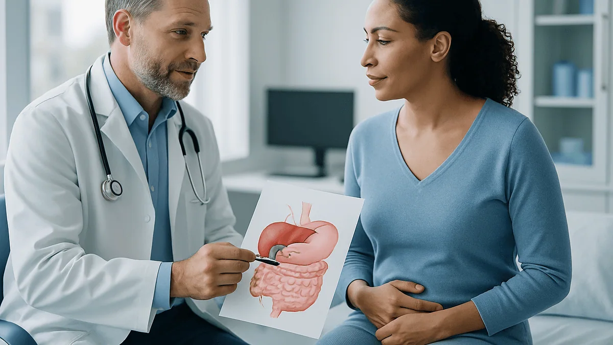
Cholecystitis is inflammation of the gallbladder, the small pear-shaped pouch tucked under the liver that stores bile between meals. Bile is the greenish digestive fluid the liver makes to help break down fat. The gallbladder concentrates it and squeezes it into the small intestine when food arrives.
Trouble starts when a gallstone lodges in the gallbladder’s exit tube, the cystic duct. Bile backs up, the wall stretches and swells, bacteria multiply in the trapped fluid, and the organ becomes hot, thick, and exquisitely tender. Cleveland Clinic notes that gallstones account for roughly 95% of acute cholecystitis cases; the remainder happen without stones, typically in people who are already critically ill.
Gallstones themselves are common. The National Institute of Diabetes and Digestive and Kidney Diseases estimates that 10 to 15 percent of adults in the United States have them, and most never know. The minority who develop cholecystitis face a different calculation. An inflamed gallbladder does not repair itself in a way that makes the next attack less likely, because the stones that caused the blockage are still there. Left alone, the wall can become gangrenous, perforate, or form an abscess.
This is why the standard treatment is removal rather than repair. The gallbladder is a convenience organ, not an essential one. Without it, bile simply drips steadily from the liver into the intestine instead of being released in bursts. People digest normally, with some adjustments discussed later. Removing the organ removes both the current inflammation and the reservoir where future stones would form. That single fact underpins everything that follows in the operating room.
How laparoscopic cholecystectomy for cholecystitis works, in plain language
Laparoscopic means the surgeon operates through small incisions using a thin camera and long instruments rather than one large cut. Cholecystectomy means surgical removal of the gallbladder. Put together, the operation removes an inflamed gallbladder using keyhole techniques while the patient is fully asleep under general anesthesia.
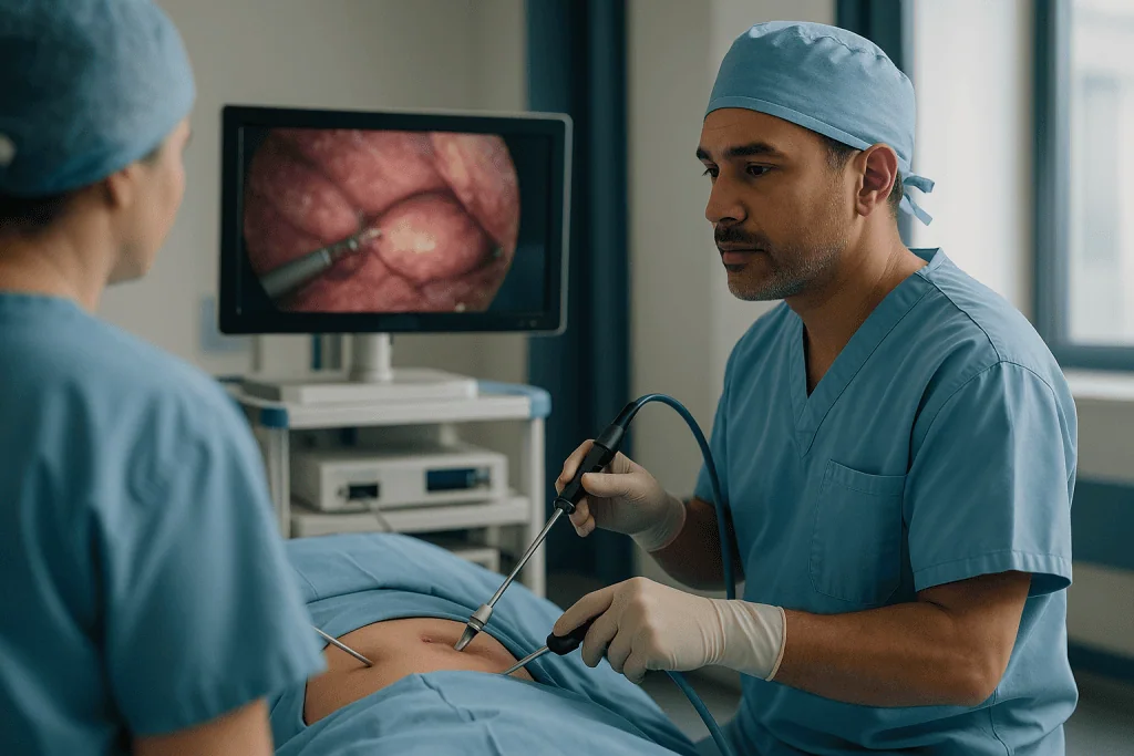
Picture the abdomen as a collapsed tent. The first job is to raise it. Carbon dioxide gas is pumped in through a small cannula until the abdominal wall lifts away from the organs, creating a dome of working space. Surgeons call this a pneumoperitoneum, literally gas in the abdominal cavity. Carbon dioxide is used because the body absorbs it quickly and breathes it out through the lungs.
Next come the ports: hollow tubes, usually three or four, each about half an inch wide according to MedlinePlus, inserted through short cuts in the skin. One carries a high-definition camera that projects onto screens above the table. The others carry graspers, a hook for cutting and sealing tissue with electrical current, and a clip applier.
The surgeon watches the screen, not the abdomen. Working with two hands and an assistant holding the camera, they lift the gallbladder, identify the two structures that connect it to the rest of the body, seal them with tiny titanium or absorbable clips, and dissect the organ free from the liver’s surface. The gallbladder goes into a small retrieval bag and exits through the widest port. The gas is released, the ports come out, and each incision is closed with a stitch or skin glue.
Everything happens on a scale of millimeters, on a screen magnified several times. Mayo Clinic describes the whole sequence as routine for experienced surgical teams, which is part of why it has replaced open surgery as the default approach.
Who is usually offered surgery, and who is asked to wait
Most adults with a confirmed diagnosis of acute cholecystitis are offered surgery, and current guidance favors doing it early. The NHS advises that gallbladder removal for acute cholecystitis should ideally take place within one week of diagnosis, during the same hospital stay if possible. Early operation avoids a second attack while waiting and, counterintuitively, is often technically easier before scar tissue matures around the inflamed organ.
Several groups are commonly asked to wait or offered a different first step:
- People who are too unwell for a general anesthetic right now, such as those with severe sepsis, uncontrolled heart or lung disease, or very advanced age combined with frailty.
- People whose inflammation has already been present for many days, where some surgeons prefer to treat with antibiotics and operate once things have settled, typically after about six weeks according to the NHS.
- People with a stone lodged further down in the main bile duct, who may need an endoscopic procedure to clear it before or alongside gallbladder surgery.
- People in the later stages of pregnancy, where timing is weighed against the risk of another attack.
- People on blood-thinning medicines that need careful planning by their prescribing clinician before any operation.
For those who cannot safely have an operation, a radiologist can place a drainage tube through the skin into the gallbladder under imaging guidance. This is called a percutaneous cholecystostomy. It relieves pressure and infection but leaves the organ and its stones in place; surgery may follow later once the person is stronger.
None of these categories is absolute. The surgical team weighs the scan findings, blood tests, and the person’s overall health, then discusses options. The decision belongs to that conversation, not to a rule of thumb.
Step 1: Anesthesia, safety checks, and inflating the abdomen
The operating room feels colder and brighter than most people expect. You are asked to confirm your name, date of birth, and what operation you are having, even though you have said it several times already. This is deliberate. The entire team pauses for a checklist before any incision, confirming allergies, blood-thinning medicines, imaging results, and equipment. The World Health Organization’s surgical safety checklist formalized this pause, and it is now standard in operating rooms worldwide.
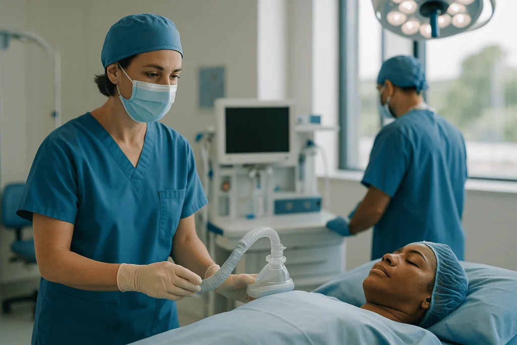
General anesthesia begins with medicine through the cannula in your hand. Most people describe a feeling of heaviness and then nothing. Once asleep, a breathing tube is placed in the windpipe and connected to a ventilator, because the muscle-relaxing medicines used during laparoscopy stop you from breathing on your own and because the inflated abdomen pushes up on the diaphragm.
You are positioned flat, then tilted head-up and rolled slightly to the left. Gravity does part of the surgeon’s work here: the intestines slide down and away from the liver, uncovering the gallbladder. Your arms are secured, pressure points are padded, and compression sleeves squeeze your calves rhythmically to reduce the chance of blood clots forming while you lie still.
The skin is cleaned with antiseptic and covered with sterile drapes, leaving only the abdomen exposed. The first incision, roughly at or just below the navel, is small. Through it the surgeon introduces a blunt cannula, confirms it sits safely inside the abdominal cavity, and connects the carbon dioxide line. Over a minute or two the abdomen rises into a firm dome. Pressure is kept at a level the anesthetist and surgeon agree on, balanced against its effect on breathing and blood return to the heart. The camera goes in, and for the first time the team sees what the scan only hinted at.
Step 2: Placing the ports and getting a first look at the gallbladder
With the camera inside, the surgeon inspects the whole abdominal cavity before touching the gallbladder. This sweep checks that the initial entry caused no injury and looks for anything unexpected: adhesions from earlier operations, fluid, or an appendix or liver that looks unusual. It takes under a minute and is one of the quiet advantages of keyhole surgery over a small open incision.
The remaining ports are placed under direct vision, watching from the inside as each one enters, so that no instrument pushes blindly toward a loop of bowel or a blood vessel. Their positions are chosen to triangulate on the gallbladder: one high in the upper abdomen below the breastbone, one or two along the right side below the ribs. Johns Hopkins describes the typical arrangement as four small cuts, though some surgeons use three and a few use a single larger incision at the navel.
Now the gallbladder is grasped. In cholecystitis this moment often reveals how much trouble it is in. A healthy gallbladder is thin-walled and bluish-green. An acutely inflamed one is swollen, tense, and dusky, sometimes wrapped in a sticky film of omentum, the fatty apron that drapes over the intestines. It may be too distended to grip, so the surgeon inserts a needle and aspirates cloudy bile or pus to decompress it, then closes the puncture with a grasper.
The assistant lifts the top of the gallbladder up and toward the right shoulder. This stretches the organ and opens the small anatomical region at its neck where the two critical structures live: the cystic duct, carrying bile out of the gallbladder, and the cystic artery, carrying blood into it. Everything that makes this operation safe or unsafe happens in that few square centimeters.
Step 3: The critical view of safety, the moment that matters most
If one idea deserves to be understood by every patient, it is this one. The cystic duct is a short tube that joins a much more important tube, the common bile duct, which carries all the liver’s bile to the intestine. Injuring the common bile duct is the most serious complication of gallbladder surgery. The NHS lists bile duct injury among the recognized, though uncommon, risks, and repairing it can require major reconstructive surgery.
To avoid it, surgeons work toward what is called the critical view of safety. In plain terms: before clipping or cutting anything, the surgeon clears away fat and inflamed tissue until exactly two structures can be seen entering the gallbladder, the lower third of the gallbladder is lifted off the liver bed, and nothing else is running through that window. Only when all three conditions are met is the anatomy considered proven rather than assumed.
In acute cholecystitis this dissection is harder. Inflamed tissue is swollen, bleeds more easily, and blurs the normal landmarks. The surgeon uses a hook with electrical current for tiny controlled cuts, blunt dissection to peel tissue apart, and suction to keep the view clear. Some teams inject a dye and take an X-ray during the operation, an intraoperative cholangiogram, to map the ducts when anatomy is uncertain.
Once the view is secured, the clip applier comes in. Two or three clips go on the duct, one or two on the artery, and each is divided between the clips with scissors. The clips stay in permanently. They are inert, do not set off airport scanners, and are compatible with MRI.
If the critical view cannot be achieved safely, the surgeon has options other than pressing on, and the next section describes them. Choosing to stop is a mark of good judgment, not failure.
Step 4: Freeing the gallbladder and lifting it out
With its duct and artery sealed, the gallbladder is attached only by a thin plane of connective tissue to a shallow groove on the underside of the liver. The surgeon now works upward along that groove, using the electrical hook to separate the two structures a few millimeters at a time while the assistant adjusts tension with the graspers. The aim is to stay exactly in the plane: too close to the gallbladder risks tearing it and spilling stones and bile; too deep risks bleeding from the liver’s surface.
In cholecystitis the wall is often friable and tears despite care. Spilled stones are retrieved one by one, the area is washed with warm saline, and the fluid is suctioned out. Small ooze from the liver bed is controlled with the electrical current or a pad of hemostatic gauze.
Before the last strands are divided, the surgeon takes a final look at the clips and the liver bed, sometimes with the gallbladder still tethered so the field can be retracted for inspection. Then the organ comes free. It is dropped into a retrieval bag, a small nylon pouch on a ring, so that the incision it exits through is never contaminated by bile or stones. The bag is drawn out through the widest port, usually at the navel, and the incision is occasionally stretched slightly if large stones make it a tight fit.
A drain, a soft tube left in the abdomen to carry away fluid, is not routine but may be placed if the field was very inflamed or bile spilled. The carbon dioxide is released, and ports are removed under vision. The navel incision is closed with a deep stitch to prevent a hernia, and skin edges are closed with absorbable sutures or glue. The gallbladder is sent to pathology for routine examination, which is standard practice and not a sign of concern.
Why some keyhole operations become open surgery
Every consent conversation for laparoscopic cholecystectomy includes the possibility of conversion, which means switching to a traditional open operation through a single incision under the right ribs. Mayo Clinic and the NHS both describe this as an expected contingency rather than a complication, and both note it is more likely when the gallbladder is acutely inflamed than when surgery is planned for uncomplicated gallstones.
Reasons a surgeon converts include:
- The anatomy cannot be clarified safely because inflammation has fused the gallbladder, duodenum, and colon into one dense mass.
- Bleeding that cannot be controlled with laparoscopic instruments.
- A suspected bile duct injury that needs direct examination and repair.
- Dense scar tissue from previous abdominal operations blocking the view.
- Unexpected findings, such as a gallbladder that looks abnormal enough to raise concern about a tumor.
There is also a middle path. If the neck of the gallbladder is too inflamed to dissect but the body is accessible, the surgeon may perform a subtotal cholecystectomy, removing most of the organ and leaving the stump closed or open with a drain. This avoids the most dangerous dissection while still removing the stones and most of the diseased wall.
Conversion changes the recovery, not the outcome the operation set out to achieve. An open incision is longer, hurts more, and the NHS estimates return to normal activity at six to eight weeks rather than around two. You will be told in the recovery room if conversion happened and why. Surgeons who convert readily when the view is unsafe are following the principle that the operation’s purpose is to remove a gallbladder without damaging anything else, and the route matters less than the destination.
Laparoscopic, open, or a drain? How the options compare
People with cholecystitis rarely get to choose freely among approaches; the surgical team recommends one based on how sick the person is and what the imaging shows. Still, understanding the landscape makes the recommendation easier to weigh. The table summarizes the three common pathways using figures from the NHS and Mayo Clinic patient guidance.
| Approach | What it involves | Typical hospital stay | Typical return to normal activity | Usually chosen when |
|---|---|---|---|---|
| Laparoscopic cholecystectomy | Three or four small incisions, camera and instruments, gallbladder removed | Same day or one night (NHS) | About 2 weeks (NHS) | Most adults fit for anesthesia |
| Open cholecystectomy | Single incision under the right ribs, gallbladder removed | Several days (NHS) | 6 to 8 weeks (NHS) | Conversion during keyhole surgery, dense scarring, complex anatomy |
| Percutaneous cholecystostomy | Drain placed through the skin into the gallbladder under imaging; organ stays | Depends on underlying illness | Not applicable; drain often stays weeks | Too unwell for anesthesia; bridge to later surgery |
Two points deserve emphasis. First, laparoscopic and open surgery are the same operation performed through different doors; the internal steps, clipping the duct and artery and freeing the gallbladder from the liver, are identical. The differences lie in pain, scarring, and time to recover, not in what is removed.
Second, the drain is not a lesser form of treatment for people who qualify for it. For someone whose heart or lungs could not tolerate anesthesia, a drain that controls infection while they recover from a separate illness may be the safer route. Whether surgery follows later is a decision revisited once the acute episode has passed and the person’s condition is clearer.
How long does gallbladder surgery take, and when do you go home?
The NHS puts the operation itself at 60 to 90 minutes. Acute inflammation tends to push toward the longer end because the dissection is slower, and conversion to open surgery adds time. From your perspective the whole event is longer: perhaps 20 to 30 minutes in the anesthetic room beforehand and one to two hours in recovery afterward while the anesthetic wears off and nurses check your pain, breathing, and wound sites.
Waking up feels different from waking from sleep. Most people surface gradually, aware of a sore throat from the breathing tube and a dull ache across the upper abdomen. A distinctive complaint after laparoscopy is pain at the tip of the right shoulder. This is referred pain from carbon dioxide irritating the diaphragm; the nerve that supplies the diaphragm also supplies the shoulder, so the brain misreads the signal. Mayo Clinic describes it as common and self-limiting over a day or two.
Discharge depends on a few practical markers: you can drink without vomiting, pass urine, walk to the bathroom, and control pain with tablets rather than injections. For a planned laparoscopic operation many people meet those markers the same day. For emergency surgery during an admission for cholecystitis, the NHS notes that a one-night stay is more usual, partly because the operation often happens later in the day and partly because people arrive already dehydrated and unwell.
You cannot drive yourself home and should not be alone the first night. Someone needs to collect you and stay, because the anesthetic affects judgment and reaction time for about 24 hours. Before leaving you will receive written instructions on wound care, which pain relievers to use and when, and a contact number for concerns. Those instructions come from your team and take precedence over anything general written here.
Keyhole gallbladder surgery recovery time: the first two weeks
The NHS estimates about two weeks to return to most normal activities after laparoscopic gallbladder removal. That figure is an average, and the shape of those two weeks tends to follow a recognizable pattern.
Days one to three are the sore ones. The incisions ache, the abdomen feels bruised and bloated from residual gas, and the shoulder tip pain may linger. Moving hurts, but moving matters: short walks around the house several times a day reduce the risk of blood clots and help gas disperse. Deep breaths and gentle coughing keep the lungs clear. Most people manage on the pain relief prescribed at discharge and taper as instructed.
Eating restarts gently, not because the gut is fragile but because anesthesia and pain medicines slow it. Small, bland, low-fat meals are easier at first. There is no evidence that a strict long-term diet is necessary after gallbladder removal; Cleveland Clinic advises reintroducing normal foods as tolerated.
By the end of the first week most people are showering normally, walking outside, and doing light housework. The NHS suggests you can drive once you can wear a seatbelt comfortably and perform an emergency stop without hesitation, often around a week, though your insurer may have its own rules. Desk-based work is frequently possible within one to two weeks. Manual work, heavy lifting, and vigorous sport wait longer, generally until the incisions have healed and lifting no longer causes a pulling sensation, which the NHS places at around two weeks for keyhole surgery.
Dressings usually come off after a few days. Absorbable stitches dissolve; glue flakes away. Mild bruising around the incisions is normal. A small firm lump under the navel incision is often just healing tissue, but any lump that enlarges or a wound that opens should be seen. Fatigue outlasts pain: expect to need more sleep than usual for a couple of weeks even once you feel physically mended.
Cholecystectomy risks and complications, honestly explained
Laparoscopic cholecystectomy is considered a low-risk operation, but low risk is not no risk, and an inflamed gallbladder raises every category slightly. The NHS and Mayo Clinic describe the following, listed roughly from most to least common.
- Wound infection, usually at the navel incision where the gallbladder exits. It presents as spreading redness, warmth, and discharge and is generally treated with antibiotics chosen by the treating team.
- Bleeding, either from the liver bed or a port site. Minor oozing is common; bleeding that requires return to the operating room is uncommon.
- Bile leak, when bile escapes from the cystic duct stump or from a small duct in the liver bed. It causes pain, fever, and sometimes jaundice in the days after surgery and may need an endoscopic procedure to stent the duct or a drain to collect the fluid.
- Injury to the bile duct, the most serious specific risk, which the NHS classes as rare. It may require specialist reconstruction.
- Injury to nearby organs such as the bowel or major blood vessels during port placement or dissection.
- Retained stones in the main bile duct, discovered after surgery when pain or abnormal liver blood tests persist, and usually cleared endoscopically.
- Blood clots in the legs or lungs, the general risk of any operation, reduced by early walking and compression stockings.
- Anesthetic complications, including reactions to medicines, breathing problems, and, rarely, heart events, which the anesthetist assesses before surgery.
Published figures for each of these vary widely between studies and depend heavily on how inflamed the gallbladder was and how the numbers were collected, so individual percentages are not quoted here. Your surgeon can give you the figures relevant to their practice and to your situation. What patient guidance from the NHS, Mayo Clinic, and Johns Hopkins agrees on is that the great majority of people have none of these problems and that serious complications are uncommon.
Life after gallbladder removal: digesting without a reservoir
The most frequently asked question at follow-up is some version of: how does my body cope without it? The answer is that the liver keeps making bile at the same rate. Instead of being stored and concentrated, bile now trickles continuously down the common bile duct into the small intestine. The duct stretches slightly over time and takes on a modest storage role of its own. Fat digestion continues because the enzymes that break down fat come mainly from the pancreas, and bile’s job is to emulsify fat so those enzymes can reach it, which a steady trickle still accomplishes.
Some people notice looser or more frequent stools in the weeks after surgery. Mayo Clinic attributes this to bile reaching the colon more continuously, where it can have a mild laxative effect. For most it settles over weeks to a few months. A smaller group has more persistent loose stools; if that happens, the treating clinician can consider whether a medicine that binds bile acids is appropriate, a decision that sits with them rather than with any article.
Very fatty meals are more likely to cause bloating or urgency early on, so a gradual reintroduction of rich foods is sensible. Beyond that, Cleveland Clinic’s guidance is that no special permanent diet is required. Alcohol, coffee, and spicy food are not forbidden; they simply may be less comfortable in the first few weeks.
A minority of people continue to have upper abdominal pain or indigestion after the gallbladder is gone. Sometimes this reflects a stone left in the duct or a bile leak; sometimes the original pain was never from the gallbladder at all. Persistent symptoms after recovery warrant reassessment rather than assumption, and your team can arrange blood tests or imaging to look for causes.
You will never form gallstones in the gallbladder again, for the plain reason that it no longer exists.
What people often get wrong about laparoscopic cholecystectomy for cholecystitis
Myth: a keyhole operation is a smaller operation. The incisions are smaller; the operation is the same. The gallbladder is entirely removed, the same structures are clipped and divided, and the same anatomical risks apply. What changes is the route in and the recovery afterward.
Myth: the gallstones are removed and the gallbladder is left. Taking stones out of an intact gallbladder was tried historically and abandoned because stones return. Every current guideline, including NHS and NIDDK patient guidance, describes removing the organ itself.
Myth: antibiotics can treat cholecystitis so surgery is optional. Antibiotics calm the infection component, and the NHS notes they are often used while awaiting surgery. They do not remove the stones causing the blockage, so the attack is likely to recur. For most people antibiotics are a bridge, not a destination.
Myth: you should wait until the inflammation has completely gone before operating. NHS guidance favors operating within about a week of diagnosis, because the tissue planes are often clearer before scarring matures and because waiting risks another episode.
Myth: you must follow a low-fat diet for life. A brief period of lighter eating helps comfort, but Cleveland Clinic and Mayo Clinic both advise that most people return to an unrestricted diet.
Myth: the clips need to come out later. They are permanent, inert, and safe in MRI scanners and at airport security.
Myth: conversion to open surgery means something went wrong. It means the surgeon judged the keyhole view unsafe and chose the route that protects the bile duct. Mayo Clinic frames it as a planned contingency.
Myth: the gallbladder is removed through the navel because that is where it sits. The gallbladder lies under the right ribs. The navel is chosen for extraction because its incision heals with the least visible scar and the skin there stretches easily.
Questions to ask your care team
A good surgical conversation leaves you knowing not just what will happen but why this plan, for you, now. These questions help you get there. Write the answers down or bring someone to listen with you; anesthetic anxiety makes memory unreliable.
- Are you recommending surgery during this admission or after the inflammation settles, and what tipped the balance for me?
- Do my scans or blood tests suggest a stone in the main bile duct, and if so, will that be dealt with before, during, or after gallbladder surgery?
- How inflamed does my gallbladder look, and how does that affect the likelihood of conversion to open surgery in my case?
- Will you use an X-ray of the bile ducts during the operation, and what would prompt it?
- If the anatomy is unsafe, would you consider a subtotal cholecystectomy, and what would that mean for follow-up?
- Which of my regular medicines should I take on the morning of surgery, and which should be paused? (Direct this to the prescribing clinician; do not adjust anything on your own.)
- Should I expect to go home the same day or stay overnight, and what criteria decide that?
- What symptoms in the first week would you want to hear about immediately, and who do I call out of hours?
- When can I drive, return to my specific job, lift my child or groceries, and exercise?
- Will I have a follow-up appointment, and how will I receive the pathology result from the gallbladder?
- If I develop loose stools that do not settle, who should I contact and when?
You are entitled to ask a surgeon how often they perform this operation and how they handle a difficult gallbladder. Most will welcome the question. Guidance from professional bodies encourages exactly this kind of shared decision, and the plan you agree together should reflect your priorities as well as the scan.
When to call your doctor
Recovery from laparoscopic gallbladder removal generally follows a steady downhill slope of discomfort. The signals that matter are the ones that break that pattern: pain that worsens after initially improving, a fever that appears after the first day, or a new symptom that was not there when you left the hospital. The NHS and Mayo Clinic advise contacting the surgical team or seeking urgent care without waiting for a scheduled appointment if you notice any of the following.
- A temperature above 100.4°F (38°C), or chills and shaking.
- Abdominal pain that is severe, spreading, or getting worse rather than better, or a belly that becomes swollen and hard.
- Yellowing of the skin or the whites of the eyes, dark urine, or pale, clay-colored stools, which can signal bile leaking or a blocked bile duct.
- Persistent vomiting or inability to keep fluids down for more than a few hours.
- Redness spreading from an incision, pus or foul-smelling discharge, or a wound that opens.
- Bleeding that soaks through a dressing.
- Swelling, warmth, or pain in one calf, which may indicate a blood clot.
- Sudden shortness of breath, chest pain, or coughing up blood, which are emergencies and warrant calling emergency services immediately.
- Feeling faint, confused, or unusually drowsy.
Shoulder tip pain, mild bloating, bruising around the incisions, and tiredness are expected and do not by themselves need a call, though it is always acceptable to ask if you are unsure. Before surgery, and during any wait for a planned operation, the same red flags apply: a fever, jaundice, or pain that becomes constant and severe means the gallbladder may have worsened and you should be reassessed promptly rather than waiting for the scheduled date. Your treating team knows your case; when in doubt, their contact number is the right one to use.
Frequently asked questions
How long does gallbladder surgery take from start to finish?
The operation itself usually takes 60 to 90 minutes according to the NHS, with acute inflammation pushing toward the longer end. Add roughly half an hour beforehand for anesthesia and one to two hours afterward in recovery. Emergency operations for cholecystitis can take longer than planned ones because swollen tissue slows dissection, and any conversion to open surgery extends the time further. Your surgeon can give a more specific estimate based on your scans.
What is the keyhole gallbladder surgery recovery time for most people?
The NHS estimates about two weeks to return to most normal activities after laparoscopic gallbladder removal. The first three days are the most uncomfortable, with incision soreness, bloating from residual gas, and shoulder tip pain. Most people are walking outside and doing light tasks within a week, and desk work is often possible by one to two weeks. Heavy lifting and vigorous exercise wait until the incisions have healed and the pulling sensation has gone.
What are the main cholecystectomy risks and complications I should know about?
The most common issues are wound infection at the navel incision, minor bleeding, and temporary loose stools. Less common are bile leaks from the duct stump, retained stones in the main bile duct, and blood clots. The rarest but most serious is injury to the common bile duct, which the NHS lists as an uncommon risk. Figures vary widely between studies, so ask your surgeon for numbers relevant to your situation.
What does life after gallbladder removal look like for digestion?
Bile flows steadily from the liver into the intestine instead of being released in bursts, and fat digestion continues because pancreatic enzymes do most of the work. Some people notice looser stools for weeks to a few months, which Mayo Clinic attributes to bile reaching the colon more continuously. Cleveland Clinic advises that no permanent special diet is needed; rich or very fatty meals are simply reintroduced gradually as comfort allows.
Why do I have pain in my shoulder after laparoscopic gallbladder surgery?
The carbon dioxide gas used to inflate the abdomen irritates the underside of the diaphragm, and the nerve supplying the diaphragm shares a pathway with nerves from the shoulder, so the brain misreads the signal as shoulder pain. Mayo Clinic describes this as common and self-limiting, usually fading within a day or two as the body absorbs the remaining gas. Walking and changing position help it disperse faster.
Can cholecystitis be treated with antibiotics instead of surgery?
Antibiotics address the infection but do not remove the gallstones blocking the cystic duct, so the attack is likely to recur. The NHS describes antibiotics as treatment given while awaiting surgery rather than as a substitute for it. People who are too unwell for an operation may instead have a drain placed through the skin into the gallbladder, with surgery reconsidered once they are stronger. The treating team decides which pathway fits.
What happens if the surgeon has to switch to open surgery?
The surgeon makes a single longer incision under the right ribs and completes the same operation by direct vision. Mayo Clinic and the NHS describe this as a planned contingency, more likely when the gallbladder is acutely inflamed or scarred. Recovery is longer, with the NHS estimating six to eight weeks to normal activity rather than about two, and the hospital stay extends to several days. You will be told why it was necessary.
Do the clips left inside after gallbladder removal need to be removed later?
No. The small clips placed on the cystic duct and artery are made of titanium or an absorbable polymer, are inert, and stay permanently. They do not set off airport security scanners and are safe in MRI machines. They occasionally show up on later X-rays or CT scans as tiny bright marks in the upper right abdomen, which radiologists recognize immediately as evidence of previous gallbladder surgery.
Will I need a special diet before or after laparoscopic cholecystectomy?
Before surgery you will fast for a set number of hours as instructed by the anesthetic team. Afterward, small, bland, lower-fat meals are easier for the first days while the gut wakes up from anesthesia and pain medicines. Beyond that, Cleveland Clinic and Mayo Clinic advise returning to a normal diet as tolerated. Very rich meals may cause bloating or urgency early on, so reintroduce them gradually rather than avoiding them for life.
Why is the gallbladder taken out through the navel when it sits under the ribs?
The navel incision heals with the least visible scar, and the skin and deeper layers there stretch to let the retrieval bag pass without tearing. The gallbladder is placed in a sealed bag inside the abdomen so no bile or stones touch the wound as it exits. A deep stitch closes the muscle layer afterward to reduce the chance of a hernia developing at that site.
References
- Gallbladder removal – laparoscopic (MedlinePlus Medical Encyclopedia)
- Gallbladder removal – NHS
- Acute cholecystitis – NHS
- Cholecystitis – Cleveland Clinic
This article is for general information only and is not a substitute for professional medical advice. Please consult a qualified doctor about your individual situation.

More from the Blog

Eating With Crohn’s Disease: How Diet Works Alongside Treatment, Not Instead of It
Diet can ease Crohn's disease symptoms and correct nutritional shortfalls, but it does not replace medical treatment. Current evidence supports using food alongside prescribed…

Flying Home After Gallbladder Surgery: How Long Cholecystitis Patients Stay in Istanbul
There is no single safe date for flying after gallbladder surgery; the wait depends on the type of operation and how recovery is going.…

The Gut-Brain Axis: How Your Microbiome Talks to Your Brain
The gut brain axis is the two-way communication network linking your digestive tract, its trillions of microbes and your brain through the vagus nerve,…

Pain or Fever After ERCP: The Warning Signs That Need a Same-Day Call
Pain that builds rather than fades in the hours after ERCP, especially pain spreading to the back with nausea or vomiting, can signal pancreatitis…

Hepatitis A Warning Signs: When Worsening Jaundice or Confusion Needs Urgent Care
Most people with hepatitis A recover with rest and fluids, but seek urgent care the same day if jaundice keeps deepening after the first…

Eating and Drinking During Hepatitis A Recovery: Fluids, Nutrition and Alcohol Rules
During hepatitis A recovery, eat small, plain meals built around easily digested carbohydrates, lean protein and cooked vegetables, drink enough fluid to keep urine…