

Lung Abscess on a CT Scan: What the Images Show and How Sputum Cultures Guide Antibiotics

Key Takeaways
- Clinicians generally call a cavity a lung abscess when it is larger than about 2 cm; multiple smaller cavities within consolidated lung are usually described as necrotizing pneumonia.
- On CT, an abscess is typically round, thick-walled, forms an acute angle with the chest wall and destroys nearby vessels, whereas an empyema is lens-shaped, forms an obtuse angle and pushes the lung aside.
- Aspiration-related abscesses favor the right lung and the gravity-dependent segments because the right main airway is wider and more vertical.
- Sputum cultures often miss the anaerobic mouth bacteria that cause most aspiration abscesses, so a negative result does not rule out infection and the clinical response guides the antibiotic plan.
- Antibiotic courses for lung abscess typically run 3–6 weeks and sometimes longer, often until follow-up imaging shows the cavity closing, because drugs penetrate the dead center of the cavity poorly.
- Most abscesses drain through the airway on their own; surgery is needed in roughly 10% of cases, usually for failure of prolonged medical therapy, suspected cancer or complications such as bronchopleural fistula.
On a CT scan, a lung abscess usually appears as a rounded, thick-walled cavity inside the lung, often with an air-fluid level where pus has partly drained into an airway. CT confirms the cavity, estimates its size and separates it from look-alikes such as empyema or a cavitating tumor. Sputum and blood cultures then help the treating team narrow initial broad-spectrum antibiotics to targeted therapy, typically continued for several weeks.
The report arrives in the patient portal before the appointment does, and one phrase stops the eye cold: “thick-walled cavitary lesion with air-fluid level, consistent with lung abscess.” Three weeks earlier it had felt like a stubborn chest infection, the kind that leaves you sweating through the sheets and coughing up something you would rather not describe. Now there is a hole in your lung with a name.
A lung abscess CT scan is one of the clearer moments in chest medicine. The image shows a pocket of dead tissue and pus that the body has walled off, and it usually shows it in enough detail that the care team can decide what to do next. The cultures that follow, grown from sputum and blood, are slower and less tidy, but they are what turn a broad first guess at antibiotics into a plan built around the actual organism.
This explainer walks through both halves of that process: what the radiologist is looking at, and how the microbiology lab shapes the weeks that follow.
What does it mean to have an abscess on your lung?
An abscess is a collection of pus, the thick fluid made of dead white blood cells, bacteria and liquefied tissue, enclosed by inflamed tissue. In the lung, that pocket forms when an infection destroys a patch of lung tissue faster than the body can clear it. The destroyed area liquefies, and once it connects to an airway, some of the contents are coughed out. Air moves in to replace them, which is why the cavity shows up on imaging.
Clinicians generally reserve the term lung abscess for a single cavity larger than about 2 cm, according to the NIH-hosted StatPearls review. Smaller, multiple cavities within an area of pneumonia are usually described as necrotizing pneumonia instead; the distinction matters and gets its own section below.
Most lung abscesses are called primary, meaning they develop in otherwise healthy lung after something is inhaled, most often a mouthful of saliva or stomach contents carrying bacteria from the mouth. Secondary abscesses develop because something else has set the stage: a blocked airway from a tumor or inhaled object, a weakened immune system, or infection carried in the bloodstream from somewhere else in the body.
Doctors also sort abscesses by time. StatPearls describes those present for less than roughly 4–6 weeks as acute and those lasting longer as chronic. Chronic abscesses tend to produce vaguer symptoms, such as weight loss, fatigue and a low-grade fever, which is one reason they are sometimes found later than anyone would like.
Having an abscess does not mean the lung is permanently damaged. It means a localized infection has reached a stage where the body needs help clearing it, and where imaging and cultures become the two tools that guide that help.
Can a CT scan show a lung infection like this, and how does it work?
Yes. A chest CT (computed tomography) scan is an X-ray examination that takes many thin cross-sectional pictures as the scanner rotates around you, then assembles them into detailed slices of the chest. MedlinePlus describes the scan itself as quick, usually a matter of minutes on the table, with the patient lying still and holding their breath briefly while images are captured.
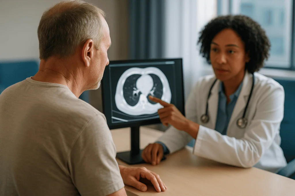
Where an ordinary chest X-ray flattens the whole chest into a single shadow, CT shows depth. A cavity hidden behind the heart or tucked against the diaphragm can be missed on a plain film and is obvious on CT. The scanner also measures how dense each small block of tissue is, which lets the radiologist tell fluid from air, pus from solid tumor, and inflamed lung from normal lung.
For a suspected abscess, the scan is often done with intravenous contrast, a dye injected through a vein that brightens blood vessels and tissues with a good blood supply. Contrast makes the wall of an abscess light up while the dead center stays dark, and it helps show whether blood vessels run through an abnormal area or have been destroyed by it. The NHS notes that most people feel only a brief warm flush when contrast is given, and that the team will ask about kidney function and allergies first.
The scan does not identify the germ. It shows the shape of the problem, where it sits, how large it is, whether fluid has gathered around the lung and whether anything is blocking an airway behind it. Those details steer every later decision: whether to sample the pleural space, whether to look inside the airways with a bronchoscope, and how closely to repeat imaging during treatment.
What a lung abscess CT scan actually shows
Radiologists describe a classic lung abscess with a handful of recurring features, and understanding them makes the report far less intimidating.
The first is the cavity itself: a rounded, often spherical hole within the lung. Its wall is typically thick and its inner edge irregular or shaggy, because it is lined with dying tissue rather than a smooth membrane. On a contrast scan, the wall enhances brightly while the center remains dark.
The second is the air-fluid level. When the cavity has drained partly into an airway, pus settles to the bottom and air sits on top, producing a flat horizontal line inside the hole. On an upright X-ray it looks like a tiny water line in a jar; on CT it appears in whichever slice cuts through it.
The third is location. StatPearls notes that aspiration-related abscesses favor the back portions of the upper lobes and the upper segments of the lower lobes, the parts of the lung that are lowest when a person lies flat. The right lung is affected more often because its main airway runs more vertically, giving inhaled material a straighter path.
The fourth is the surrounding lung. There is often a halo of consolidation, the medical word for lung that has filled with fluid and inflammatory cells, around the cavity. Blood vessels and airways may appear to stop abruptly at the cavity edge, which signals destruction rather than displacement.
Radiologists also look at how the cavity meets the chest wall. An abscess, being inside the lung, tends to form an acute angle with the pleura, like a ball resting against a wall. That single observation is one of the most useful clues in the next comparison.
Lung abscess vs empyema: how the CT tells them apart
Empyema is pus in the pleural space, the thin gap between the lung and the chest wall, rather than inside the lung tissue. The two conditions can look similar on a plain X-ray, both producing a fluid-and-air pocket near the edge of the chest, but they are treated differently. An abscess usually drains on its own through the airways and is managed primarily with antibiotics; an empyema almost always needs a drainage tube or procedure because pus in the pleural space has no natural exit, as the NHS explains.
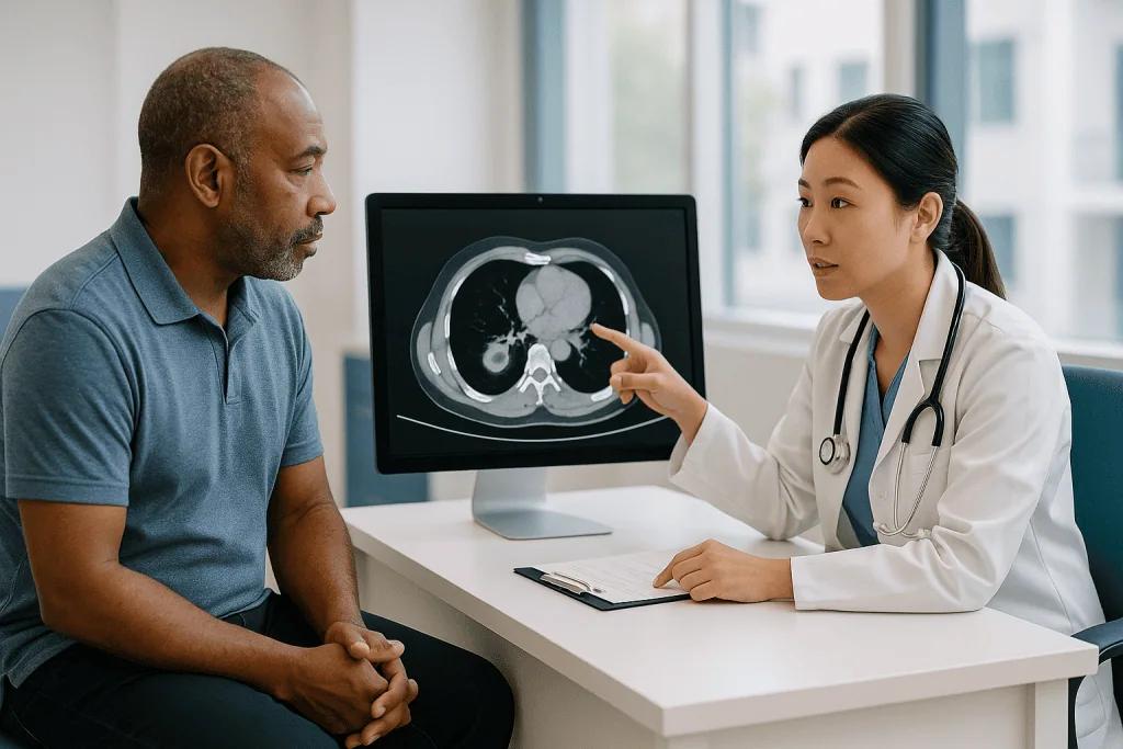
CT separates them in most cases by asking a few geometric questions.
| Feature on CT | Lung abscess | Empyema |
|---|---|---|
| Shape | Round or spherical | Lens-shaped, elongated along the chest wall |
| Angle with the chest wall | Acute, like a ball against a wall | Obtuse, tapering smoothly into the pleura |
| Wall | Thick, irregular inner edge | Thin, smooth; the two pleural layers may appear separated and enhancing (“split pleura”) |
| Effect on nearby lung | Destroys vessels and airways, which end abruptly | Compresses and pushes lung away; vessels bend around it |
| Change with position | Shape stays the same | Fluid may shift or layer |
No single feature is decisive, and the two can coexist: an abscess near the lung surface can rupture into the pleural space and create an empyema, a complication called bronchopleural fistula when air also escapes. When the picture is ambiguous, the team may sample pleural fluid with a needle (thoracentesis) or repeat the scan after a short interval. The decision rests with the treating clinicians, who weigh the images alongside how the person is actually doing.
Necrotizing pneumonia vs lung abscess: what is the difference?
People searching this topic often meet both terms in the same report and reasonably wonder whether they describe different diseases. They are better understood as points on one spectrum of lung tissue destruction.
Necrotizing pneumonia is a severe pneumonia in which parts of the infected lung die and liquefy, producing multiple small cavities, usually each under about 2 cm, scattered through an area of consolidation. The StatPearls review frames lung abscess as the larger, more organized end of that process: typically a single dominant cavity larger than 2 cm, with a defined wall. In practice, a patient can start with necrotizing pneumonia and, over days to weeks, see small cavities coalesce into one abscess.
Why the label matters is mostly about expectations and monitoring. Necrotizing pneumonia tends to be the more acutely ill picture, often tied to aggressive organisms such as Staphylococcus aureus, Klebsiella pneumoniae or certain streptococci, and it carries a higher chance of complications such as empyema or bronchopleural fistula. A mature abscess, especially a primary one from aspiration, often follows a slower, grumbling course.
The antibiotic approach overlaps substantially, since both conditions call for drugs that reach necrotic tissue and cover anaerobic bacteria, the oxygen-avoiding organisms from the mouth that dominate aspiration infections. Where they differ is in how the team watches the lung: with necrotizing pneumonia, repeat imaging is often arranged sooner to catch spreading destruction, and drainage decisions can arise earlier if a collection forms against the chest wall.
For the person reading the report, the practical message is this: the two names describe degree, not a different diagnosis, and the treatment plan will be built on the organism and the clinical picture rather than on which word the radiologist chose.
Could the cavity be something other than infection?
A hole in the lung is not automatically an abscess, and good radiologists say so in their reports. Several conditions can cavitate, and the treating team keeps them in mind from the first scan onward.
Lung cancer is the most consequential mimic. Certain tumors, particularly squamous cell carcinomas, outgrow their blood supply, die in the center and form a cavity. Features that raise suspicion include a very thick or nodular wall, a cavity without the surrounding inflamed lung that infection usually produces, enlarged lymph nodes, and a person whose fever and cough do not match the size of the lesion. A tumor can also block an airway and cause a true abscess behind it, so the two can be present together.
Tuberculosis classically cavitates in the upper lobes and may be accompanied by small scattered nodules in a branching pattern. Fungal infections, septic emboli (infected clots that travel from an infected heart valve or an intravenous line and lodge in the lung, often forming several cavities at once), and some non-infectious inflammatory conditions of the blood vessels can all produce similar pictures.
This is why a CT finding of “cavitary lesion” is a starting point rather than an answer. The team typically combines the images with the history (aspiration risk, smoking, travel, immune status), sputum tests that include stains for tuberculosis and fungi, and, when the picture does not fit infection, a closer look through bronchoscopy or a needle biopsy guided by CT.
Repeat imaging also helps. An infectious abscess treated with appropriate antibiotics should shrink over weeks; a cavity that stays the same or grows despite therapy prompts a search for another cause. When a report hedges, that hedge usually reflects honest uncertainty rather than alarm.
Sputum culture for lung abscess: what the test can and cannot tell you
A sputum culture is a laboratory test in which mucus coughed up from the lungs is spread on nutrient plates and incubated so that any bacteria present can grow and be identified. MedlinePlus describes the collection as straightforward: a deep cough into a sterile cup, ideally first thing in the morning, after rinsing the mouth with water to reduce contamination. Preliminary results often appear within a few days, with full identification and antibiotic sensitivity testing taking somewhat longer.
For a lung abscess, the test is useful but imperfect, and it helps to know why. The mouth is home to hundreds of bacterial species, and any sample that passes through it picks up passengers. The lab often grades the sample’s quality under a microscope first, looking for many white blood cells and few cells from the cheek lining, which suggests the mucus came from deep in the lung rather than the throat.
Anaerobic bacteria add a second complication. These organisms, which thrive without oxygen and are the most common culprits in aspiration-related abscesses, die quickly in air and are hard to grow from an ordinary coughed sample. A negative culture therefore does not mean no bacteria; it may mean the main organism simply did not survive the trip to the plate.
Because of these limits, the team usually casts a wider net. Blood cultures, taken before antibiotics when possible, can catch bacteria that have spilled into the bloodstream. Stains and specialized cultures for tuberculosis and fungi are often added. If the person cannot produce sputum, or the initial results are unhelpful, bronchoscopy allows fluid to be washed from the affected segment and collected directly, bypassing much of the mouth’s contamination. Each step is a judgment call made by the treating clinicians based on how sick the person is and how the first results look.
How sputum cultures guide antibiotics: from broad guess to targeted plan
Antibiotic treatment for a lung abscess happens in two phases, and the culture results are the hinge between them.
The first phase is empiric: treatment begun before any organism is identified, chosen to cover the bacteria most likely to be responsible. For a suspected aspiration abscess, that means agents active against anaerobes and the common mouth streptococci. Guideline-level reviews, including the StatPearls summary, describe antibiotics that combine a penicillin-type drug with a beta-lactamase inhibitor, or alternatives from the clindamycin or carbapenem classes, as typical starting points. If the setting or history raises concern for resistant staphylococci or hospital-acquired gram-negative bacteria, coverage is broadened accordingly. None of this is a recommendation; the choice depends on local resistance patterns, allergies, kidney and liver function, and the prescribing clinician’s judgment.
The second phase is targeted. When cultures grow an organism and the lab reports which antibiotics it is sensitive to, the team can often narrow therapy to a drug that hits that organism precisely. Narrowing matters for more than tidiness. It reduces side effects, lowers the chance of resistant bacteria or Clostridioides difficile infection in the gut, and may allow a switch from intravenous to oral treatment once fever settles and the person is eating and drinking normally.
When cultures are negative or grow only mixed mouth flora, which is common, the team usually continues broad anaerobic coverage and watches the clinical response: temperature, oxygen levels, the volume and character of sputum, and repeat imaging. Improvement within several days to a week is the usual signal that the empiric choice was right.
The mechanism is simple to state and slow to deliver. Antibiotics reach the abscess wall through its blood supply but penetrate the dead center poorly, which is why treatment runs for weeks rather than days and why the body’s own drainage through the airway does much of the work.
Who usually develops a lung abscess, and who is asked to wait
The common thread in primary lung abscess is aspiration, and the risk factors are mostly about what makes aspiration more likely. StatPearls lists reduced consciousness from alcohol or sedatives, seizures, stroke or other neurological conditions that weaken swallowing, general anesthesia, poor dental health with a heavy load of mouth bacteria, and conditions such as reflux or esophageal disorders that bring stomach contents upward. MedlinePlus adds that older adults and people with difficulty swallowing are at particular risk of aspiration pneumonia, the precursor state.
Secondary abscesses point to a different list: a tumor or inhaled object blocking an airway, a weakened immune system from chemotherapy, transplant medicines or HIV, intravenous drug use that seeds the bloodstream, and infected heart valves. Any of these shifts the balance of likely organisms and may change the antibiotic approach.
Who is asked to wait? Two groups, in different senses. The first are people whose first scan is ambiguous and who are clinically stable. Rather than rushing to an invasive biopsy, the team may begin antibiotics and repeat imaging after a short interval to see whether the lesion behaves like an infection. The second are people for whom drainage or surgery is being considered. Because most abscesses drain on their own through the airway and respond to antibiotics alone, interventional procedures are typically held in reserve until medical therapy has had a fair trial, usually measured in weeks, unless the person is deteriorating.
Children form their own category. Pediatric lung abscess is uncommon, often follows a severe pneumonia, and is approached with the same imaging logic, with extra attention to minimizing radiation exposure and to comfort during scanning and sample collection. Decisions about children’s treatment, including every aspect of medication, belong entirely to the pediatric team.
Lung abscess treatment duration: what the following weeks usually look like
Few things surprise people more than the length of treatment. The StatPearls review describes antibiotic courses for lung abscess typically running 3–6 weeks and sometimes longer, often continued until the cavity has closed or shrunk to a small, stable scar on follow-up imaging. That is a long time to take any medicine, and knowing the usual rhythm helps.
The first days are often spent in hospital, particularly if fever is high, oxygen is low or the person cannot keep fluids down. Antibiotics usually start intravenously while cultures are pending. Many people notice the fever beginning to settle within the first several days to a week; the cough may temporarily become more productive as the cavity drains, which can be unsettling but is generally a good sign.
The middle weeks are about consolidation of gains. Once fever has gone and the person is eating, drinking and walking, the team often switches to an oral antibiotic matched to the culture results. Follow-up imaging, usually a chest X-ray and sometimes a repeat CT, checks that the cavity is shrinking. Imaging improvement lags clinical improvement; feeling better by week two and still having a visible cavity at week four is the expected pattern, not a failure.
Fatigue and weight loss are common companions of a chronic infection and can take weeks to reverse after the infection itself is controlled. Good nutrition, gradual return to activity and attention to the original cause (dental care, swallowing assessment, management of reflux or seizures) are part of the recovery plan.
Completing the full course exactly as prescribed is the single most important thing a person can do during this period. Stopping early because symptoms have resolved risks relapse into a half-treated cavity, and any thought of shortening or changing treatment should go straight to the prescribing clinician.
When antibiotics alone are not enough: drainage and surgery
Most lung abscesses heal with antibiotics and the body’s own drainage through the airways. A minority do not, and the care team watches for the signs: persistent fever beyond the first one to two weeks of appropriate treatment, a cavity that is not shrinking, a very large abscess, or evidence that the abscess has ruptured into the pleural space.
Several procedural options exist, and they are considered in roughly escalating order.
- Bronchoscopy: a thin flexible camera passed through the mouth or nose into the airways. It can clear a blockage, retrieve an inhaled object, collect uncontaminated samples for culture and, in selected cases, help the cavity drain.
- Percutaneous drainage: a thin tube placed through the chest wall into the abscess under CT or ultrasound guidance. It is used mainly for large cavities that will not drain through the airway, and the team weighs the risk of contaminating the pleural space when choosing it.
- Pleural drainage: if an empyema has formed alongside the abscess, a chest tube into the pleural space is usually needed, as the NHS describes for empyema generally.
- Surgery: removal of the affected segment or lobe. StatPearls notes that surgical treatment is required in roughly 10% of cases, typically for abscesses that fail prolonged medical therapy, suspected cancer, major bleeding or a bronchopleural fistula that will not close.
Each option carries its own risks, including bleeding, air leak, spreading infection and the ordinary risks of anesthesia, and each has alternatives, including simply continuing antibiotics for longer with closer monitoring. The team’s choice depends on the person’s overall health, the size and position of the abscess, and what the cultures and imaging show. There is no fixed threshold that mandates a procedure, and the decision always rests with the treating clinicians in discussion with the patient.
What people often get wrong about a lung abscess CT scan
“A cavity on CT means I have lung cancer.” Infection is the far more common cause of a new cavity in a person with fever and productive cough, and radiologists describe features that favor one or the other. When the picture is uncertain, repeat imaging after treatment usually settles it. Worry is understandable; certainty from a single scan is not realistic in either direction.
“The scan found the germ.” CT shows shape, size and location. It cannot identify a bacterium. That job belongs to sputum and blood cultures, and even they frequently come back negative or mixed, because anaerobic mouth bacteria are hard to grow.
“A negative culture means the antibiotics are not working.” A negative culture most often means the organism did not survive sampling. The real test of the antibiotic choice is clinical: falling fever, improving oxygen levels, shrinking cavity.
“If I feel better, the infection is gone.” Symptoms improve weeks before the cavity closes on imaging. Stopping antibiotics early because of improved symptoms is one of the recognized paths to relapse, which is why courses typically run 3–6 weeks or longer per the StatPearls review.
“An abscess always needs to be drained with a needle or tube.” That is true of empyema, which has no natural exit, but most lung abscesses drain into the airway on their own. Procedures are reserved for those that do not respond.
“The right lung was affected because I sleep on my right side.” The right lung is more often involved because its main airway is wider and more vertical, offering inhaled material a straighter path, not because of sleeping position in any simple way.
“Coughing up more phlegm means it is getting worse.” In the first days of treatment, increased drainage can mean the cavity is emptying. Rising fever or blood in the sputum are different matters and warrant a call.
Questions to ask your care team
A lung abscess diagnosis arrives with a lot of vocabulary and a long treatment horizon. Having a few questions written down before a ward round or clinic visit helps the conversation stay useful.
- How large is the abscess, and where exactly is it? Does the scan show fluid around the lung as well, or anything blocking an airway?
- What did the radiologist think caused the cavity, and were any features mentioned that need a second look, such as those that could suggest tuberculosis or a tumor?
- Which cultures were sent, and when should results be expected? Were blood cultures taken before antibiotics started?
- If cultures come back negative or mixed, how will you decide whether the antibiotic is working?
- What is the planned total duration of treatment, and what will tell you it can stop?
- When do you expect to repeat imaging, and will it be an X-ray or another CT?
- What would make you consider bronchoscopy, a drainage procedure or surgery, and what are the alternatives to each?
- What was the likely reason the aspiration happened, and should I have a swallowing assessment, dental review or other follow-up to lower the chance of it recurring?
- Which symptoms should prompt me to call during treatment, and whom exactly do I call?
- Are there interactions between the antibiotic and my other medicines, alcohol or supplements that I should know about?
Write down the answers, or ask a family member to. Weeks-long treatment means several different clinicians may be involved, and a shared record of what was said keeps everyone aligned. If something in the plan is unclear, asking for it to be explained again is reasonable, not a nuisance.
When to call your doctor
Treatment of a lung abscess is a weeks-long process, and the care team expects to hear from you during it. Some symptoms simply need a phone call; others mean emergency care without waiting.
Seek emergency help immediately for any of the following red-flag signs: coughing up more than a small streak of blood, especially if it is bright red or increasing; sudden worsening shortness of breath or chest pain, which can signal rupture into the pleural space or a collapsed lung; confusion, fainting or a very fast heartbeat; blue or gray lips or fingertips; or a fever with shaking chills that returns after it had settled.
Call your doctor the same day if fever persists beyond the first week of antibiotics or comes back after improving; if sputum becomes foul-smelling, markedly bloodier or much more abundant after an earlier decline; if you develop severe diarrhea, a new rash, or swelling of the face or throat on the antibiotic; if you cannot keep the medicine or fluids down; or if you feel generally worse rather than steadily better.
Between those extremes, the ordinary course includes fatigue that lifts slowly, a cough that improves unevenly and imaging that lags behind how you feel. Report what you notice at each follow-up, bring your list of medicines, and never stop or alter the antibiotic without speaking to the prescribing clinician. If the diagnosis itself is still being clarified, keep every scheduled scan and appointment: a cavity that fails to shrink is the signal the team is watching for, and it can only be seen if the follow-up happens. Every decision about changing treatment, adding a procedure or extending the course belongs with the treating team, who have the full picture of your scans, cultures and progress.
Frequently asked questions
What does it mean to have an abscess on your lung?
It means an infection has destroyed a pocket of lung tissue, leaving a cavity filled with pus and dead material walled off by inflamed lung. Most form after inhaling bacteria-laden saliva or stomach contents. The cavity usually connects to an airway, so some contents are coughed out and air fills the space, which is why it shows on imaging. It is treated with weeks of antibiotics and, in a minority of cases, drainage.
Can a CT scan detect an abscess in the lung?
Yes, CT is the most reliable routine imaging test for it. The scan shows the cavity, its thick wall, any air-fluid level inside, and its exact position, including areas a chest X-ray can hide behind the heart or diaphragm. Contrast dye highlights the enhancing wall against the dark center. CT cannot identify the bacterium responsible; that requires sputum, blood or bronchoscopy cultures.
What are the differences between necrotizing pneumonia and lung abscess?
They are stages of the same destructive process. Necrotizing pneumonia describes multiple small cavities, usually under 2 cm each, scattered through an area of pneumonia and often linked to aggressive organisms. A lung abscess is a single, larger, well-walled cavity, typically over 2 cm, that may evolve from those smaller cavities. Treatment overlaps heavily; the main difference is how closely and how soon imaging is repeated.
How does a lung abscess vs empyema show up differently on CT?
An abscess sits inside the lung, so it looks round, has a thick irregular wall, meets the chest wall at an acute angle and cuts off nearby vessels. An empyema sits between the lung and chest wall, so it is lens-shaped, has thin smooth walls that may show the two pleural layers separated, meets the chest wall at an obtuse angle and compresses the lung rather than destroying it.
How is a sputum culture for lung abscess collected and how long do results take?
You rinse your mouth with water, take a deep breath and cough mucus from deep in the chest into a sterile cup, ideally first thing in the morning. The lab checks sample quality, then grows bacteria on nutrient plates. MedlinePlus notes preliminary results often appear within a few days, with full identification and antibiotic sensitivity testing taking longer. Anaerobic organisms frequently fail to grow from coughed samples.
What is the typical lung abscess treatment duration?
Guideline-level reviews such as the StatPearls summary describe antibiotic courses typically lasting 3–6 weeks and sometimes longer, often continued until follow-up imaging shows the cavity closed or reduced to a small stable scar. Treatment usually starts intravenously in hospital and switches to oral medicine once fever settles. The exact length is decided by the prescribing clinician based on cultures, imaging and clinical response.
Why do antibiotics take so long to work on a lung abscess?
Antibiotics travel in blood, and the center of an abscess has no blood supply; it is dead tissue and pus. Drugs reach the inflamed wall but penetrate the core poorly, so the body’s own drainage through the airway does much of the clearing while antibiotics stop the infection spreading. Fever usually falls within days, but the cavity itself closes over weeks, which sets the length of treatment.
Can a lung abscess be mistaken for lung cancer on CT?
Yes, some tumors, especially squamous cell carcinomas, outgrow their blood supply and cavitate, and a tumor can also block an airway and cause a true abscess behind it. Features favoring cancer include a very thick nodular wall, enlarged lymph nodes and little surrounding inflammation. When the picture is unclear, the team may repeat imaging after antibiotics or arrange bronchoscopy or a CT-guided biopsy.
Do I need a chest tube or surgery for a lung abscess?
Usually not. Most abscesses drain through the airway and respond to antibiotics alone. Procedures are reserved for abscesses that fail prolonged medical therapy, very large cavities, suspected cancer, significant bleeding or rupture into the pleural space. StatPearls notes surgery is needed in roughly 10% of cases. Empyema, pus around the lung rather than in it, is different and nearly always requires a drainage tube.
Will the hole in my lung close completely after treatment?
Many cavities shrink and close over weeks to months, sometimes leaving a small scar or thin-walled space visible on later imaging. Imaging improvement lags behind feeling better, so a visible cavity partway through treatment is expected rather than alarming. The treating team uses follow-up X-rays or CT to confirm the cavity is resolving and to decide when antibiotics can stop; no outcome can be guaranteed in advance.
References
- Lung Abscess (StatPearls), NIH National Library of Medicine
- Chest CT, MedlinePlus Medical Encyclopedia
- Routine sputum culture, MedlinePlus Medical Encyclopedia
- Aspiration pneumonia, MedlinePlus Medical Encyclopedia
This article is for general information only and is not a substitute for professional medical advice. Please consult a qualified doctor about your individual situation.

More from the Blog

After Pleural Effusion Drainage: Normal Soreness vs the Breathing Changes That Need Urgent Care
After thoracentesis, most people notice easier breathing within hours, along with a dull ache or pinching at the needle site that fades over one…

Pulmonary Edema: How the Heart, Lung or Kidney Cause Dictates the Treatment Plan
Pulmonary edema is fluid leaking into the lungs' air sacs. The most common cause is a heart that cannot pump or relax properly, which…

Traveling With Cystic Fibrosis: Flights, Therapy Equipment and Planning Care Away From Home
Most people with cystic fibrosis can travel, including by air, when their lung disease is stable and the trip is planned with their CF…

Which Tests Guide Cystic Fibrosis Care? Sweat Test, Genetic Testing and Sputum Cultures
Cystic fibrosis care relies on three kinds of tests. The sweat chloride test measures how much salt is in sweat and remains the main…

Who Benefits Most From a Structured Smoking Cessation Program? What Doctors Consider
A structured smoking cessation program tends to help most when nicotine dependence is strong, earlier quit attempts have not lasted, or a health condition…

The First 24 Hours After a Bronchoscopy: Sore Throat, Swallowing Checks and Driving Rules
After a bronchoscopy, most people spend one to several hours in a recovery area while sedation wears off, then go home the same day…