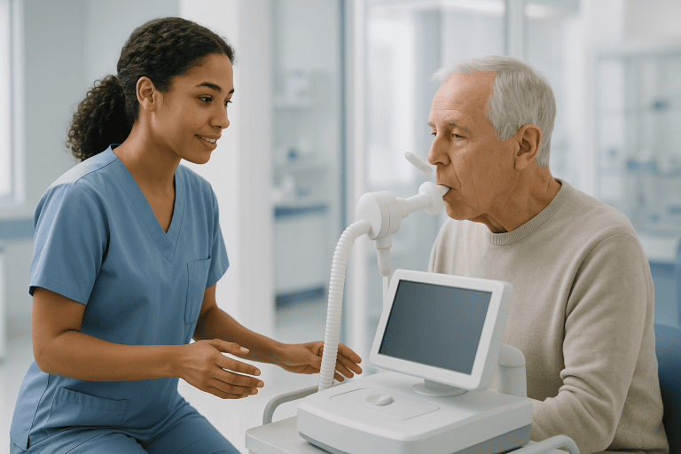
Lung Abscess Treatment
Lung abscess treatment focuses on clearing a pus-filled lung cavity caused by infection, usually with prolonged antibiotics and supportive care. Drainage or surgery may be needed if medication is not enough.

Quick answer
A lung abscess is a pus-filled cavity that forms in lung tissue when infection destroys a localised area of the lung. Treatment centres on a prolonged course of antibiotics, usually lasting weeks, alongside oxygen, fluids and nutrition support where needed. Large or non-responding abscesses may need image-guided drainage, and surgery is reserved for a small minority of complicated cases.
What Is a Lung Abscess?
A lung abscess is a pus-filled cavity that forms inside the lung when infection destroys a localised area of lung tissue. Bacteria, very often organisms that normally live harmlessly in the mouth and throat, take hold in the lung, the immune system walls the infected area off, and the tissue at its centre breaks down into pus. Treatment aims to clear that cavity, usually with a prolonged course of antibiotics, to support your breathing and strength while the lung heals, and to identify why the abscess developed in the first place. The diagnosis is made in a wide range of people: otherwise healthy adults after severe pneumonia, older patients with swallowing difficulties, and people whose immune defences are weakened by illness or its treatment.
For many patients, the diagnosis of a lung abscess comes after days or weeks of fever, cough, exhaustion, night sweats, weight loss, chest discomfort, or sputum that smells unpleasant or contains blood. It can be unsettling to hear that an infection has created a cavity in the lung, particularly if you are seeking a second opinion on whether antibiotics alone are enough. The encouraging reality is that most lung abscesses respond to a carefully chosen antibiotic plan, close monitoring and good supportive care. Treatment usually takes longer than treatment for ordinary pneumonia, because the antibiotics must penetrate an infected cavity and keep working until the abscess shrinks and symptoms settle. Some patients also need the cavity drained, a bronchoscopy to look for a blocked airway or, less commonly, surgery when infection persists despite appropriate medical therapy.
At Acibadem, lung abscess treatment is approached as a diagnostic and therapeutic process, not simply a prescription. Physicians work to identify the organism causing the infection, understand why the abscess formed, assess your breathing and general condition, and select a plan suited to your age, immune status, other illnesses and imaging findings. Because treatment can involve several weeks of antibiotics and follow-up imaging, this planning is laid out clearly from the start, so that every stage, from the first scan to the final follow-up, has a defined purpose.
Is a pulmonary abscess the same as a lung abscess?
Yes. A pulmonary abscess and a lung abscess are exactly the same condition; pulmonary is simply the medical adjective for anything relating to the lung. The term appears in several forms, which can be confusing when you are reading reports or searching for information. Older textbooks and hospital index systems sometimes reverse the words to abscess pulmonary, search results are full of the common misspelling lung abcess, and the frequent mistyping lung access leads to the same pages. All of these refer to one thing: a pus-filled cavity inside lung tissue. One distinction is genuinely important, though. An abscess on chest wall tissue, a pocket of pus in the skin or muscle overlying the ribcage, is a separate and usually far less serious problem than a cavity inside the lung itself, and it is treated quite differently.
What are lung abscesses, compared with ordinary pneumonia?
Lung abscesses are areas of destroyed, liquefied lung tissue, whereas pneumonia is inflammation of lung tissue that usually recovers its structure once the infection clears. In pneumonia, the air sacs fill with inflammatory fluid but the underlying architecture of the lung generally survives. In an abscess, part of that architecture has broken down, leaving a cavity that typically contains both pus and air. This structural difference explains why an abscess needs longer antibiotic courses than uncomplicated pneumonia, why the imaging appearance takes longer to normalise, and why a small proportion of patients need drainage or surgery when medication alone cannot empty the cavity. It also explains why doctors treat a cavity on a scan as a finding that must be fully explained, not just observed.
What Causes a Lung Abscess?
The most common cause of a lung abscess is aspiration: breathing small amounts of bacteria-laden secretions from the mouth or throat into the lung, usually during sleep or a period of reduced consciousness. In a healthy person, cough reflexes and immune defences clear these tiny aspirations without any illness developing. When larger volumes are aspirated, or when those defences are impaired, the bacteria can establish an infection that progresses to tissue destruction and cavity formation. Situations that raise the risk include poor dental health, heavy alcohol use, sedative use, general anaesthesia, seizures, stroke and other neurological disease, swallowing disorders and severe reflux, anything that weakens the protective reflexes of the airway or increases the bacterial load in the mouth.
Doctors describe an abscess as primary when it develops in a person without another major lung problem, most often after aspiration of oral bacteria. It is secondary when another condition sets the stage: an airway blocked by a tumour, foreign body or scar tissue; immune suppression from medicines or disease; an underlying structural lung abnormality; or bacteria arriving through the bloodstream as septic emboli, sometimes from an infected heart valve or an infected intravenous line. Almost any severe lung infection can, in the wrong circumstances, break down tissue and cavitate. That is why finding the underlying cause is as important as treating the pus itself: it is the underlying cause that determines whether the abscess is likely to come back.
Can TB cause a lung abscess?
Yes, tuberculosis can produce cavities in the lung that look very similar to a bacterial lung abscess on a scan. The distinction matters enormously, because TB cavities are not treated with standard abscess antibiotics; they need specific anti-tuberculous therapy taken over many months, under structured supervision. This is one of the main reasons physicians test for tuberculosis whenever a lung cavity is found, particularly in patients who have lived in or travelled through regions where TB is common, who have had known contact with TB, or whose symptoms include prolonged night sweats, weight loss and a cough lasting many weeks. Sputum studies, specialised laboratory tests and sometimes bronchoscopy samples are used to make the distinction, because getting it wrong means weeks of ineffective treatment.
Can a lung abscess be cancerous?
An abscess itself is an infection, not a cancer, but the two can be connected in ways that matter for your care. A cavitating lung cancer can mimic an abscess on imaging, showing a similar cavity, often with thicker or more irregular walls. Separately, a tumour that blocks an airway can trap secretions behind it and cause a genuine infection, a post-obstructive abscess, so that a cancer and an abscess exist together in the same patient. This is why radiologists study the wall of the cavity carefully on CT, why bronchoscopy is sometimes recommended to inspect the airway directly, and why follow-up imaging continues until the cavity has resolved. A cavity that fails to shrink despite appropriate antibiotics is a signal for further investigation, not simply for a longer prescription.
What is necrotizing pneumonia?
Necrotizing pneumonia is a severe lung infection in which areas of infected lung tissue die and break down, sometimes forming one or several cavities. It sits on the same disease spectrum as a lung abscess: an abscess is usually a single dominant cavity with a defined wall, while necrotizing pneumonia involves more widespread tissue destruction, often with multiple smaller cavities scattered through the affected lung. Both are managed with prolonged antibiotics and close monitoring, and both can be complicated by infected fluid collecting around the lung. The distinction matters mainly for judging how unwell a patient is likely to become, how intensively they need to be monitored in the early phase, and how urgently drainage or surgical opinions should be sought if the infection does not settle.
Symptoms: When a Lung Infection Becomes an Abscess
The typical story is a chest infection that refuses to behave like one. A patient may have fever that persists beyond the expected course, a cough producing thick sputum, deepening fatigue, loss of appetite, unintended weight loss, chest pain that worsens with breathing, or growing shortness of breath. Some people notice that their sputum has a distinctly foul odour, which can point towards infection with anaerobic bacteria: the organisms most often involved in aspiration-related abscesses. Others begin coughing up blood, which may range from small streaks in the sputum to heavier bleeding that needs urgent assessment.
Not every patient has dramatic symptoms. Older adults, people with diabetes, patients receiving chemotherapy or immune-suppressing medicines, and those with chronic lung conditions may run little or no fever and may present mainly with weakness, confusion, worsening breathlessness or a general decline in health. In these patients the picture can look like frailty rather than infection, which is exactly why timely imaging and laboratory evaluation matter: the abscess is often found only when someone looks for it.
When an abscess has been present for weeks before diagnosis, additional signs of chronic infection can appear: continuing weight loss despite eating, anaemia found on blood tests, and in long-standing cases clubbing: a gradual change in the shape of the fingertips and nails that clinicians recognise as a marker of prolonged lung disease. These slower signs are easy to attribute to age or stress, which is one reason a chronic abscess can smoulder undetected for a surprisingly long time.
How serious is a lung abscess?
A lung abscess is a serious condition that always needs medical treatment, but with an accurate diagnosis and the right antibiotics most patients improve steadily. How serious a particular abscess is depends on its size and location, the organism involved, the patient’s overall health and immune status, whether an underlying problem such as a blocked airway is present, and how early treatment begins. Left untreated or treated inadequately, an abscess can enlarge, rupture into the space around the lung, cause empyema or sepsis, provoke significant bleeding, or leave lasting damage to lung tissue. Treated promptly and followed through to completion, the outlook for most patients is good, which is why the emphasis in modern care falls on early recognition and a properly finished course of treatment.
How a Lung Abscess Is Diagnosed
Diagnosis begins with a careful medical history and physical examination. Physicians ask about recent pneumonia, choking episodes, swallowing difficulty, dental infections, alcohol or sedative use, neurological disease, reflux, recent anaesthesia, travel history, immune status and previous lung problems. Each answer narrows the likely cause. The examination may reveal fever, abnormal breath sounds over the affected area, signs of dehydration, low oxygen levels or visible weight loss, findings that also help judge how urgently treatment should begin and whether hospital admission is needed.
Chest imaging is central. A plain chest X-ray may show the classic appearance of a cavity with an air-fluid level, but computed tomography provides far more detail. CT defines the size and wall of the abscess, shows whether there are multiple cavities, maps its relationship to airways and blood vessels, and helps distinguish a lung abscess from conditions that can mimic it, cavitating lung cancer, tuberculosis, fungal infection, an infected cyst, or a pulmonary embolism that has caused an area of infarcted lung. Because each of these mimics needs a different treatment, the imaging review is not a formality; it shapes everything that follows.
Laboratory tests usually include a complete blood count, inflammatory markers, kidney and liver function tests, blood cultures when fever is present, and sputum studies when a good sample can be obtained. Depending on the history, samples may also be tested for tuberculosis and fungal organisms. These results guide the initial antibiotic choice and provide a baseline against which improvement can be measured.
In selected cases, physicians perform bronchoscopy: a procedure in which a thin, flexible camera is passed into the airways under sedation. Bronchoscopy can collect deeper samples for culture when sputum is inadequate, evaluate the airway for obstruction, clear thick secretions, and investigate a possible foreign body or tumour. It is not needed for every patient, but when the cause of an abscess is unclear or the response to treatment is poor, it often provides the missing answer.
Patient Situations That Commonly Lead to Treatment
Lung abscess treatment may begin after an emergency visit for severe pneumonia, during a hospital stay for persistent fever, or after outpatient imaging reveals a suspicious cavity. Some patients seek a further specialist opinion because an abscess has not improved despite initial antibiotics. Others seek evaluation because imaging raises the possibility of an obstruction or a cancer, and the case needs joint review by pulmonology, thoracic surgery, infectious diseases, radiology and, where appropriate, oncology.
Patients usually need clarity on a specific set of questions: whether antibiotics alone are likely to be sufficient, whether drainage is advisable, how long inpatient care may be needed, and how follow-up will be organised. The honest answer is that these decisions depend on clinical stability, culture results, oxygen requirements, the size of the abscess, the speed of response to therapy and the risk of complications, which is why they are made stepwise, not all at once on day one.
Conditions and Indications Lung Abscess Treatment Addresses
Lung abscess treatment is used for infections that create a localised cavity of pus within lung tissue. The main situations in which it is needed are:
- Aspiration-related lung abscess: often associated with swallowing difficulty, reduced consciousness, alcohol or sedative use, neurological disease, reflux, dental infection or choking episodes.
- Necrotizing pneumonia with cavity formation: a severe lung infection in which part of the lung tissue breaks down, sometimes producing one or more cavities.
- Post-obstructive infection: infection trapped behind a blocked airway, which may be caused by a tumour, a foreign body, scar tissue or thick secretions.
- Abscess in an immunocompromised patient: infection in people receiving chemotherapy, transplant medicines, long-term steroids or immune-modifying therapy, or in those with certain blood disorders.
- Infection related to septic emboli: multiple areas of lung infection caused by bacteria travelling through the bloodstream, sometimes from infected heart valves or infected intravenous lines.
- Complicated or non-resolving lung infection: persistent fever, stubbornly elevated inflammatory markers, or imaging findings that fail to improve despite appropriate initial treatment.
Because a cavity in the lung has several possible explanations, physicians must first confirm that the diagnosis really is an abscess and not another disease requiring different care. Tuberculosis, fungal infection, autoimmune disease, pulmonary infarction and malignancy can all mimic a lung abscess on imaging. Comprehensive imaging, microbiology and specialist review are therefore often needed before the treatment plan is finalised: a step that occasionally feels slow to patients but prevents weeks spent treating the wrong condition.
How Lung Abscess Treatment Is Performed
Preparation and Initial Assessment
Preparation begins with stabilising the patient and clarifying the diagnosis. If there are low oxygen levels, dehydration, significant fever, confusion, sepsis or laboured breathing, hospital admission is usually needed. The care team reviews previous imaging, prior antibiotic use, allergies, culture results and the full medical history. Previous CT scans, discharge summaries and an accurate medication list allow physicians to avoid repeating tests and to make decisions faster, so gathering these records early is genuinely worthwhile.
Blood tests assess inflammation, organ function, anaemia and the body’s response to infection. Sputum or airway samples may be sent for bacterial culture, tuberculosis testing, fungal studies or other analyses depending on the history and the scan appearance. If aspiration is suspected, the team may assess swallowing function and dental health, since both influence the risk of recurrence. If airway obstruction is a possibility, bronchoscopy is planned early rather than late.
Antibiotic Therapy
Antibiotics are the main treatment for most lung abscesses. Initial therapy is chosen to be broad enough to cover anaerobic bacteria, the organisms typical of aspiration, along with the other likely culprits. In hospitalised patients, antibiotics are commonly started intravenously, particularly when symptoms are severe or oral medication cannot be reliably absorbed. As fever settles, breathing improves, inflammatory markers fall and appetite returns, treatment can usually transition to oral antibiotics that continue at home.
The regimen is refined as information arrives. When cultures identify a specific organism or a resistance pattern, the treating physician narrows or adjusts the antibiotics accordingly. The aim is a treatment strong enough to clear the infection without exposing you to medicines you do not need. Every change of this kind is a decision for the treating doctor, made against culture results, imaging and your clinical progress, not something judged on symptoms alone.
How long does it take to treat a lung abscess?
Treating a lung abscess takes weeks rather than days. Many patients need several weeks of antibiotics, and some require longer courses depending on the size of the cavity, the organism involved, immune status and how quickly the imaging improves. Symptoms usually recover well before the scan does: fever and energy often improve within the first days to weeks of effective treatment, while the cavity itself shrinks gradually over the following weeks and months. Physicians decide when treatment can safely end using follow-up imaging and clinical review together, improvement in how you feel is necessary but not, on its own, sufficient.
Supportive Care During Treatment
Supportive care carries real weight in recovery. Depending on need, patients receive oxygen, intravenous fluids, medicines to control fever and pain, nutrition support and respiratory physiotherapy. Nutrition deserves particular attention because a prolonged infection burns through reserves, causing weight loss and muscle weakness that slow everything else down. Patients are encouraged to stop smoking, since smoking impairs lung healing, and the care team reviews anything that blunts airway protection, including heavy alcohol use and sedating medicines, as part of the overall plan.
If aspiration risk is identified, the plan may add a formal swallowing assessment, dietary modification, positioning strategies, reflux management, dental treatment, and input from neurology or gastroenterology where relevant. These measures are not secondary details. They protect the lung from re-contamination while it heals, and they are the main defence against the abscess returning after treatment ends.
Imaging and Diagnostic Technology
Modern imaging lets physicians understand the abscess in three dimensions: its size, wall thickness, relationship to airways and blood vessels, and whether further areas of infection exist elsewhere in the lung. CT is used both to confirm the diagnosis and to guide decisions about drainage or bronchoscopy. When percutaneous drainage is considered, ultrasound or CT guidance allows interventional specialists to plan a safe route into the cavity that avoids injuring surrounding structures.
Bronchoscopy adds direct visualisation of the airways and access to deeper samples when sputum is insufficient. In selected patients it can clear secretions, locate a source of bleeding, or reveal a narrowing or blockage that explains why the infection took hold. Behind both technologies sits the microbiology laboratory, which identifies the bacteria, tests antibiotic sensitivity and screens for less common infections when the clinical picture calls for it.
Drainage When Antibiotics Are Not Enough
Some abscesses do not shrink adequately on antibiotics alone. Drainage is considered when the cavity is large, symptoms remain severe, fever persists, cultures point to organisms that are difficult to treat, or complications develop. The decision is individualised, because drainage helps the right patient but carries its own risks, bleeding, air leak, spread of infection into the pleural space, or injury to nearby tissue. Weighing these risks against the likely benefit is precisely why the timing of drainage is a specialist judgement rather than a fixed rule.
When percutaneous drainage goes ahead, the sequence is straightforward: the interventional team maps a safe route on imaging, a thin tube is placed through the chest wall into the abscess under image guidance, pus drains while antibiotics continue, and the team monitors drainage volume, symptoms, blood tests and follow-up imaging before removing the tube. In carefully selected situations where the cavity communicates with an airway and the anatomy is suitable, a bronchoscopic approach to drainage may be considered instead.
Surgery in Selected Cases
Surgery is uncommon in modern lung abscess care, but it remains important for a small, carefully selected group. Thoracic surgery may be considered when infection persists despite antibiotics and drainage, when there is life-threatening bleeding, when a tumour is suspected or confirmed, when an abscess has ruptured and contaminated the pleural space, or when a portion of lung is so damaged that it cannot recover. The operation ranges from a limited resection to removal of a larger section of affected lung, depending on the location and extent of disease.
Before any operation, the team evaluates lung function, heart health, infection control, nutritional status and overall surgical risk. For many patients the sensible sequence is to control the infection first, so that surgery, if still needed, can happen under safer conditions. A multidisciplinary discussion balances the benefit of removing diseased tissue against the priority of preserving as much working lung as possible.
Does a lung abscess go away on its own?
Rarely, and it is not something to rely on. Occasionally a small abscess drains itself through a connecting airway, the patient coughs up the pus and the cavity gradually collapses, but far more often an untreated abscess enlarges, damages more tissue, or spills infection into the space around the lung. Even in the unusual cases where symptoms fade without treatment, the underlying cause remains unaddressed, and a cavity that appears to have resolved on its own still needs imaging follow-up to be sure it was truly an abscess and not something that requires different care. The practical answer to this question is that a lung abscess is treated, not waited out.
Typical Duration and Early Recovery
The length of hospital stay varies widely. Patients with mild disease and stable breathing may be treated largely as outpatients, while those with sepsis, oxygen needs, large cavities or complex medical backgrounds need inpatient care until they stabilise. Fever and energy levels typically improve before the imaging normalises: this is expected, and it is why scans continue after you feel better.
Recovery continues well after discharge. Patients typically complete their oral antibiotics, attend follow-up visits, repeat blood tests and undergo imaging to confirm that the cavity is shrinking. Ending treatment early because symptoms have improved is one of the commonest reasons an abscess relapses; the decision to stop is made by the treating physician, guided by imaging and clinical review rather than by how you feel on a given day.
Why Acting Early Matters
A lung abscess is treatable, but delay makes it more complicated. As pus accumulates, bacteria continue to destroy lung tissue and inflammation can spread. The abscess may enlarge, rupture into the pleural space around the lung, or cause empyema: a collection of infected fluid around the lung that usually needs its own drainage. Persistent infection can also lead to sepsis, respiratory failure, significant bleeding or chronic scarring that permanently affects breathing capacity.
Early treatment also gives physicians the best chance to find the cause. If aspiration is the underlying problem, prevention strategies can begin immediately. If a blocked airway is present, early bronchoscopy can diagnose and address the obstruction before it causes a second infection. And if the cavity turns out not to be a typical bacterial abscess, earlier testing for tuberculosis, fungal infection, malignancy or inflammatory disease prevents weeks of treatment aimed at the wrong target.
There is also a purely biological reason timing matters. An abscess treated in its first weeks usually has a thin, immature wall that antibiotics can penetrate and that collapses readily as the pus clears. An abscess that has smouldered for months develops a thick fibrous capsule; medication reaches its interior poorly, the surrounding lung becomes scarred, and the likelihood that drainage or surgery will eventually be needed rises. In other words, the same infection becomes a progressively harder problem the longer it is left, not because the bacteria change, but because the anatomy around them does.
Benefits of Lung Abscess Treatment
Effective treatment addresses both the infection itself and the conditions that allowed it to develop.
| Benefit | What It Means for You |
|---|---|
| Control of infection | Appropriate antibiotics reduce fever, inflammation and cough, and lower the risk that infection spreads beyond the lung. |
| Improved breathing and strength | As infection settles, oxygen levels, energy, appetite and daily function begin to recover. |
| Clearer diagnosis | Imaging, cultures and bronchoscopy when needed confirm whether the cavity is an abscess or another condition requiring different care. |
| Reduced risk of complications | Timely treatment lowers the likelihood of empyema, sepsis, persistent lung damage and repeated hospitalisation. |
| Prevention of recurrence | Addressing aspiration, dental disease, airway blockage, immune factors or other causes reduces the chance of another abscess. |
Recovery Timeline After Lung Abscess Treatment
Recovery varies with abscess size, the organism, immune status and whether drainage or surgery is needed, but many patients follow a broadly similar pattern.
| Time Period | What Patients Can Expect |
|---|---|
| Day 1 | Evaluation focuses on oxygen level, infection severity, imaging review, cultures, and starting or adjusting antibiotics. Hospital care may be needed if symptoms are significant. |
| First Week | Fever, cough, chest discomfort and inflammatory markers are monitored closely. Some patients improve on antibiotics alone; others may need bronchoscopy or drainage if the response is limited. |
| First Month | Many patients continue oral antibiotics and gradually regain strength. Follow-up testing and imaging confirm that the abscess is shrinking. |
| Longer Term | Imaging changes take time to resolve fully. Care focuses on preventing recurrence through aspiration management, smoking cessation, dental care, or treatment of an underlying airway or immune problem. |
Can you recover from a lung abscess?
Yes, most people who receive an accurate diagnosis and complete an appropriate course of treatment recover well and return to their usual activities. The lung has a considerable capacity to heal once the infection is cleared, although a healed abscess sometimes leaves a small area of scarring visible on later scans, usually without any noticeable effect on breathing. Recovery of strength and stamina follows its own timetable and often lags a few weeks behind the infection itself, particularly in patients who lost significant weight during the illness. Good nutrition, gradually increasing activity and completing follow-up appointments all shorten that tail of the recovery.
What is the mortality rate for lung abscesses?
Published mortality figures for lung abscess vary so widely between studies, eras and patient populations that quoting a single number would be misleading, and we deliberately do not. What the evidence consistently shows is a pattern: outcomes are markedly better in patients who are diagnosed early, treated with appropriate antibiotics and followed through to resolution, and markedly worse when treatment is delayed, when the patient is elderly or immunocompromised, when the organism is aggressive or resistant, or when an underlying obstruction or malignancy goes unrecognised. In practical terms, the risk attached to a lung abscess is not a fixed statistic: it is something clinicians actively reduce through prompt diagnosis, correct treatment and honest follow-up.
Factors That Influence Outcomes
The outlook after lung abscess treatment is generally favourable when the diagnosis is accurate, the antibiotics are appropriate and the underlying cause is addressed. Several factors shape the individual course. The size and location of the abscess matter: larger cavities take longer to heal and are more likely to need drainage. The organism matters too, some bacteria are more aggressive or resistant to standard antibiotics, while fungal or mycobacterial infections require entirely different treatment strategies.
Overall health strongly affects recovery. Patients with diabetes, chronic lung disease, neurological conditions, malnutrition, cancer, immune suppression or swallowing disorders often need closer monitoring and longer treatment. Smoking slows lung healing, and poor dental health increases the load of bacteria available to be aspirated. An unrecognised blocked airway, especially from a tumour or foreign body, can prevent antibiotics from resolving the infection at all until the obstruction itself is diagnosed and managed.
Adherence to the plan is another decisive factor. Because symptoms usually improve well before the cavity has healed, stopping antibiotics early is tempting and genuinely risky: relapse after an incomplete course tends to be harder to treat than the original infection. Follow-up imaging and clinical review are what allow the treating physician to end treatment at the right moment. Clear written medication instructions, defined follow-up milestones and a named point of contact within the treating team keep the plan intact once care moves back into daily life.
A good result is not measured only by the disappearance of fever. It includes meaningful symptom improvement, a shrinking cavity on imaging, recovery of physical strength, a confirmed explanation for why the abscess occurred, and a prevention plan that fits the patient’s life. In complex cases, a multidisciplinary discussion determines whether continued antibiotics, drainage, bronchoscopy, surgery or further diagnostic testing is the most appropriate next step, and revisits that judgement as new information arrives.
How Acibadem Organises Lung Abscess Care
Patients with a lung abscess usually arrive carrying the same urgent questions: Is this truly an abscess? Are the antibiotics correct? Do I need drainage? Could this be cancer or tuberculosis? How long will treatment take? Acibadem approaches these questions through organised specialist evaluation rather than a single consultation: the diagnosis is verified, the cause is pursued, and the treatment intensity is matched to the actual clinical risk.
Care can involve pulmonologists, infectious disease specialists, thoracic surgeons, radiologists, interventional radiology teams, anaesthesiology, intensive care physicians, physiotherapists and nutrition specialists, with oncology or gastroenterology joining when the case requires it. Multidisciplinary review is particularly valuable when imaging is complex, when an abscess is not improving, or when there is concern about an underlying obstruction or malignancy. Treatment plans are shaped by international protocols, microbiology results, imaging findings and the patient’s overall condition.
Coordination matters as much as clinical expertise when treatment spans several weeks. Appointments, imaging, laboratory reviews and specialist consultations are scheduled so that each step informs the next, and when part of the course continues under another physician, the team prepares complete documentation, imaging, culture results, the antibiotic plan and follow-up recommendations, so that care remains coherent from the first day of treatment to the final scan.
Personalised planning is essential because no two cases are identical. A young, otherwise healthy patient with an aspiration-related abscess needs a different plan from an older patient with diabetes and a large cavity, or from a patient whose CT raises the possibility of a blocked airway. Some patients are managed medically throughout; others need procedures. Some require inpatient care; others continue safely as outpatients once stable. Where a second opinion is sought, the review typically covers prior imaging, cultures, antibiotic history and any procedure reports, asking one central question: is the current strategy adequate, or does something in the picture still need explaining?
Moving Forward With a Clear Treatment Plan
A lung abscess sounds alarming, and it deserves to be taken seriously, but it is, in most cases, a treatable condition with a well-understood pathway. The essential steps are consistent: confirm that the cavity really is an abscess, begin appropriate antibiotics, monitor the response closely, identify the reason the abscess formed, and reserve drainage or surgery for the situations that genuinely call for them.
The patients who do best are those whose treatment is completed rather than merely started: the full antibiotic course, the follow-up imaging that confirms the cavity has closed, and the prevention work, dental care, swallowing assessment, smoking cessation, management of any airway or immune problem: that stops the story repeating itself. Understood that way, lung abscess treatment is less a single intervention than a supervised journey from an infected cavity back to a healed lung, with each decision along the way grounded in evidence about your particular case.
Preparation
- Evaluation usually includes a chest X-ray or CT scan, blood tests, sputum culture, and assessment of breathing status. Patients should share all medications, allergies, smoking history, and any swallowing or aspiration problems. Fasting may be required only if bronchoscopy, drainage, or surgery is planned.
Aftercare
- Antibiotics must be completed exactly as prescribed, even if symptoms improve. Follow-up imaging and blood tests may be needed to confirm that the abscess is shrinking. Patients should avoid smoking, maintain hydration and nutrition, and seek urgent care for worsening fever, chest pain, coughing blood, or shortness of breath.
Frequently Asked Questions
What affects the cost of lung abscess treatment?
Cost is influenced by the severity of infection, diagnostic imaging, laboratory tests, hospital stay, antibiotic route, specialist consultations and whether drainage, bronchoscopy or surgery is required.
How can I get a personalised quote from Acibadem?
You can request a free consultation by sharing recent medical reports, imaging results, medication history and a summary of symptoms. The medical team reviews the case and prepares an individual estimate based on the likely care plan.
Is lung abscess treatment usually included in a package?
For international patients, a package may include specialist evaluation, planned diagnostics, hospital services, interpreter support and care coordination. Items such as advanced procedures, intensive care or unexpected complications may be quoted separately depending on the case.
Can the quote change after arrival?
Yes. Lung abscess care can change if new imaging, cultures or clinical findings show a need for different antibiotics, longer monitoring, drainage or surgery. The team should explain any recommended change before proceeding whenever clinically possible.
Do I need to travel immediately for treatment?
Some lung abscess cases need urgent care, while others can be planned after review of medical records. A specialist should decide based on symptoms, imaging, oxygen needs, fever, general condition and response to current antibiotics.
Medically reviewed by the Acıbadem International Medical Board September 13, 2026
See our medical review board →
Update history
- PublishedJune 8, 2026
- Medical review approvedSeptember 13, 2026
- Last content updateSeptember 12, 2026
References1
- Lung Abscess ncbi.nlm.nih.gov
Trusted care for international patients
From the Acibadem Blog

How Long Does Lung Abscess Treatment Take? Why Antibiotic Courses Run for Weeks
Lung abscess treatment usually takes several weeks, not days. Published reviews describe antibiotic courses of roughly three to six weeks, sometimes extended to eight…
Lung Abscess After Aspiration: Why Swallowing Problems Matter in Your Treatment Plan
A lung abscess after aspiration is a pus-filled cavity that forms when food, saliva or stomach contents slip into the lungs and infection destroys…
When Does a Lung Abscess Need Drainage or Surgery? What Your Team Watches For
Most lung abscesses are treated with several weeks of antibiotics and do not need a procedure. A care team usually considers drainage or surgery…

Lung Abscess Treatment: What Is Normal During Recovery and When to Call Your Doctor
During lung abscess recovery, a lingering cough, foul-tasting sputum that gradually lessens, tiredness and mild chest soreness are usually expected for several weeks while…