

Orthodontic Assessment Explained: Bite Analysis, X-Rays and Impressions Before a Plan
Key Takeaways
- MedlinePlus notes that most malocclusion is mild and needs no treatment, so an assessment ending in “monitor” is a valid clinical conclusion rather than a missed opportunity.
- Bite analysis describes the teeth in three dimensions, molar class front to back, overjet and overbite at the front, and midlines side to side, and distinguishes dental from skeletal problems, which drives the whole plan.
- A panoramic X-ray shows every tooth including unerupted buds and roots, while a cephalometric side film judges the jaw relationship; MedlinePlus describes the radiation from dental films as very low.
- The “poor man’s cephalometric analysis” is a two-finger check of the upper and lower jaw concavities that estimates skeletal pattern without radiation, useful for screening but not for final planning.
- The NHS Index of Orthodontic Treatment Need grades bites from 1 to 5, with grade 4 and 5 (and grade 3 with a marked appearance component) qualifying children for NHS treatment.
- The NHS gives eighteen months to two years as a typical span for a full course of treatment, followed by retainers; the assessment can offer a range but never a personal guarantee.
An orthodontic assessment is the fact-finding visit that comes before any braces or aligner plan. A dentist or orthodontist examines the face, jaws and how the teeth meet (bite analysis), usually takes X-rays such as a panoramic or side-profile film, and records the teeth with putty impressions or a digital scan. Those records, not a first glance, determine whether treatment is advised, when it should start and which options are realistic.
The waiting room has a fish tank and a basket of retainers in colors no adult would choose. A father sits with his twelve-year-old, who has spent the morning practicing a closed-mouth smile in the car mirror. Two chairs over, a woman in her forties scrolls through a photo of herself laughing at a wedding, one front tooth turned like a door left ajar. Neither of them is here for braces today. They are here for an orthodontic assessment, and most people have only a hazy idea of what that involves.
The hazy idea is usually this: someone looks at your teeth, says a number and hands you a leaflet. The reality is closer to a structured investigation. Faces get measured. Jaws get watched as they open and close. Films are taken that show roots and buds no mirror can reveal. Only after all of that does anyone talk seriously about a plan.
Here is what actually happens in that room, why each step exists, and which questions are worth asking before you sign anything.
What an orthodontic assessment actually is, and why no honest plan skips it
Orthodontics is the branch of dentistry concerned with how teeth and jaws line up, and a malocclusion (from the Latin for “bad bite”) is any way in which they do not. MedlinePlus describes malocclusion as common, mostly inherited, and in the majority of people mild enough to need no treatment at all. That last point matters, because it tells you what an assessment is for. It is not a sales pitch for braces. It is the process of working out whether a bite problem exists, how much it matters for health and function, and whether the moment is right to do anything about it.
A full orthodontic assessment has three layers. The first is history: what bothers you, previous dental work, habits such as thumb-sucking, any jaw clicking, and your general health. The second is the clinical examination, done with the naked eye, a mirror, a ruler and, in some cases, fingers on the face. The third is records, meaning the X-rays, impressions or scans and photographs that let the clinician study your case away from the chair.
The NHS frames the purpose plainly: treatment aims to improve the appearance of the teeth, the way they work together, and the long-term health of teeth, gums and jaw joints. Reaching a decision on any of those requires evidence, and the evidence lives in the records. Some findings, such as a tooth stuck under the gum or a root already shortened, are invisible without a film.
Which is why a plan offered before records have been studied deserves a raised eyebrow. A confident clinician can often guess the broad direction on day one. A careful one waits to confirm it.
Orthodontic consultation: what to expect, step by step
Most first visits follow a recognizable sequence, whether the patient is nine or forty-nine. You will be asked what you have noticed and what, if anything, you want changed. Clinicians pay close attention here, because the concern that brought you in (a gap, a tooth behind the others, a jaw that feels tired) shapes what counts as a good result later.
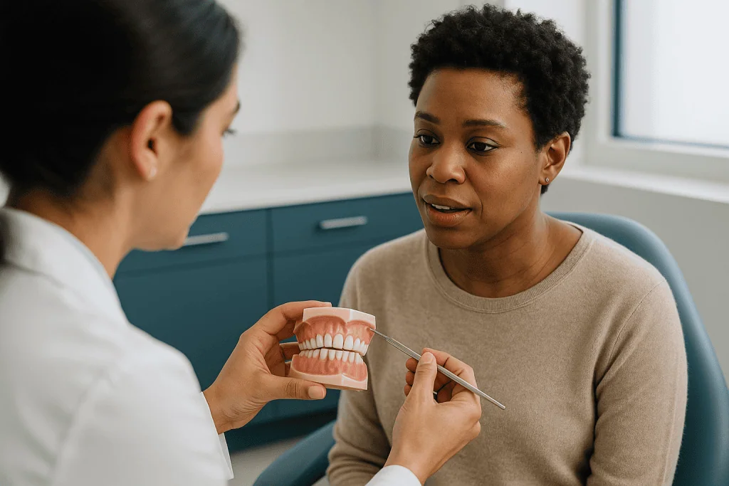
Then comes the look from the outside. The clinician studies your face from the front and the side, at rest and smiling, checking symmetry, how much tooth shows when your lips are relaxed, and whether the lips close together without strain. You may be asked to swallow, open wide, slide your jaw side to side and bite down while a finger rests on the joint just in front of each ear.
Inside the mouth, the count begins. Which teeth are present, which baby teeth remain, which adult teeth have not yet appeared, and how crowded or spaced the arches are. The clinician measures how far the upper front teeth sit ahead of the lower ones and how much they overlap vertically. Gum health and any decay are noted, because both can pause an orthodontic plan.
Records usually follow in the same appointment: photographs of the face and teeth, X-rays if they are justified, and either putty impressions or a digital scan. In children, a hand may be placed on the head to check the bite from above.
You should leave with a clear sense of what was found, what records were taken and when you will hear the conclusions. Ask if that is not offered. The consultation ends with a conversation, and in many cases a second visit to present the plan, never with a signature on the spot.
How does bite analysis work?
Occlusion is the technical word for how the upper and lower teeth meet. Bite analysis is simply the disciplined description of that meeting, in three dimensions.
Front to back, the clinician notes where the first molars sit relative to each other, a system introduced over a century ago and still in daily use. Class I means the molars meet in the textbook position, with any problem confined to crowding or spacing. Class II means the lower teeth sit further back than ideal, often with prominent upper front teeth. Class III means the lower teeth sit further forward. Cleveland Clinic notes that these classes describe position, not severity, so a Class I bite can still be badly crowded and a Class II bite can be mild.
Two measurements at the front are taken with a small ruler. Overjet is the horizontal distance the upper front teeth sit ahead of the lower ones. Overbite is the vertical overlap. When the upper teeth fail to overlap at all and a gap remains with the back teeth closed, the term is open bite. When some upper teeth bite inside the lower ones, that is a crossbite.
Side to side, the midlines of the upper and lower teeth are compared to each other and to the center of the face. A shift can point to a jaw asymmetry, a missing tooth or simply a habit.
Finally the clinician watches the bite in motion. Do the teeth slide before they settle? Does the jaw deviate on opening? Does anything click? Movement tells a story that a still photograph cannot, and it can change whether a problem is judged dental (the teeth), skeletal (the jaws) or a mix of both, which is the single most consequential distinction in the whole assessment.
What is “poor man’s cephalometric analysis”?
The phrase sounds like a joke, and it started as one among dental students, but it describes something clinicians do at almost every assessment. A cephalometric radiograph is a side-on X-ray of the skull on which angles and distances are measured to judge how the jaws relate to each other and to the face. The “poor man’s” version is the same judgement made without the film: by looking at the profile and by feeling two landmarks with the fingertips.
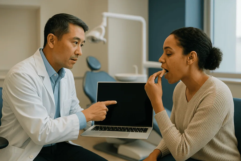
Here is the maneuver. With the head level and the eyes looking straight ahead, the clinician places one index finger at the deepest point of the curve beneath the nose, on the upper jaw, and the other at the deepest point of the curve above the chin, on the lower jaw. In an average skeletal relationship the lower finger sits a few millimeters behind the upper one. If it sits well behind, the lower jaw is likely set back (a skeletal Class II pattern). If it sits level with or ahead of the upper finger, the lower jaw is likely forward (a skeletal Class III pattern).
It is quick, free of radiation and surprisingly informative for screening. It is also imprecise. Soft tissue thickness varies from person to person, lips and chins can disguise the bone beneath, and the finger test says nothing about the vertical dimensions of the face or the angle of the front teeth. That is why it complements rather than replaces the real film in cases where the jaw relationship, growth prediction or possible surgery will decide the plan.
If you hear a clinician murmur “mild Class II skeletal” after touching your chin, this is what has just happened. It is a first estimate, and honest clinicians treat it as one.
Orthodontic x rays: which films are taken and what each shows
Not every assessment needs an X-ray, and a clinician should be able to explain why each one is being taken. MedlinePlus describes the radiation dose from dental X-rays as very low, with modern digital sensors and lead aprons or collars used to keep it lower still, but the guiding principle remains that a film is justified only when the information will change a decision.
The workhorse is the panoramic radiograph, a single sweeping image of both jaws that shows every tooth, including those still developing under the gum, the roots, the jaw joints and the sinuses. It answers questions such as: are all the adult teeth present, is any of them heading in the wrong direction, and are there extra or missing teeth?
The cephalometric film described above adds the skeletal picture and the tilt of the front teeth. Small periapical or bitewing films may be requested to check a single root or hidden decay. Cone-beam computed tomography (CBCT), a three-dimensional scan, is reserved for specific problems such as a tooth trapped in the palate or planning around an implant, because its dose is higher than the plain films.
| Record | What it shows | When it is typically used |
|---|---|---|
| Panoramic X-ray | All teeth, developing buds, roots, jaw joints | Most assessments where treatment is being considered |
| Cephalometric X-ray | Jaw relationship, facial proportions, incisor angle | Skeletal problems, growth questions, possible surgery |
| Periapical / bitewing | Detail of one or two teeth and their roots | Suspected root damage or decay |
| CBCT (3D scan) | Bone and tooth position in three dimensions | Buried or displaced teeth, complex surgical planning |
| Photographs | Face, smile, teeth in color; a baseline | Nearly all assessments |
Ask which films are planned and what question each will answer. A clear reply is a good sign.
Dental impressions for braces: putty trays, digital scans and study models
Impressions are the part most people remember, usually for the wrong reasons. The traditional method loads a horseshoe-shaped tray with alginate, a seaweed-derived putty that sets in the mouth within a couple of minutes and pulls away as a rubbery negative of the teeth. Plaster poured into that negative gives a study model: a stone replica the clinician can turn in the hand, measure and, in complex cases, cut apart to simulate tooth movement.
Gagging is the common complaint. Sitting upright, breathing slowly through the nose and focusing the eyes on a fixed point genuinely helps, and staff take these impressions many times a day. Tell them if you have a strong gag reflex; tray size, material consistency and the order of upper and lower can all be adjusted.
Increasingly, a digital intraoral scanner replaces the tray. A wand roughly the size of an electric toothbrush is moved slowly over the teeth while a camera stitches thousands of images into a three-dimensional model on a screen within minutes. There is nothing to swallow and no set time, and the file can be measured, stored and shared without a shelf of plaster. Scans and putty impressions serve the same purpose; the choice usually reflects the practice’s equipment rather than anything about your teeth.
The bite registration completes the set. You bite gently into a wafer of soft wax or silicone so the upper and lower models can be related exactly as they meet in your mouth. Without it, two perfect casts are just two casts.
Models and scans do quiet, essential work later on. They record where you started, allow space calculations to the millimeter, and give the clinician something to compare against when treatment ends and questions of stability arise.
How orthodontists grade the need for treatment
Deciding that a bite is “bad enough” to treat is not a matter of taste, at least not entirely. In the United Kingdom, the NHS uses the Index of Orthodontic Treatment Need (IOTN), a five-grade scale that ranks a malocclusion by its likely effect on dental health and, separately, by its effect on appearance. Grade 1 means essentially no need; grade 5 covers problems such as teeth that cannot erupt because they are blocked, several missing teeth, or a large overjet. The NHS explains that treatment is provided free of charge to children with grade 4 or 5 need, and to those at grade 3 with a marked appearance component, while children with milder findings and most adults fall outside that threshold.
Whatever the local system, the logic travels. Features that raise the grade are those with functional consequences: front teeth prominent enough to be at higher risk of injury, a deep bite where lower teeth strike the gum behind the upper ones, a crossbite that shifts the jaw on closing, or adult teeth with nowhere to go. Features that lower it are cosmetic and stable: a slight rotation, a small gap, mild crowding that cleans easily.
The appearance component is assessed with a standardized set of ten photographs against which a patient’s own front teeth are matched. It sounds crude, and clinicians acknowledge its limits, but it forces a consistent, evidence-based conversation about how noticeable a problem really is.
Grading also protects patients from the opposite error: treatment of a bite that would have caused no harm. MedlinePlus notes that most malocclusion is mild and requires no intervention. When a clinician says your child’s bite is “grade 2, keep an eye on it,” that is a finding, not a brush-off.
Who an orthodontic assessment is usually for, and who is asked to wait
Anyone with a concern about how their teeth meet or look can have an orthodontic assessment, and general dentists routinely refer for one. The more useful question is who is usually told to proceed and who is usually told not yet.
Children are typically ready for comprehensive treatment once most adult teeth have come through, which the NHS places at around age twelve, though it varies with each child’s dental development. Before that, an assessment may still be valuable for spotting a developing problem, but the plan is often to review rather than to start.
Adults have no upper age limit. Bone remodels around teeth throughout life, and the NHS confirms that treatment is available to adults, usually privately. What changes with age is the list of things to check first.
Several findings commonly lead a clinician to postpone or decline treatment until they are resolved:
- Active gum disease or untreated decay, because moving teeth through inflamed bone risks harm
- Oral hygiene that is not yet consistent, since brackets and wires make cleaning harder
- Adult teeth that have not finished erupting, when the plan depends on them
- Roots that are already short or damaged on the films
- Some medical circumstances, including uncontrolled diabetes or a history of certain bone-modifying medicines used for osteoporosis or cancer, which can alter how bone responds to tooth movement
- Jaw growth that is still incomplete when the plan would involve surgery
Few of these are permanent disqualifications. Most are “first do this, then we reassess” instructions, and the treating team will say which applies. A referral back to the general dentist for a filling or a gum treatment is one of the most common, and most sensible, outcomes of an assessment.
Orthodontic evaluation for kids: when is “early” too early?
Parents often arrive convinced they have either waited too long or come far too soon. Usually neither is true. The purpose of an early look is to identify the small number of problems that benefit from action while the jaws are still growing, and to reassure everyone else that watching is enough.
What the clinician is watching for in a younger child is different from the teenage checklist. Baby teeth lost too early or held too long. An upper jaw so narrow that the back teeth bite inside the lower ones. A lower jaw forced sideways to find a comfortable close. Habits such as prolonged thumb-sucking that MedlinePlus lists among the causes of malocclusion. Front teeth so prominent they are vulnerable to a playground fall. Adult teeth visible on the panoramic film but pointing the wrong way.
When one of these is found, a short course of interceptive treatment (a limited, targeted intervention during growth) may be discussed, such as a removable plate or a device to widen the upper arch. When none is found, the honest recommendation is a review in six to twelve months and a note in the chart, and the NHS position that comprehensive treatment usually waits until most adult teeth are present applies.
Preparation is mostly about language. Tell your child that someone will count their teeth, take pictures and possibly a “mouth selfie” with a small camera, and that nothing will hurt. Impressions can feel strange, so mention the putty in advance. Bring the name of any medicine your child takes and the details of any dental injury, however minor it seemed at the time.
The outcome you want from a pediatric assessment is clarity, not a start date. A child told “nothing to do yet” has had a successful visit.
After the assessment: what the following weeks usually look like
The chair is not where the thinking happens. Once you leave, the clinician sits with your films, models or scan, and photographs, and works through a problem list: skeletal pattern, tooth alignment in each arch, the bite front to back and side to side, the vertical relationship, and any teeth that are missing, extra or misplaced. Space is calculated: how many millimeters the teeth need versus how many the arch provides. That arithmetic often decides whether extractions, expansion or accepting some compromise will be discussed.
In most practices you return for a second appointment to hear the conclusions, or receive a written report through your referring dentist. Expect a summary of the diagnosis, the treatment need grade if one is used, the options considered (including doing nothing), the appliances proposed, the likely duration expressed as a range, and the risks. The NHS gives eighteen months to two years as a typical span for a full course of treatment, with retainers worn afterward; your own figure will depend on complexity and cooperation, and no one can promise it.
This gap between visits is useful. Use it to book the dental check-up or hygiene visit the assessment may have recommended, to settle any fillings, and to think about practicalities: sports, musical instruments, school or work schedules for adjustment appointments.
Consent comes at the end of this process, not the beginning. A proper consent conversation covers the alternatives, what the treatment cannot fix, what happens if you stop early, and the lifelong role of retainers. It should feel unhurried. If a child is the patient, both the child and the parent should understand what is being agreed, because a twelve-year-old who has not bought in to the plan will struggle with elastics and cleaning.
What the assessment cannot tell you
An honest assessment has edges, and knowing where they are prevents disappointment later.
It cannot tell you exactly how long treatment will take. Duration depends on how far the teeth must move, how bone responds, how growth behaves in a child, and how consistently appliances are worn. The NHS range of eighteen months to two years is a population-level typical span, not a personal forecast. A clinician who quotes a precise month is guessing.
It cannot predict growth with certainty. Two children with identical films at twelve can develop very differently by fifteen, and the cephalometric analysis gives probabilities, not guarantees. This is why some plans deliberately build in a review before committing to a second phase.
It cannot guarantee stability. Teeth have a tendency to drift back toward where they started, which is why retention is described as indefinite. No set of records at the start can promise how a bite will look a decade after appliances come off.
It cannot see inside soft tissue or measure how a jaw joint will feel under load. Clicking noted at assessment may or may not change with treatment, and the evidence on orthodontics and jaw joint pain is mixed enough that cautious clinicians avoid promising relief.
And it cannot decide for you. The same records could reasonably support treatment now, treatment later or no treatment, depending on what you value. The assessment’s job is to make sure that whichever choice you and the treating team reach is made with the facts in front of you. That is a smaller promise than “we’ll fix it,” and a far more reliable one.
What people often get wrong
“If they didn’t offer braces, they missed something.” Most malocclusion is mild and MedlinePlus is explicit that most people need no treatment. A recommendation to monitor is a clinical conclusion, not an oversight.
“X-rays at the dentist are dangerous, so refuse them.” MedlinePlus describes the radiation from dental films as very low, and clinicians justify each one. Refusing a panoramic film can mean a hidden misdirected tooth goes unnoticed until it damages a neighbor. Ask why a film is needed rather than declining reflexively.
“Straight front teeth mean a good bite.” Bite analysis looks at how the back teeth meet, how deep the overlap is and whether the jaws are aligned. A camera-ready smile can hide a crossbite or a deep bite that wears teeth over decades.
“Adults are too old to be assessed.” Bone around teeth remodels throughout life, and the NHS notes treatment is available to adults. The relevant question is gum health, not age.
“Digital scans are more accurate, so a practice using putty is behind.” Both methods produce models suitable for planning. The scanner is more comfortable and easier to store; it is not a marker of a better clinician.
“A crooked bite is a cosmetic problem.” Cleveland Clinic lists chewing difficulty, speech effects, tooth wear and gum trauma among the consequences of significant malocclusion. Appearance is one factor among several in the grading systems.
“Once the assessment is done, the plan is fixed.” Plans are revised as children grow and as teeth respond. Records are repeated at key points precisely so that changes can be made on evidence rather than habit.
Questions to ask your care team
Bring these to the second visit, or ask them at the first if the plan is being presented straight away. You are entitled to clear answers in ordinary language.
- What exactly did you find, and which parts are dental (the teeth) and which are skeletal (the jaws)?
- How would you grade the need for treatment, and on what scale?
- Which X-rays or scans did you take, and what did each one tell you?
- What would happen if we did nothing for now? Is monitoring a reasonable choice?
- What are all the options, including ones you would not pick first, and why do you prefer the one proposed?
- Will any teeth need to be removed, and what is the alternative to that?
- What is the typical range of treatment time for a case like this, and what could make it longer?
- What are the specific risks for my (or my child’s) teeth and roots, given what the films show?
- Is there anything, such as gum treatment or a filling, that needs to happen before we start?
- How will retainers work afterward, and for how long?
- If growth changes the picture, at what point would you review the plan?
- Who do I contact between appointments if something breaks or hurts?
Write down the answers. Assessments generate a lot of information in a short time, and the calmer decision is the one made at home a week later, with the notes in front of you and the treating team’s reasoning fresh in your mind. A team that welcomes these questions is showing you how it will behave for the next two years.
When to call your doctor
An orthodontic assessment itself is low risk, but it can uncover conditions that need attention sooner than a routine follow-up, and the weeks between assessment and plan are not a time to ignore symptoms. Contact your dentist, orthodontist or doctor promptly if any of the following occur.
- Swelling of the face, gums or jaw, particularly with fever, which can signal a dental infection needing urgent treatment
- Severe or worsening toothache or pain on biting that keeps you awake or does not settle
- A permanent tooth that becomes loose, or any tooth knocked out or displaced by a fall or blow, which should be seen the same day
- Bleeding gums that persist, or gums pulling away from the teeth, which suggest active gum disease
- Numbness or tingling in the lip, chin or tongue
- A jaw that locks open or shut, or joint pain that limits eating or speaking
- An allergic-type reaction after impressions, such as hives, lip swelling or breathing difficulty (breathing difficulty requires emergency care)
- In a child, a baby tooth lost early through decay or injury, or an adult tooth visibly emerging in the wrong place
Findings on X-rays that were incidental to the orthodontic question, such as a cyst-like shadow or an unexpected root problem, should also be followed up as the clinician advises rather than left until the treatment plan appointment.
None of this is intended as a checklist for diagnosing yourself. Every decision about whether, when and how to treat sits with the treating team, who have the films and the history in front of them. If something feels wrong and you are unsure whether it counts, call and ask. Dental teams would far rather answer a cautious question than treat a neglected problem.
Frequently asked questions
How long does an orthodontic assessment take?
Most first orthodontic assessments are completed in a single visit, and the practice will tell you how long to allow when you book. The examination itself is fairly brief; the record-taking, photographs, X-rays where justified, and putty impressions or a digital scan, usually fills most of the appointment. Analysis of those records happens afterward, so the treatment plan is often presented at a second visit or in a written report rather than on the day.
How much is an orthodontic assessment?
This article does not give prices, because they vary widely by country, funding system and what records are taken. In the United Kingdom, the NHS explains that assessment and treatment are free for children who meet the Index of Orthodontic Treatment Need threshold, while adults are usually treated privately. Elsewhere, ask the practice what the assessment includes, whether X-rays and scans are separate, and whether a plan presentation visit is part of it.
What disqualifies you from braces?
Few things disqualify someone permanently; most are reasons to wait. Active gum disease, untreated decay and inconsistent oral hygiene are the common ones, because moving teeth through unhealthy tissue risks harm. Adult teeth not yet erupted, roots already shortened on X-ray, uncontrolled medical conditions and a history of certain bone-modifying medicines can also change the plan. The treating team decides which applies, and often the instruction is “resolve this first, then we reassess.”
What is poor man's cephalometric analysis?
It is a clinical shorthand for estimating the jaw relationship without an X-ray. The clinician places one fingertip in the deepest curve beneath the nose (upper jaw) and another in the deepest curve above the chin (lower jaw), then compares their positions in profile. A lower finger set well back suggests a Class II skeletal pattern; level or ahead suggests Class III. It is a screening estimate, refined by a true cephalometric film when the plan depends on it.
Orthodontic consultation: what to expect if I am an adult?
Expect the same core steps as for a child: a conversation about your concerns, an examination of your face, jaws and bite, then photographs, any justified X-rays and impressions or a scan. The difference lies in emphasis. Gum health and existing dental work such as crowns or implants are examined closely, because they influence which teeth can move and how. The NHS notes treatment is available to adults, usually privately.
Which orthodontic x rays are usually needed, and are they safe?
A panoramic film showing both jaws is the most common, sometimes with a cephalometric side view when jaw relationship or growth matters. Small periapical films check individual roots, and a three-dimensional CBCT scan is reserved for specific problems such as a buried tooth. MedlinePlus describes the radiation from dental X-rays as very low, and clinicians take a film only when it will change a decision. Ask what question each one is answering.
Are dental impressions for braces painful?
They are not painful, though many people find them uncomfortable for the two minutes or so the putty takes to set, mainly because of gagging. Sitting upright, breathing through the nose and fixing your eyes on one point helps, and staff can adjust tray size and material. Many practices now use a digital scanner instead, moving a small camera wand over the teeth with nothing to swallow. Both produce models suitable for planning.
When should an orthodontic evaluation for kids happen?
The NHS notes comprehensive treatment usually starts once most adult teeth have come through, at around age twelve, though this varies with each child. An earlier look can still be useful when a dentist notices a crossbite, very prominent front teeth, early loss of baby teeth or a persistent thumb habit, because a few problems benefit from a short intervention during growth. For most children, the outcome of an early assessment is reassurance and a review date.
Does an orthodontic assessment mean I will definitely be offered braces?
No. MedlinePlus states that most malocclusion is mild and needs no treatment, and grading systems such as the NHS Index of Orthodontic Treatment Need exist partly to identify bites that are best left alone. A recommendation to monitor, or to fix a dental problem first and return later, is a common and legitimate outcome. The assessment’s purpose is an evidence-based decision, whichever direction it points.
What happens between the assessment and the treatment plan?
The clinician studies your films, models or scan and photographs away from the chair, builds a problem list and calculates how much space the teeth need versus what the arches provide. You then return for a plan presentation or receive a report through your dentist covering diagnosis, options including doing nothing, typical duration as a range, risks and retention. Consent comes at the end of that conversation, never at the first glance.
References
- NHS – Orthodontics
- MedlinePlus Medical Encyclopedia – Malocclusion of teeth
- MedlinePlus Medical Encyclopedia – Dental x-rays
- MedlinePlus Health Topic – Orthodontia
This article is for general information only and is not a substitute for professional medical advice. Please consult a qualified doctor about your individual situation.

More from the Blog
Impacted Canine vs Impacted Wisdom Tooth: Why One Is Often Saved and the Other Removed
An impacted canine is usually saved because it is a cornerstone tooth with the longest root in the mouth, guides the bite and anchors…
Do Root Canals Make You Sick? Old Myths About Treated Teeth, Corrected
No. Current evidence does not show that a properly treated root canal tooth causes cancer, heart disease, arthritis or other illnesses. The idea traces…
Do Teeth Need Straightening Before Porcelain Veneers? When Orthodontics Comes First
Not everyone needs braces before veneers, but many people benefit from straightening first. Porcelain veneers can mask mild rotations, small gaps, and uneven edges,…
Sinus Lift: Why Upper-Jaw Implants Sometimes Need It, and How It Heals
A sinus lift is a surgical procedure that adds bone between the upper jaw and the maxillary sinus so a dental implant has enough…
How 3D Imaging Changes Oral and Maxillofacial Surgery Planning Around Nerves and Sinuses
3D imaging in oral surgery planning, most often a cone beam CT scan, gives surgeons a layered view of the jaw so they can…

Oil Pulling: The Ancient Practice vs the Actual Evidence
Oil pulling, swishing edible oil in the mouth for 10 to 20 minutes, may modestly reduce plaque and mouth bacteria, according to a handful…