

How 3D Imaging Changes Oral and Maxillofacial Surgery Planning Around Nerves and Sinuses
Key Takeaways
- A cone beam CT shows whether a lower wisdom tooth root truly touches the nerve canal or only overlaps it on a flat X-ray, and on which side the canal runs.
- The NHS describes numbness after wisdom tooth removal as usually temporary but occasionally permanent, which is the main reason surgeons map the nerve canal in 3D before difficult lower extractions.
- For implants, a 3D scan gives bone width and density as well as height, letting a surgeon test a virtual fixture against the sinus floor and nerve before drilling.
- Studies show 3D imaging frequently changes the surgical approach in high-risk cases, but evidence that it lowers the overall rate of permanent nerve injury remains inconclusive.
- Professional guidance supports CBCT only when it answers a specific question the panoramic image cannot, with the smallest field of view that covers the region.
- Metal restorations and patient movement degrade a 3D scan, so the same equipment can deliver very different image quality from one person to the next.
3D imaging in oral surgery planning, most often a cone beam CT scan, gives surgeons a layered view of the jaw so they can measure exactly where a tooth root, planned implant or cyst sits relative to the mandibular nerve canal and the floor of the maxillary sinus. Evidence shows it frequently changes the surgical approach; whether it lowers nerve injury rates overall is still being studied.
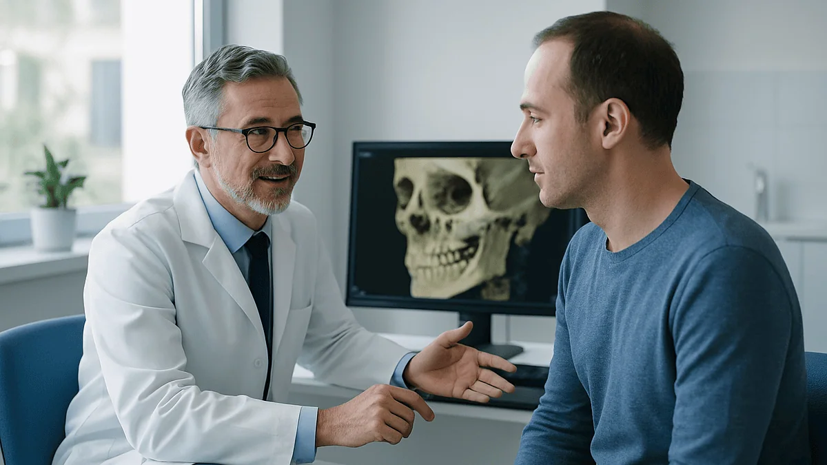
Maria had come in expecting a lecture about her wisdom teeth. Instead the surgeon swung a monitor toward her, tapped the screen, and a grey tunnel appeared, curving under the root of her lower molar like a garden hose pinned beneath a paving stone. That tunnel carried the nerve that gives feeling to her lip and chin. On the flat X-ray she had seen years earlier, it had been a faint line, easy to miss.
This is what 3D imaging oral surgery planning looks like from the chair. A scan that takes a single sweep around the head becomes a stack of thin slices, and a surgeon can scroll through them the way you flip through pages, pausing at the millimeter that matters.
Nerves and sinuses are the two structures that keep oral and maxillofacial surgeons awake at night. Both are invisible in the mouth, both sit close to teeth people need removed or replaced, and both punish guesswork. The question this article answers is how a 3D view changes the plan, and where the evidence stops.
What is 3D imaging in dentistry?
Ask a dentist what 3D imaging is and you will usually hear three letters: CBCT. Cone beam computed tomography is a type of X-ray scan in which a cone-shaped beam rotates once around the head, and a computer rebuilds the captured data into a three-dimensional model of bone, teeth and air spaces. Traditional dental radiographs, the small bitewing films and the wide panoramic view, flatten everything onto a single plane. A 3D scan keeps depth.
That difference sounds academic until you picture the anatomy. A lower molar root and the nerve canal may appear to overlap on a panoramic image simply because one sits in front of the other, the way a lamppost can seem to touch a distant building in a photograph. Slice the same region into cross-sections and the true gap, or the true contact, becomes measurable.
Medical CT scanners in hospitals work on a related principle, described in detail by the National Institute of Biomedical Imaging and Bioengineering: many X-ray projections from different angles are combined into cross-sectional images. Dental cone beam machines are smaller, are usually built into a unit the patient sits or stands in, and are tuned for the high-contrast job of separating bone, tooth and air rather than distinguishing soft tissues.
Two other technologies get folded under the 3D label. Intraoral optical scanners create a digital impression of the teeth and gums without radiation, and software can merge that surface map with the CBCT volume so the surgeon sees crowns and roots together. Neither replaces the other; they answer different questions, and for the nerves and sinuses this article is about, CBCT does the heavy lifting.
How does a cone beam CT dental scan actually work?
In plain terms, you sit or stand still while an arm circles your head once, and a computer does the rest. That is the patient experience. What happens inside is worth a paragraph, because it explains both the strengths and the limits of the scan.
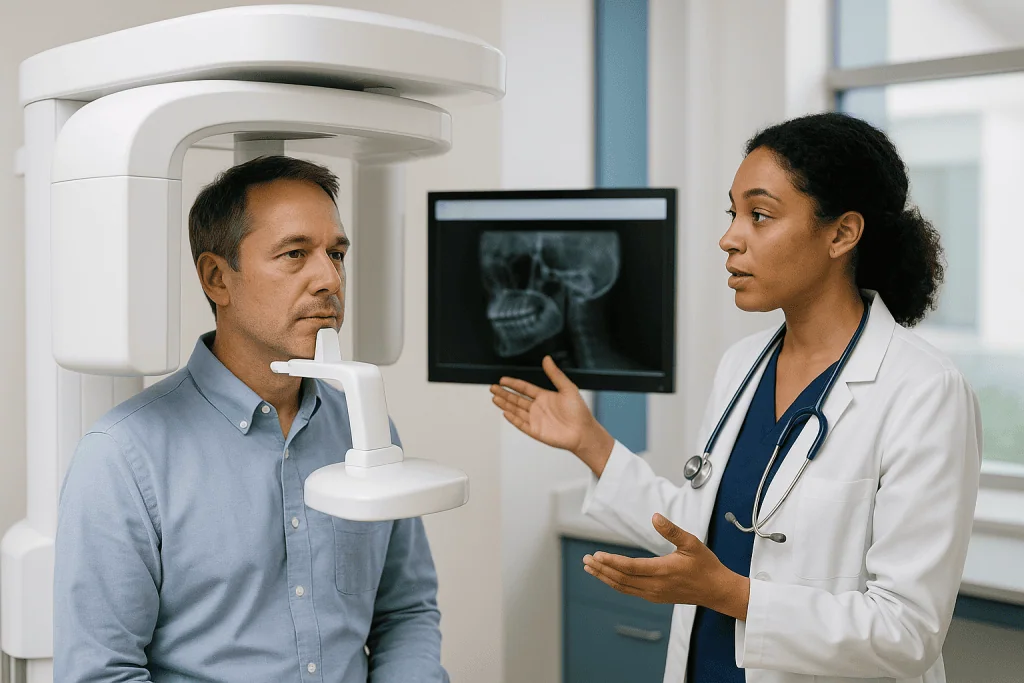
An X-ray source and a flat detector sit opposite each other on the rotating arm. As it turns, the detector captures several hundred snapshots from slightly different angles. Software then works backward from those projections to calculate how much radiation each tiny cube of tissue absorbed. Those cubes are called voxels, the 3D equivalent of pixels, and the smaller they are, the finer the detail. Dense bone absorbs a lot and appears bright; air in the sinus absorbs almost nothing and appears black; the nerve canal shows up as a dark tube because it holds soft tissue rather than bone.
Field of view is the phrase that governs much of the planning conversation. A small field might cover only a few teeth; a large one can capture both jaws, the sinuses and the base of the skull. Larger fields gather more information and more radiation exposure, so surgeons typically choose the smallest region that answers the clinical question, in line with the general dental X-ray guidance MedlinePlus describes.
Movement is the enemy. A person who swallows, shifts or talks during the rotation produces a blurred volume, which is why staff spend a moment positioning the chin and asking you to hold still. Metal fillings, crowns and implants also scatter the beam and create streaks, called artefacts, that can hide the very margin a surgeon wants to see. Reading a CBCT is a skill, not a glance at a picture.
Why 3D imaging oral surgery planning matters around the inferior alveolar nerve
The inferior alveolar nerve runs through a bony canal inside the lower jaw and exits near the chin, supplying sensation to the lower lip, chin and lower teeth on that side. When it is bruised or cut during surgery, the result is numbness, tingling or altered feeling. The NHS notes that this numbness after wisdom tooth removal is usually temporary but can occasionally be permanent, which is exactly why surgeons want to know where the canal lies before the first cut.
Here is what 3D imaging oral surgery planning adds. On a panoramic film, a surgeon sees the root and the canal in one plane and infers their relationship from indirect clues: a darkened band across the root, a break in the canal outline, a sudden narrowing. Those signs raise suspicion but cannot settle the question. A CBCT settles it. The surgeon can see whether the canal passes on the cheek side of the root, the tongue side, between two roots, or through a notch the root has grown around.
That geometry changes technique. If the canal sits on the tongue side, the surgeon may section the tooth and lift fragments toward the cheek. If the root wraps the canal, the surgeon might discuss leaving the root tip in place deliberately, an approach known as coronectomy, rather than dragging it past the nerve. The decision belongs to the treating surgeon, who weighs infection risk, the condition of the tooth and the patient’s health.
Honesty about the evidence matters. Studies consistently show that CBCT changes what surgeons decide to do in high-risk cases. Whether it reduces the overall rate of permanent nerve injury across all patients is less certain, partly because permanent injury is uncommon to begin with. The scan sharpens judgment; it does not remove risk.
How 3D imaging oral surgery planning protects the maxillary sinus
Above the upper molars and premolars sits the maxillary sinus, an air-filled cavity lined with a thin membrane. In some people a plate of bone several millimeters thick separates root tips from sinus floor. In others, the roots poke up into the sinus like tent poles under canvas, covered by little more than membrane. Nothing in the mouth reveals which pattern you have.
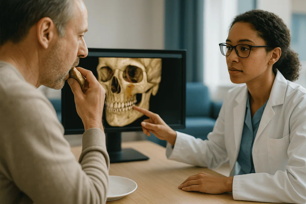
A perforation into the sinus during an extraction or implant placement is not a catastrophe, but it complicates matters: air and fluid can pass between mouth and nose, and the opening may need to be closed. Chronic sinus problems can follow if the communication persists. Surgeons would rather know in advance.
With 3D imaging oral surgery planning, the sinus floor becomes a measurable line. The surgeon can read the bone height beneath each root, check whether the membrane is thickened or the sinus is filled with fluid or a polyp, and see whether a bony wall or septum divides the cavity. Each finding shifts the plan. A thickened membrane may prompt a referral to an ear, nose and throat colleague before dental surgery. Low bone height under a planned implant may mean a shorter or angled fixture, or a sinus lift, a grafting procedure that raises the sinus floor to create room, which Johns Hopkins describes as one of the preparatory steps some implant patients need.
Implant planning is where the sinus view earns its keep. Software lets the surgeon place a virtual implant of a given length and diameter into the scan and rotate the model, checking the tip against the sinus floor from every angle. If the virtual fixture breaches the floor, the surgeon adjusts before anything touches the patient.
What does a CBCT before wisdom tooth removal show that an X-ray cannot?
Wisdom teeth are the commonest reason a healthy young adult meets an oral surgeon, and lower wisdom teeth are the commonest reason a surgeon reaches for a 3D scan. The NHS explains that removal is offered when impacted teeth cause repeated infection, decay, cysts or damage to neighbouring teeth, and that the difficulty of the procedure depends on how the tooth sits in the jaw.
A panoramic X-ray answers the first questions well: is the tooth present, which way does it tilt, is there a shadow suggesting a cyst. A CBCT before wisdom tooth removal answers the second set, the ones that shape the operation itself. How many roots are there, and do they curve? Is the root tip touching the nerve canal or separated from it by bone? Is the tooth positioned toward the cheek or the tongue? Is the bone over the crown thick or eggshell-thin? Has the tooth eroded the root of the molar in front of it?
Root curvature deserves special mention. A hooked root that curls around the canal cannot be lifted straight out without dragging across the nerve. Seeing the hook in advance lets the surgeon plan to divide the tooth and remove the pieces in a sequence that respects the anatomy.
Not every wisdom tooth needs this. Guidelines from radiology bodies consistently recommend CBCT only when the panoramic image raises a specific concern that the 3D view would resolve, such as signs of overlap between root and canal. An upper wisdom tooth well clear of the sinus, or a lower one whose roots sit far above the canal, is usually planned from the 2D image alone. Extra imaging is justified by the question it answers, not by habit.
How is a 3D scan for dental implants used to place a fixture safely?
A dental implant is a titanium post placed into the jawbone to hold a replacement tooth. Mayo Clinic describes the process as staged: the post is placed, bone grows onto it over months, and a crown is fitted once healing is complete. Every stage depends on the bone the surgeon started with, and a 3D scan for dental implants is how that bone is measured.
Three numbers dominate implant planning. Bone height tells the surgeon how long a fixture the site can accept before its tip reaches the nerve canal below or the sinus floor above. Bone width tells them whether the post will be surrounded by bone on the cheek and tongue sides or will sit exposed. Bone density, judged from how bright the bone appears, hints at how firmly the implant will grip on the day of placement. A flat X-ray gives height only, and even that is distorted by magnification.
Planning software takes this further. The surgeon loads the CBCT, often merged with a surface scan of the teeth, and positions a virtual implant so its future crown lines up with the neighbouring teeth while its body stays inside safe bone. The program displays the distance to the canal and the sinus in real time. Once satisfied, the surgeon can export the plan to a surgical guide, a custom resin template that fits over the teeth or gums with a sleeve directing the drill to the planned angle and depth.
Guides reduce the difference between plan and placement, but they do not eliminate it. Guide fit, bone quality and the surgeon’s experience all matter. Johns Hopkins and Mayo Clinic both frame implant success as depending on healthy bone, good oral hygiene and factors such as smoking, none of which a scan can change.
Who is usually offered a 3D scan, and who is usually asked to wait?
Radiation carries no benefit on its own, so the principle guiding every dental X-ray, as MedlinePlus puts it, is that the image should be taken only when it will change care. Applied to CBCT, that produces a fairly consistent pattern of who gets scanned and who does not.
Surgeons commonly request a 3D scan when a planned lower wisdom tooth extraction shows signs on the panoramic film that the roots and nerve canal may touch; when an implant is planned in a region where bone height near the sinus or nerve is uncertain; when a cyst, tumour or unusual bony lesion needs its extent mapped; when a jaw fracture or facial trauma requires assessment of displaced fragments; when corrective jaw surgery is being planned and the bones will be repositioned; and when a previous procedure left a retained root, a displaced implant or an unexplained sinus problem.
Others are usually asked to wait, or simply do not need the scan. A routine filling, a straightforward upper extraction, or a wisdom tooth clearly separated from the canal on the panoramic image can be managed without it. Children and adolescents are scanned more sparingly because developing tissues are more radiation-sensitive, and the field of view is kept small when a scan is genuinely needed. Pregnancy prompts the same caution, and non-urgent imaging is typically deferred, with the decision resting with the dentist and the person’s obstetric team.
Acute infection with swelling is a special case. A surgeon may prioritise draining the infection and managing pain, then image once the swelling has settled and the anatomy is easier to read. A person who feels a scan has been withheld can ask what the panoramic image already showed and what additional question a 3D view would answer.
2D X-ray vs 3D imaging: what each one can and cannot tell a surgeon
Neither technique is better across the board. The panoramic radiograph remains the workhorse of oral surgery because it is quick, familiar and low in radiation. The CBCT is the specialist tool that resolves specific uncertainty. Seeing their strengths side by side clarifies why a surgeon might order one, both or neither.
| Planning question | Panoramic or intraoral X-ray | Cone beam CT |
|---|---|---|
| Is the tooth present and which way does it tilt? | Answers clearly | Answers clearly |
| Do the root and nerve canal truly touch, or only overlap? | Suggests, cannot confirm | Shows the actual relationship in cross-section |
| Is the canal on the cheek or tongue side of the root? | Cannot show | Shows directly |
| How much bone lies between root tip and sinus floor? | Approximate, affected by magnification | Measurable to a fraction of a millimeter |
| How wide is the bone for an implant? | Cannot show | Shows directly |
| How large is a cyst or lesion in all directions? | Two dimensions only | Three dimensions |
| Radiation exposure | Lower | Higher, varies with field of view and settings |
| Sensitivity to patient movement | Moderate | High; blurring degrades the whole volume |
| Effect of metal fillings and crowns | Minor | Streak artefacts can obscure nearby detail |
The final two rows are the ones patients rarely hear about. A person who cannot hold still, or whose mouth is full of metalwork near the area of interest, may get less from a CBCT than the technology promises, and an experienced surgeon factors that in before ordering it.
What is virtual surgical planning and does it change outcomes?
Virtual surgical planning means rehearsing an operation on a computer model built from the patient’s own scan before performing it in the operating room. In oral and maxillofacial surgery it has moved from research curiosity to routine for complex cases, especially corrective jaw surgery, reconstruction after tumour removal and fractures involving the eye socket or cheekbone.
The workflow runs like this. The CBCT or medical CT is converted into a digital model of the skull. The surgeon, sometimes with an engineer, cuts and repositions the bones on screen, checks the new bite against a scan of the teeth, and confirms that nerve canals and sinus walls stay intact in the planned position. From that plan, a 3D printer can produce cutting guides that mark exactly where bone should be divided, and pre-bent or custom-milled plates that fit the new position. Some teams print a physical model of the jaw so the surgeon can handle it before the day.
What does the evidence say? Reviews of virtual planning in jaw surgery generally report that the final bone position matches the plan more closely than with conventional model surgery, and that time in the operating room is often reduced once a team is experienced. Evidence that these gains translate into fewer complications or better long-term function is more mixed, and studies are frequently small or single-centre.
For the reader planning a wisdom tooth or implant procedure, the relevance is proportion. Full virtual planning with printed guides is reserved for reconstruction and repositioning of whole jaw segments. A simpler version, placing a virtual implant and printing a drill guide, is the everyday application. Both rest on the same foundation: a clean, well-positioned 3D scan and a surgeon who knows how to read it.
How much radiation does a 3D dental scan involve, and is it safe?
People ask this question more than any other, and it deserves a straight answer rather than reassurance. A cone beam CT exposes you to more radiation than a single dental X-ray, and usually less than a medical CT of the same region, because it uses a narrower beam and settings tuned for bone rather than soft tissue. The exact figure depends heavily on the field of view, the resolution chosen and the machine itself, which is why responsible practices do not quote a single number for every scan.
MedlinePlus describes dental X-rays as low-dose and notes that modern equipment and protective measures keep exposure small. The same logic governs CBCT: operators select the smallest field that covers the clinical question, use the lowest resolution that still shows the detail needed, and avoid repeating scans when a recent one exists. Lead shielding of the thyroid is commonly used where it will not block the beam.
Risk from a single scan is small, and the NIBIB points out that the benefit of a CT that answers a genuine clinical question generally outweighs that risk. What professional guidance rejects is scanning by routine, for example imaging every new patient in 3D regardless of need. Children are treated with particular restraint because their tissues are more sensitive and they have more years ahead in which any effect could emerge.
A sensible approach for a patient is to ask two things: what specific question the scan will answer, and whether a recent scan from another clinician could be used instead. Digital volumes can be shared between practices, and a surgeon can often plan from an existing scan if its field and quality suit the purpose. That single question avoids more unnecessary radiation than any lead apron.
What happens on scan day and in the days and weeks after surgery?
Scan day is short. You remove earrings, glasses, removable dentures and hairpins, because metal near the head creates streaks. Staff position your chin on a rest, ask you to bite lightly on a marker or close your lips gently, and step behind a screen. The arm circles once. You hold still, then it is over. Some machines ask you to stand, others to sit; neither is better. Results are usually reviewed with you at the planning appointment rather than on the spot, because the surgeon needs time to scroll through the volume and take measurements.
Surgery follows its own timeline, and the scan does not change the biology of healing. After wisdom tooth removal, the NHS advises that swelling, bruising and jaw stiffness are expected and that it can take up to two weeks to recover fully, with the first few days the most uncomfortable. Mild numbness of the lip or tongue that resolves over days to weeks is described as common; longer-lasting altered sensation is the outcome the nerve mapping was intended to reduce, and any persisting numbness should be reported at follow-up.
Implant timelines run in months, not days. Mayo Clinic explains that after the post is placed, bone must grow onto its surface before a crown can be fitted, a process that may take several months and varies with bone quality, the site and the person’s health. Where a sinus lift was needed, the graft may need its own healing period first.
Follow-up appointments matter more than most people expect. A surgeon will check the wound, confirm that sensation is returning where it was affected, and, for implants, may take a further image to confirm the fixture sits where the plan intended.
What people often get wrong about 3D imaging in oral surgery
The first misunderstanding is that a 3D scan guarantees a nerve will not be damaged. It does not. Imaging shows position; it cannot make a root that wraps around the canal let go of it. What the scan does is let the surgeon choose a technique suited to that position, and discuss with you in advance the option of leaving a root tip in place when full removal would mean stretching the nerve. Risk falls; it does not vanish.
A second myth runs the other way: that 3D scans are radiation-heavy and best avoided. In context, a small-field CBCT ordered for a specific question sits well within the range of medical imaging people accept without hesitation, and skipping a justified scan can mean operating on anatomy the surgeon has to guess at. The problem is unjustified scanning, not the technology.
Third, people assume the scan shows the nerve itself. It shows the bony canal that houses the nerve, which appears as a dark tube. In some jaws the canal wall is thin or incomplete and the tube fades in and out, so the surgeon reads the course from the segments that are visible. Soft tissue detail is not CBCT’s strength; that is why a medical MRI, which images soft tissue well, is sometimes chosen for tumours or joint problems instead.
Fourth, a practice advertising 3D imaging is not thereby a safer practice. The scanner is a tool; interpretation and surgical skill determine outcomes, and no professional body ranks practices by their equipment list.
Finally, an implant placed with a printed guide is not immune to failure. Bone quality, gum health, smoking and general health still decide whether the fixture integrates, as Johns Hopkins and Mayo Clinic both make clear.
Questions to ask your care team before the scan and before surgery
A planning appointment goes better when you arrive with questions rather than trying to remember them afterwards. These are the ones surgeons say they wish patients asked more often, grouped by stage. None of them has a right answer; the value is in hearing your surgeon’s reasoning.
- What did my panoramic X-ray already show, and what specific question will the 3D scan answer that it could not?
- Do I have a recent scan from another dentist or hospital that could be used instead of a new one?
- How close is the nerve canal to the roots of the tooth you plan to remove, and on which side does it run?
- If the root wraps the nerve, would you consider leaving the root tip in place, and what are the trade-offs of that approach for me?
- How much bone lies between my planned implant site and the sinus floor or nerve, and does that change the length or angle of the implant you would choose?
- Will a surgical guide be used, and what happens if the guide does not fit well on the day?
- What signs of nerve or sinus involvement should I watch for after surgery, and at what point would you want to see me again?
- Roughly how long should I expect swelling and numbness to last based on my anatomy, and when would numbness become a concern?
- Are there alternatives to this procedure, including monitoring rather than treating, and what does the scan tell you about them?
Write the answers down, or ask whether the practice can send a summary. Complex anatomy is easier to accept when you have seen it on screen and heard, in plain language, how it shapes the plan. A good team will welcome the conversation; the decision about how to proceed rests with them and with you.
When to call your doctor after oral surgery planned with 3D imaging
Careful planning lowers the odds of trouble but does not remove the need to watch for it. Most recovery from wisdom tooth removal or implant placement is uneventful, and the NHS lists swelling, bruising, stiffness and mild discomfort as expected in the first days. Some symptoms sit outside that expected range and warrant a call the same day.
Contact your surgeon or dental team promptly if you notice any of the following:
- Numbness, tingling or a burning sensation in the lip, chin or tongue that has not begun to improve after the first few days, or that appears for the first time after initially normal sensation.
- Air, fluid or food passing between your mouth and nose, a nasal-sounding voice you did not have before, or bubbling at the wound when you breathe out through your nose, which can signal a communication with the sinus.
- Nosebleeds or one-sided nasal discharge after upper jaw surgery.
- Swelling that keeps increasing after the third day, or that spreads toward the eye, the neck or the floor of the mouth.
- Difficulty swallowing, difficulty breathing or trouble opening your mouth that is getting worse rather than better.
- Fever, a foul taste or pus from the wound.
- Bleeding that does not stop with firm pressure on a clean gauze pad for the time your team advised.
- Pain that escalates after an initial improvement, particularly a throbbing ache radiating to the ear several days after a lower extraction.
Seek emergency care immediately, rather than waiting for a callback, if swelling threatens your airway, you cannot swallow your own saliva, you develop a high fever with rapidly spreading facial swelling, or you feel faint from bleeding. These are rare, but they are the situations in which minutes matter.
For anything less urgent that still worries you, calling is always the right choice. Surgeons would far rather hear about a symptom that turns out to be normal healing than learn late about one that was not.
Frequently asked questions
What is 3D imaging in dentistry and how is it different from a normal dental X-ray?
3D imaging in dentistry usually means cone beam computed tomography, a scan in which an X-ray arm rotates once around your head and software builds a three-dimensional volume of bone, teeth and air spaces. A normal dental X-ray flattens everything onto one plane, so overlapping structures can appear to touch. The 3D volume can be sliced in any direction, letting a surgeon measure real distances.
Do I need a cone beam CT dental scan before every extraction?
No. Most extractions are planned from a panoramic or small intraoral X-ray. Surgeons typically order a cone beam CT when the 2D image raises a specific concern, such as a lower wisdom tooth whose roots appear to overlap the nerve canal, an upper root close to the sinus floor, or an unusual lesion. If a scan is proposed, it is reasonable to ask what question it will answer.
Can a 3D scan for dental implants tell whether I have enough bone?
It can measure the height, width and approximate density of bone at the planned site, which are the main factors a surgeon uses to judge whether an implant will fit safely between the sinus floor above or the nerve canal below. If bone is short, the scan helps the surgeon consider a shorter or angled implant, or a grafting procedure. Healing and integration still depend on your general and oral health.
Is a CBCT before wisdom tooth removal painful or long?
The scan itself is painless and quick. You remove metal items from your head and neck, rest your chin on a support, and stay still while the arm makes a single rotation around you. There is no injection and no contrast dye. The main requirement is not moving, since even a small shift blurs the whole volume. Results are usually discussed at your planning appointment rather than immediately.
What is the most difficult oral surgery?
There is no official ranking, because difficulty depends on the individual’s anatomy rather than the procedure name. Among common operations, deeply impacted lower wisdom teeth with roots wrapped around the nerve canal are widely regarded as technically demanding, as are implants placed into thin bone beneath the sinus. Reconstruction after tumour removal and repositioning of both jaws are more complex still, which is why virtual planning is routine for them.
Does 3D imaging guarantee my nerve will not be damaged?
No. Imaging shows the position of the nerve canal relative to the roots; it cannot change that position. What it does is let the surgeon choose a technique suited to the anatomy, and discuss options such as leaving a root tip in place when full removal would risk the nerve. Evidence shows the scan changes decisions in high-risk cases, while its effect on overall injury rates is still being studied.
Is a 3D dental scan covered by insurance, and how much does it cost?
Coverage varies widely between insurers, plans and countries, and depends on whether the scan is judged medically necessary for planning surgery rather than routine screening. This magazine does not publish prices, because they differ by region and setting and change over time. The administrative team at the practice or hospital can tell you what your plan covers and what any personal contribution would be before the scan is booked.
How much radiation does a cone beam CT use compared with a medical CT?
A dental cone beam CT usually delivers less radiation than a medical CT of the same region, because it uses a narrower beam and settings tuned for bone, though it delivers more than a single dental X-ray. The dose depends heavily on the field of view and resolution chosen. Operators keep exposure low by selecting the smallest field that answers the clinical question and avoiding repeat scans.
Can a scan from another dentist be used for my oral surgery planning?
Often, yes. Cone beam volumes are stored digitally and can be transferred between practices. A surgeon will check whether the existing scan covers the region of interest, whether its resolution is adequate, and whether anything has changed since it was taken, such as new decay or a cyst that has grown. Reusing a suitable scan avoids unnecessary radiation, so it is worth mentioning any recent imaging.
What does a sinus perforation during oral surgery mean for me?
A perforation is an opening between the mouth and the maxillary sinus created during an upper extraction or implant placement. Small openings often close on their own with care; larger ones may need to be closed surgically to prevent air, fluid and infection passing between the spaces. Mapping the sinus floor in 3D beforehand helps the surgeon anticipate thin bone, but it cannot eliminate the possibility entirely.
References
- MedlinePlus: Dental X-rays
- NHS: Wisdom tooth removal
- NIH National Institute of Biomedical Imaging and Bioengineering: Computed Tomography (CT)
This article is for general information only and is not a substitute for professional medical advice. Please consult a qualified doctor about your individual situation.

More from the Blog
Impacted Canine vs Impacted Wisdom Tooth: Why One Is Often Saved and the Other Removed
An impacted canine is usually saved because it is a cornerstone tooth with the longest root in the mouth, guides the bite and anchors…

Do Root Canals Make You Sick? Old Myths About Treated Teeth, Corrected
No. Current evidence does not show that a properly treated root canal tooth causes cancer, heart disease, arthritis or other illnesses. The idea traces…

Do Teeth Need Straightening Before Porcelain Veneers? When Orthodontics Comes First
Not everyone needs braces before veneers, but many people benefit from straightening first. Porcelain veneers can mask mild rotations, small gaps, and uneven edges,…
Sinus Lift: Why Upper-Jaw Implants Sometimes Need It, and How It Heals
A sinus lift is a surgical procedure that adds bone between the upper jaw and the maxillary sinus so a dental implant has enough…

Oil Pulling: The Ancient Practice vs the Actual Evidence
Oil pulling, swishing edible oil in the mouth for 10 to 20 minutes, may modestly reduce plaque and mouth bacteria, according to a handful…

How Laminate Veneers Are Placed: Impressions, Minimal Preparation and Bonding the Shells
A laminate veneers procedure usually unfolds over two or three dental visits spread across a few weeks. The dentist first assesses the teeth and…