

Plates, Screws, Rods and Pins: What Internal Fixation Actually Does for a Healing Fracture

Key Takeaways
- Internal fixation holds a fracture still so the body can heal it; the plate or rod does not create new bone itself.
- A rod runs down the hollow center of a long bone and often allows earlier weight-bearing than a plate laid on the outer surface.
- Many fractures heal enough for routine use in roughly 6–8 weeks, but muscle strength takes months to return after the bone is solid.
- In adults, plates and screws are usually left in permanently and removed only when they cause irritation, infection or mechanical problems.
- Growth plate fractures in children are graded by whether they cross into the joint or crush the plate, and are monitored for months for growth disturbance.
- Smoking, poorly controlled blood sugar and disrupted blood supply are the biggest modifiable threats to a fracture healing around hardware.
Plates, screws, rods and pins are internal fixation devices that hold the pieces of a broken bone in the correct position and keep them still so the bone can knit back together. The metal does not heal the bone; it stabilizes the fracture while the body does the repair. Most hardware is left in place permanently unless it causes problems, and the decision to use it rests with the treating surgeon.
The X-ray goes up on the screen and there it is: a clean white line through the shin bone, with the two halves sitting a few millimeters out of true. The surgeon traces it with a fingertip and says the phrase that most people hear for the first time in an emergency department: plates and screws. The patient nods, then asks the question everyone asks. Will that metal be in my leg forever?
Plates and screws fracture surgery is one of the most common operations in orthopedics, and one of the least understood by the people who have it. The hardware looks dramatic on a scan, so it is easy to assume it is doing the healing. It is not. Its job is quieter than that: to act as an internal splint that keeps bone ends exactly where they need to be while cells do the slow work of rebuilding.
This explainer walks through what the metal does, who usually needs it, what the first weeks feel like, and the honest downsides nobody mentions in the waiting room.
What does plates and screws fracture surgery actually do?
Start with the word itself. Internal fixation means placing metal inside the body to hold a broken bone steady; it is called internal to distinguish it from a cast or a frame worn on the outside. The formal name for the most common version is open reduction and internal fixation, usually shortened to ORIF. Open reduction means the surgeon makes an incision and lines the bone pieces up by hand and by sight, rather than through the skin.
Think of a fracture as a snapped broom handle. If you tape the two halves together tightly and leave them alone, wood glue sets and holds. Bone works on the same principle, except the glue is living tissue that the body produces itself. The plate is the tape. It spans the break, screws anchor it to healthy bone on either side, and the whole arrangement stops the ends from shifting, rotating or pulling apart every time the leg bears weight or the arm swings.
Why does stillness matter so much? Because a fracture heals through a bridge of new tissue, and that bridge is fragile for weeks. Each small movement at the break tears the first strands of repair, delays the next stage and can shift the bone into a position that will not function well later. According to Cleveland Clinic, many fractures take roughly 6–8 weeks to heal, and complex breaks take longer; the hardware is there to protect that window.
Two further points are worth fixing in mind. First, the metal carries load only until the bone can carry it again; once healing is complete the implant is mostly along for the ride. Second, ORIF is a means to an end. The goal is a bone that is straight, the right length, correctly rotated, and strong enough to move a joint through its full range. Where a cast can achieve that, a cast is used. Where it cannot, metal steps in.
Plates, screws, rods and pins: which device is used for which break?
Surgeons choose hardware the way a carpenter chooses fasteners: by the shape of the pieces and the forces acting on them. A quick tour of the four main tools helps the X-ray make sense.
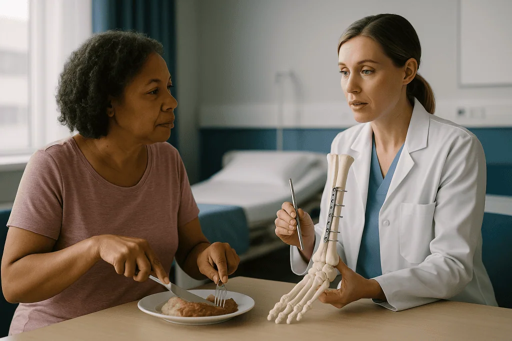
A plate is a flat strip of metal, usually titanium or stainless steel, laid along the outside of the bone and fastened with screws. Plates suit fractures near joints and in bones such as the forearm, where exact alignment matters for rotation. A screw on its own can pull two fragments together, compressing them so they knit; a lag screw across a slanted break is a classic example. A rod, also called an intramedullary nail, is a long metal tube passed down the hollow center of a long bone such as the femur or tibia and locked in place with screws at each end; it works like a dowel inside a broken table leg and lets the bone share load early. A pin, often a thin wire, is a temporary hold used mainly in small bones of the hand, in wrist fractures and in children, where the surgeon wants minimal disturbance to growing bone.
| Device | Typical location | How it holds | Usually permanent? |
|---|---|---|---|
| Plate and screws | Wrist, forearm, ankle, collarbone, around joints | Bridges the break from the outside surface of the bone | Often, unless symptomatic |
| Lone screws | Oblique breaks, small fragments, some ankle and hip fractures | Compress fragments together | Often |
| Rod (intramedullary nail) | Femur and tibia shafts | Runs inside the bone canal, locked at both ends | Often |
| Pins and wires | Hand, wrist, children’s elbows | Skewer fragments in place | Usually removed in weeks |
The pattern in the last column explains a lot of confusion. Pins are routinely taken out once the bone is stable. Plates, screws and rods are usually left alone, a topic covered in its own section below. Johns Hopkins Medicine describes these broad categories; the specific choice for any one fracture belongs to the surgeon looking at the actual X-ray.
How a broken bone heals, and why movement at the break is the enemy
Bone is the only tissue in the adult body that heals without a scar. It rebuilds itself as bone, which is remarkable, but the process is choreographed in stages and each stage needs the stage before it to have gone well.
In the first hours a blood clot forms between the fragments. Over the next days inflammatory cells clear debris and release signals that summon repair cells. Within a week or two a soft callus appears: a cuff of cartilage-like tissue around the break that is rubbery rather than rigid. Callus is the medical word for this temporary bridge. Over the following weeks mineral is laid into the callus and it hardens into woven bone, visible on X-ray as a cloudy band. Months later, remodeling slowly replaces that rough patch with organized, strong bone shaped to the loads it carries.
Movement interferes at every step. Too much shearing at the fracture prevents blood vessels from growing across it, and without blood supply the callus starves. Repeated micromotion can leave the body stuck producing cartilage that never mineralizes, a state called nonunion, which means the fracture has stopped progressing toward healing. Misalignment produces malunion: healing in the wrong position, so a wrist cannot rotate fully or a leg carries weight unevenly.
Here is the subtle part that makes fixation an art rather than a formula. When a plate squeezes two fragments together with no gap at all, bone can heal directly across the line with almost no callus, a quiet process sometimes called primary healing. When a rod holds a shaft steady but allows tiny amounts of compression, the body builds a generous callus around it. Both routes work. The surgeon’s job is to choose a device that produces the right amount of stability for that bone, that break and that patient, as MedlinePlus outlines in its overview of fracture care.
Who is usually offered plates and screws fracture surgery, and who is asked to wait?
The most common misunderstanding about fracture surgery is that worse breaks get operations and milder ones get casts. Severity matters, but the real question is whether the bone will stay in an acceptable position on its own.
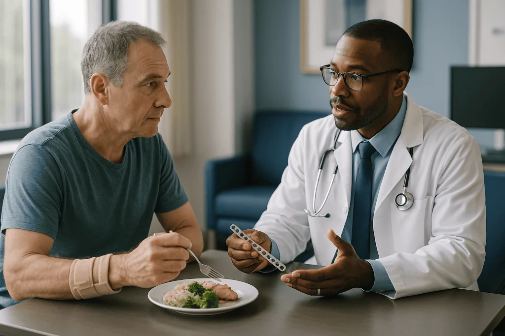
Surgery is usually considered when a fracture is displaced, meaning the pieces have moved out of line and cannot be held straight by a cast; when the break runs into a joint surface, where even a small step can wear cartilage for decades; when the fracture is unstable and keeps slipping after a reduction; when the bone has broken into several pieces; when the skin is torn and the bone exposed, an open fracture that carries infection risk; or when a cast would keep someone in bed for so long that the stillness itself becomes dangerous. Hip fractures in older adults are the clearest example of that last point. NHS guidance notes that surgery is usually carried out within 48 hours of admission because prolonged immobility raises the risk of chest infection, pressure sores and blood clots.
Who is asked to wait, or steered away from surgery altogether? People whose fracture sits in a good position and is likely to stay there. People with soft tissue so swollen or bruised that an incision would be at high risk of breaking down; surgeons often wait several days for swelling to settle before operating on badly swollen ankles and heels. People whose heart, lung or kidney health makes anesthesia a greater risk than a slower nonoperative recovery. And children, whose bones remodel so vigorously that angles adults would never accept can straighten on their own within months.
Age, bone density, smoking status, diabetes control and what a person needs the limb to do all feed into the decision. A displaced wrist fracture in a concert pianist and the same fracture in someone who is bedbound may be treated quite differently, and both choices can be right. The treating team weighs these factors with the patient; no checklist replaces that conversation.
What happens during ORIF surgery, step by step
Most people are surprised by how structured the operation is. Here is the usual sequence in plain terms, with the caveat that details vary by bone and by surgeon.
Before the incision, anesthesia is chosen: a general anesthetic, where the patient is asleep, or a regional block that numbs the whole limb, sometimes combined with sedation. Antibiotic is given through a drip shortly before the first cut to reduce infection risk, a standard step in bone surgery. The limb is cleaned and a tourniquet may be inflated on an arm or leg to keep the field clear of blood.
The surgeon then makes an incision over the fracture, moving muscles and tendons aside rather than cutting through them where possible. The fragments are cleaned of clot and gently maneuvered back into alignment under direct vision, often confirmed with a live X-ray machine in the room. Temporary clamps or wires hold the position. The plate is shaped to the bone’s contour, laid across the break, and screws are drilled and placed through holes in the plate into solid bone on both sides. For a rod, the approach is different: a small incision is made near the end of the bone, the hollow canal is prepared, the nail is passed down inside, and locking screws are inserted through the skin at each end.
Final X-rays check alignment, length and rotation. The wound is closed in layers and dressed, and the limb may be placed in a splint for comfort. Operations range from under an hour for a simple wrist to several hours for a shattered joint.
Minimally invasive plating, sometimes called percutaneous or bridging plating, slides the plate under the skin through small incisions away from the break so the fracture’s blood supply is disturbed as little as possible. It is one technique among several, not a superior product, and the choice depends on the fracture pattern. Cleveland Clinic’s overview of fracture treatment describes these broad surgical approaches.
ORIF surgery recovery time: what the first days and weeks usually look like
Nobody warns you that the first thing you feel after fracture surgery is often relief. The grinding instability of a loose break is gone, replaced by the duller ache of a surgical wound. That ache peaks in the first two or three days and then recedes, though swelling can linger for weeks.
Day one is about elevation, circulation and getting out of bed. Keeping the limb above heart level reduces swelling; moving the fingers or toes keeps blood flowing; and for leg and hip fractures, physiotherapists usually have patients standing with a frame within a day. NHS hip fracture guidance describes getting up the day after surgery as a normal expectation, not an ambitious one.
The first two weeks revolve around the wound. Stitches or staples come out at roughly 10–14 days in many protocols, and this is when the surgical team checks for redness, discharge or gaping. A splint may be swapped for a removable boot or brace. Gentle movement of the joints above and below the fracture usually begins now, because stiffness, not weakness, is the complaint that follows people for longest.
Weeks two to six are the plateau that tests patience. The bone is doing invisible work. X-rays at around six weeks look for callus and confirm nothing has shifted. Cleveland Clinic gives a typical range of 6–8 weeks for many fractures to heal enough for routine use, while NHS guidance on a broken leg notes that severe leg fractures can take considerably longer and that returning to sport may take several months.
Beyond that, strength rebuilds over months. Muscle wastes quickly in a resting limb and returns slowly. People commonly notice that the fracture site feels fine before the surrounding muscles stop aching after exertion. These ranges are typical, not guarantees; the surgeon will set your timeline from your own X-rays.
Weight-bearing, physiotherapy and the moment the bone takes over
The single most important instruction you will receive after plates and screws fracture surgery is how much weight to put through the limb, and it is the instruction most often misremembered.
The vocabulary is precise. Non-weight-bearing means the foot does not touch the floor; the limb is held clear using crutches or a frame. Touch-down or toe-touch weight-bearing means the foot may rest on the ground for balance but carries no load, roughly the pressure you would use to avoid crushing an egg. Partial weight-bearing allows a stated fraction, often judged by standing with the affected foot on a bathroom scale. Full weight-bearing means no restriction. Which category you fall into depends on the bone, the device and how well the screws grip. A rod in a femur often allows early weight because it shares load along the bone’s center; a plate on a small, crushed ankle may not, because screws in thin bone can loosen under load before healing is advanced.
Physiotherapy begins earlier than most people expect. In the first phase it is about swelling control and keeping neighboring joints mobile: a fractured wrist stiffens the fingers and shoulder if they are left alone. The second phase, once X-rays show healing, adds strength and balance. The third phase, often months in, rebuilds the specific movements a person needs, from kneeling to pushing off for a run.
Why not simply rest until everything is solid? Because bone is adaptive. Controlled load through a stable fracture stimulates mineralization and strengthens the new bone along the lines of stress; prolonged unloading does the opposite and leaves the limb weaker and the surrounding bone thinner. The balance between protection and stimulation is exactly what the fixation makes possible, and it is why the surgeon’s weight-bearing plan is tailored to the X-ray rather than to a calendar. MedlinePlus summarizes this rehabilitation sequence in general terms; your own plan supersedes it.
Disadvantages of fracture plating: the risks nobody mentions in the waiting room
Every fracture clinic has a patient who assumed surgery was the safe option and a cast the risky one. Both carry risks; they are simply different risks, and a fair conversation lays them side by side.
Infection is the complication surgeons think about most. Any incision can become infected, but infection around metal is harder to clear because bacteria form a film on the implant surface that antibiotics penetrate poorly. Deep infection may require further surgery to wash out the wound or remove the hardware. Open fractures, diabetes and smoking raise the risk considerably.
The plate itself can cause trouble. Where bone sits close under the skin, as at the collarbone, the outer ankle and the wrist, a plate may be felt or seen, catch on tendons or rub when kneeling. Screws placed near a joint can irritate soft tissue; screws that are too long can abrade tendons on the far side of the bone. Hardware can also loosen or break if the bone fails to heal, because metal fatigues under repeated load that it was never meant to carry indefinitely.
There are deeper biological costs. Opening a fracture strips some of the blood supply from the bone surface and disturbs the fracture hematoma, the early clot that seeds healing. That is one reason nonunion rates are not zero even with perfect fixation. Nerve branches run close to many surgical approaches, and numbness over or beyond a scar is common and sometimes permanent. Stiffness in the nearest joint is frequent after surgery around the elbow, wrist and ankle.
Then there are the risks of any operation: bleeding, anesthetic reactions and blood clots in the leg veins, the latter a particular concern after lower limb and hip surgery. Johns Hopkins Medicine lists these broad complication categories. None of this argues against surgery; it argues for choosing it when the alternative is worse, which is the judgment the treating team is making.
Do plates and screws need to be removed, or do they stay for life?
Here is the answer most people do not expect: in adults, plates, screws and rods are usually left in for life. They are made of titanium or surgical stainless steel, which are inert in body tissue, do not corrode meaningfully and do not need replacing. Modern implants do not set off most airport metal detectors, and they are compatible with MRI scanners, though you should always tell the radiology team you have hardware so they can adjust the scan.
Removal is a second operation with its own risks: another anesthetic, another wound, another infection chance, and a short period when the bone, having healed around and under the plate, is briefly weaker where the screw holes remain empty. Those holes fill in over weeks, but a fall in that window can refracture the bone through them. Removing a well-healed plate from a deep bone such as the femur can be more difficult than putting it in, because bone grows over the edges and screw heads can strip.
So when is removal reasonable? When the hardware is causing a problem rather than merely existing. Common reasons include a plate that is prominent and painful under thin skin, screws irritating a tendon or joint, confirmed infection, hardware that has broken or loosened, or a clear preference in a young person whose surgeon agrees the bone is fully healed and the benefits outweigh the risks. In children, pins and wires are routinely removed within weeks, and plates are more often removed because growing bone can envelop them and because decades of life remain.
The decision is individual. Some surgeons offer elective removal of ankle or collarbone plates once healing is complete; others advise leaving well alone unless symptoms demand action. The NHS notes that metalwork is often left permanently and removed only if it causes problems, and that framing is the mainstream position across guidelines. Ask what your surgeon’s usual practice is and why.
How serious is a growth plate fracture in a child?
Parents of a child with a broken wrist often hear two words that frighten them more than the fracture: growth plate. A growth plate is a band of soft cartilage near each end of a child’s long bones where new bone is produced, allowing the bone to lengthen; it is the weakest part of the growing skeleton and hardens into solid bone when growth finishes. Mayo Clinic notes that growth plates typically close in the mid-teens, somewhat earlier in girls than boys, and that these injuries are common, making up a meaningful share of childhood fractures.
How serious a growth plate fracture is depends mostly on the pattern. Surgeons grade them by whether the break runs along the plate, through it into the bone shaft, through it into the joint, or crushes the plate itself. Fractures that run along or just above the plate usually heal uneventfully and rarely affect growth. Those that cross the plate into the joint surface need exact alignment, because a step in the cartilage can trouble a joint for life, and these are the ones most often fixed with pins or small screws. Crush injuries to the plate are the rarest and the most concerning, because damaged cartilage can stop producing bone on one side, leading to a limb that grows crooked or short.
Fixation in children has a particular logic. Surgeons aim to hold fragments without driving hardware across the growth plate itself; when a device must cross it, smooth pins are preferred over threaded screws, and they are removed early. Children also heal fast, so casts are kept shorter and surgery is reserved for displaced or joint-involving breaks.
The honest part is the follow-up. Growth disturbance may not show for months, so children with growth plate injuries are usually seen in clinic at intervals, sometimes for a year or more, with X-rays comparing the injured and uninjured sides. Most grow normally; the monitoring exists to catch the exceptions early, when corrective options are widest. Mayo Clinic’s page above describes this surveillance in detail.
Casts, braces and external frames: the alternatives to internal fixation
Metal inside the body is not the default; it is one option on a spectrum, and understanding the others clarifies why a surgeon chooses it.
A cast is a rigid shell of plaster or fiberglass molded around the limb. It holds a fracture that is already in acceptable position and resists bending, but it cannot stop fragments sliding past one another inside the bone, and it immobilizes the joints above and below, which is why casted joints stiffen. A functional brace is a removable version that permits some joint motion while still supporting the bone, often used for the upper arm and for later-stage healing.
Traction, once common for leg fractures, uses weights to pull the limb into alignment and was largely abandoned for adults because it required weeks in bed. It survives mainly as a short-term measure before surgery and in some pediatric settings.
External fixation is the halfway house. Pins are drilled into bone above and below the break and connected to a rigid bar or ring frame outside the skin. It can be applied quickly, disturbs the fracture zone very little and allows wound care to continue around it, so it is the usual first step for badly contaminated open fractures, for limbs too swollen to cut into safely, and for severely injured patients who cannot tolerate a long operation. Pin sites need daily cleaning and can become infected, and frames are cumbersome, so many are converted to internal fixation once the soft tissues recover.
Then there is the option of doing less. Many fractures of the collarbone, the upper arm, the fifth metatarsal and the fingers heal perfectly well with a sling, a boot or buddy taping, and surgery adds risk without adding benefit. NHS guidance on a broken arm or wrist describes this range of approaches, from a simple cast to operative fixation. A good surgeon can explain not just why surgery is being recommended but why each alternative is less suitable for this particular break.
Smoking, blood sugar and nutrition: what you control while the metal does its job
Once the hardware is in, the surgeon’s part is largely done. What happens over the next three months depends heavily on the biology of the person wearing the plate, and some of that biology can be influenced.
Smoking is the factor with the strongest evidence. Nicotine narrows small blood vessels, and carbon monoxide reduces the oxygen that blood can carry, starving the fracture callus at exactly the moment it needs the most supply. Smokers heal more slowly on average, have higher rates of nonunion and wound breakdown, and more infections around implants. Orthopedic teams increasingly ask patients to stop or pause smoking before and after fixation; the same applies to vaping nicotine. Support for quitting is available through most health systems, and the surgical team can point you to it.
Diabetes matters for similar reasons. Persistently high blood sugar impairs the white blood cells that clear bacteria from a wound, damages small vessels and slows collagen formation. Good glucose control around surgery is part of the plan, and anyone with diabetes should expect their team to ask about recent readings.
Nutrition is less glamorous but real. Bone repair needs protein, calcium and vitamin D, and people recovering from injury often eat less than usual just as demand rises. The NIH Office of Dietary Supplements summarizes calcium’s role in bone and the dietary sources that supply it. Whether supplements are appropriate is a question for the treating team, especially since some medicines and conditions change what is safe.
Finally, certain anti-inflammatory painkillers have been studied for a possible slowing effect on bone healing, with mixed and mostly low-quality evidence. Many surgeons prefer to limit their use after fracture surgery; others consider the evidence too weak to change practice. Follow the specific advice given at discharge rather than a general rule, and never stop or start a medicine on the strength of an article.
What people often get wrong about plates and screws
Fracture clinics hear the same misconceptions week after week. Clearing them up makes recovery easier and the follow-up conversations shorter.
The metal heals the bone. It does not. The plate holds position; the body produces the new bone. A fracture that is not healing will not heal faster because the hardware is strong, and sometimes the hardware fails precisely because the bone has not taken over its share of the load.
Once the surgery is done, the bone is fixed. Fixed in position, yes. Healed, no. For the first weeks the plate alone is bearing forces it was never designed to carry indefinitely. The weight-bearing restrictions after surgery are about protecting the screws until the bone can help them, not about protecting the incision.
Metal in the body sets off airport scanners and rules out MRI. Modern titanium and stainless implants are nonmagnetic or weakly magnetic, rarely trigger walk-through detectors, and are compatible with standard MRI, though you should tell staff about them. There is no card you need to carry, though some patients choose to keep a note of what was implanted.
Plates must come out eventually. In adults they usually stay unless they cause a problem. Removal is a separate decision with its own risks.
Plates make the bone stronger than before. The healed bone is strong, but the plated segment is stiffer than natural bone, and the transition zone at the plate’s end is a place where stress concentrates. That is one reason surgeons care about how long a plate is and where it ends.
A plated bone cannot break again. It can, especially from a high-energy fall, and sometimes through screw holes after hardware removal. Healed bone is robust, not indestructible.
Cold weather makes the plate ache. Many patients report it, the mechanism is unclear, and the research is thin. Reporting the symptom is reasonable; attributing it to the weather with certainty is not something the evidence supports. Cleveland Clinic covers several of these points in its patient material on fractures.
Questions to ask your care team before and after fixation
A fracture consultation moves fast, often in an emergency department with a surgeon who has several other patients waiting. Having questions ready turns a monologue into a conversation, and the answers belong in your notes for the weeks ahead.
Before surgery, the questions that matter most are about the alternative and the goal. What would happen if this fracture were treated without an operation, and why is surgery better for me? What position is the bone in now, and what are you aiming to achieve? Which device do you plan to use and why that one for this break? Will the hardware be felt under the skin? What are the specific risks for this location: nerves, tendons, joint stiffness? What happens to my other medicines around the operation, particularly blood thinners?
Immediately afterward, the practical questions dominate. Exactly how much weight may I put through this limb, and when will that change? Which joints should I be moving now, and which should stay still? How do I care for the wound, and what should the dressing look like when it is time to change it? When is my first clinic appointment and X-ray? What will the physiotherapy plan involve, and who arranges it? Which symptoms should bring me back before that appointment?
For the longer term, ask about the hardware itself. Do you usually leave this type of plate in, or do you expect to remove it, and what would prompt that? How long until I can drive, return to work, kneel, or play my sport? Will this fracture affect the joint in later years, and is there anything I can do about that now? If I have a growing child, how long will the growth plate be monitored?
Write the answers down. Pain and anesthesia erode memory, and the people who do best are usually the ones who leave the clinic with a plan on paper rather than an impression in their head. MedlinePlus offers general preparation guidance that complements these questions.
When to call your doctor after plates and screws fracture surgery
Most recoveries are uneventful, but a few complications are time-critical and need to be recognized at home, often at night, by someone who is tired and sore. These are the signs that should prompt a call to the surgical team or an emergency department, not a wait for the next appointment.
Pain that is escalating rather than easing, especially pain out of proportion to what you were told to expect, that is not relieved by elevation and prescribed medicine, and that worsens when the toes or fingers are passively stretched. Together with a tense, tight-feeling limb, numbness or tingling, and pale or cool digits, this pattern raises concern for compartment syndrome, a build-up of pressure inside a muscle compartment that can cut off blood supply and must be treated within hours. Mayo Clinic flags this as an urgent complication of leg fractures and their treatment.
Fever, chills, spreading redness or warmth around the wound, pus or cloudy discharge, a wound edge that has opened, or a smell from the dressing. These suggest infection, which is far easier to treat early than once it reaches the implant.
Calf pain, swelling or warmth in the leg, particularly if one leg is noticeably larger than the other; or sudden breathlessness, chest pain or coughing up blood. These can indicate a blood clot in the leg or lung and are emergencies.
A sudden crack, a change in the shape of the limb, new grinding, or a return of the instability you felt before surgery, particularly after a fall. These may mean the hardware has shifted or broken.
Loss of movement or sensation in the hand or foot that is new since surgery, a splint or cast that feels far tighter than it did, or a limb that turns blue or white.
If in doubt, call. Surgical teams would far rather hear about a false alarm than miss a real one, and the number to ring should have been on your discharge paperwork.
Frequently asked questions
How long does it take to recover from bone fracture plate surgery?
Bone healing around a plate commonly takes about 6–8 weeks for many fractures, with complex or lower limb breaks taking longer, according to Cleveland Clinic and NHS guidance. Full recovery is a separate question: strength, balance and joint range often take several months to rebuild, and return to sport or heavy work may take longer still. Your surgeon sets the timeline from your own X-rays, not from averages.
Do plates stay in a fracture for the rest of your life?
Usually, yes, in adults. Titanium and stainless steel implants are inert, do not need replacing and are compatible with MRI scanning. Removal is a second operation with its own risks, including refracture through empty screw holes, so most surgeons leave hardware alone unless it is prominent, painful, infected, broken or irritating a tendon. In children, pins and wires are routinely removed within weeks.
What are the disadvantages of fracture plating?
Plating opens the fracture, which disturbs blood supply and the early healing clot and introduces infection risk around metal, where bacteria are harder to clear. Plates near the skin can be felt or rub tendons, screws can irritate joints, nerves near the incision may be bruised, and the nearest joint often stiffens. Hardware can loosen or break if the bone does not heal. These risks are weighed against the risks of leaving a fracture poorly aligned.
How serious is a fractured growth plate?
It depends on the pattern. Fractures running along or just above the growth plate usually heal without affecting growth. Those crossing into the joint surface need precise alignment and are often pinned. Crush injuries to the plate are the rarest and carry the highest risk of uneven or stunted growth. Mayo Clinic notes that children with these injuries are followed with X-rays for months to catch any disturbance early.
What is ORIF surgery recovery time for an ankle compared with a wrist?
Ankles generally take longer because they bear weight. Many ankle fixations involve weeks of protected or non-weight-bearing before gradual loading, whereas a plated wrist can often begin gentle motion within days to a couple of weeks. In both cases, bone healing is commonly in the 6–8 week range per Cleveland Clinic, but functional recovery of the ankle for walking distances and uneven ground tends to lag behind the wrist.
Do plates and screws need to be removed before an MRI or at airports?
No. Modern orthopedic implants are MRI-compatible, though you should tell radiology staff about them so the scan can be planned and images interpreted correctly, since metal distorts nearby detail. Most plates and screws do not set off walk-through airport detectors; larger rods occasionally do. Keeping a brief note of what was implanted and where is useful but not required.
Can a bone break again after it has been plated?
Yes, though healed bone is strong. Refracture most often follows a significant fall or impact, and the junction at the end of a plate is a point where stress concentrates. After hardware removal there is a window of several weeks when empty screw holes weaken the bone until they fill in. Surgeons advise avoiding high-risk activities during that period.
Why can I not put weight on my leg if the metal is holding it?
Because the metal alone is not designed to carry your full body weight indefinitely. In the first weeks the screws grip bone that has not yet healed, and repeated full load can loosen or bend them before new bone takes over. Weight-bearing instructions protect the hardware during that window. As X-rays show healing, the surgeon progressively increases the load you are allowed.
What is the difference between a rod and a plate for a broken leg?
A rod, or intramedullary nail, passes down the hollow center of the bone and is locked with screws at each end; it shares load along the bone’s axis and is favored for shaft fractures of the femur and tibia. A plate sits on the outer surface and is better for breaks near joints where exact alignment of the joint surface matters. The surgeon chooses based on the fracture’s location and pattern.
Is numbness around the scar after fracture surgery normal?
Some numbness is common. Small skin nerves are stretched or cut in most surgical approaches, and sensation may return over months or remain reduced in a patch around the incision. What is not expected is new loss of movement or sensation in the hand or foot, worsening numbness with severe pain, or a pale, cool limb; those need urgent review by the surgical team.
References
This article is for general information only and is not a substitute for professional medical advice. Please consult a qualified doctor about your individual situation.

More from the Blog

Joint Hypermobility: When Being Bendy Is a Problem
Joint hypermobility means one or more joints move beyond the usual range, and for most people it causes no trouble at all. It becomes…

Knee Injections Compared: Cortisone, Hyaluronic Acid and PRP
The three main knee injections for osteoarthritis are cortisone (a corticosteroid), hyaluronic acid (gel) and platelet-rich plasma (PRP). Cortisone has the strongest evidence for…

How Long a Slipped Disc Takes to Heal, and Whether It Heals on Its Own
Most slipped (herniated) discs settle without surgery. Mainstream guidance says pain typically eases within about six weeks, and roughly nine in ten people improve…

Hand Arthritis: Osteoarthritis vs Rheumatoid, and What Protects Grip
Hand arthritis is joint damage or inflammation in the fingers, thumb or wrist. Osteoarthritis wears down cartilage, usually at the fingertip joints and thumb…

Local, Regional or General? Anesthesia Options for Finger and Toe Surgery Explained
Most finger and toe operations can be done under local anesthesia, usually a digital block that numbs only the affected digit while you stay…

Who May Be a Candidate for Proximal Fibular Osteotomy When Inner-Knee Arthritis Is Mild?
Proximal fibular osteotomy is usually considered for adults whose knee osteoarthritis is confined mainly to the inner (medial) compartment, who have mild to moderate…