

Posterior Fossa Decompression Explained: What Chiari Malformation Surgery Removes and Enlarges

Key Takeaways
- The operation removes a window of occipital bone and usually the back of the C1 vertebra; the cerebellum itself is not removed in a standard decompression.
- Radiologists generally use about 5 mm of tonsillar descent below the foramen magnum to label Chiari type I, but symptoms and fluid flow, not millimeters, drive the surgical decision.
- Duraplasty, patching the brain's outer membrane with a graft, expands the fluid space more reliably but raises the risk of cerebrospinal fluid leak compared with bone-only decompression.
- A syrinx, a fluid cavity inside the spinal cord, is the finding that most often shifts a Chiari malformation from monitoring toward surgery and toward opening the dura.
- The cough- or strain-provoked headache at the back of the head is the symptom most consistently relieved by decompression; migraine-pattern headaches are far less predictable.
- People whose Chiari was found incidentally without symptoms are usually monitored rather than operated on, because many never develop problems.
Posterior fossa decompression is surgery for symptomatic Chiari malformation. A surgeon removes a small piece of bone from the lower back of the skull, often part of the top vertebra, and frequently opens and patches the brain's outer membrane with a graft. The aim is to enlarge the crowded space at the skull base so cerebrospinal fluid can flow freely again. Results vary, and the decision rests with the treating team.
The MRI report arrives before the appointment does, and one line keeps catching the eye: “cerebellar tonsils extend 8 mm below the foramen magnum.” A search of that phrase leads to a diagram of the skull base, a word that looks like a pasta dish, and a surgeon’s name attached to something called a posterior fossa decompression procedure. Most people at this point have two questions, and they are the honest ones: what exactly gets taken out, and what does that actually fix?
Both deserve straight answers. The operation is one of the more misunderstood in neurosurgery, partly because the condition it treats was mislabeled for a century as vanishingly rare, and partly because online forums mix triumph and heartbreak without much context. The anatomy is not complicated once someone draws it for you.
So let’s draw it. Think of the bottom of the skull as a bowl with a drain hole. In Chiari malformation, the bowl is a little too small and the drain is partly blocked. Surgery makes the bowl bigger.
What is Chiari malformation, and why does the back of the skull matter?
The posterior fossa is the compartment at the lower back of the skull that holds the cerebellum, the part of the brain responsible for balance and coordination, and the brainstem, the stalk that controls breathing, heart rate, and swallowing. At its floor sits the foramen magnum, the large opening through which the brainstem becomes the spinal cord.
In Chiari malformation, the posterior fossa is smaller than average, so the lowest parts of the cerebellum, called the tonsils, are pushed down through that opening into the upper spinal canal. Radiologists generally use a threshold of about 5 mm of descent below the foramen magnum to call it a Chiari malformation type I, according to Cleveland Clinic, though the number matters less than whether the crowding is causing trouble.
The trouble comes from two things. The tonsils can press directly on the brainstem and upper spinal cord. They can also act like a cork, blocking the normal to-and-fro movement of cerebrospinal fluid, the clear liquid that bathes the brain and spinal cord and cushions them with every heartbeat. When that flow is obstructed, pressure differences build between the head and the spine, and fluid can pool inside the spinal cord itself.
The classic symptom is a headache at the back of the head that flares with coughing, sneezing, laughing, or straining, because those actions briefly spike pressure. Neck pain, dizziness, unsteadiness, numbness in the hands, and trouble swallowing are also described by the NIH’s National Institute of Neurological Disorders and Stroke. Many people with the anatomy on MRI have no symptoms at all, which is why the scan alone never decides anything.
What does the posterior fossa decompression procedure actually remove?
Two pieces of bone, in most cases, and neither of them is brain.
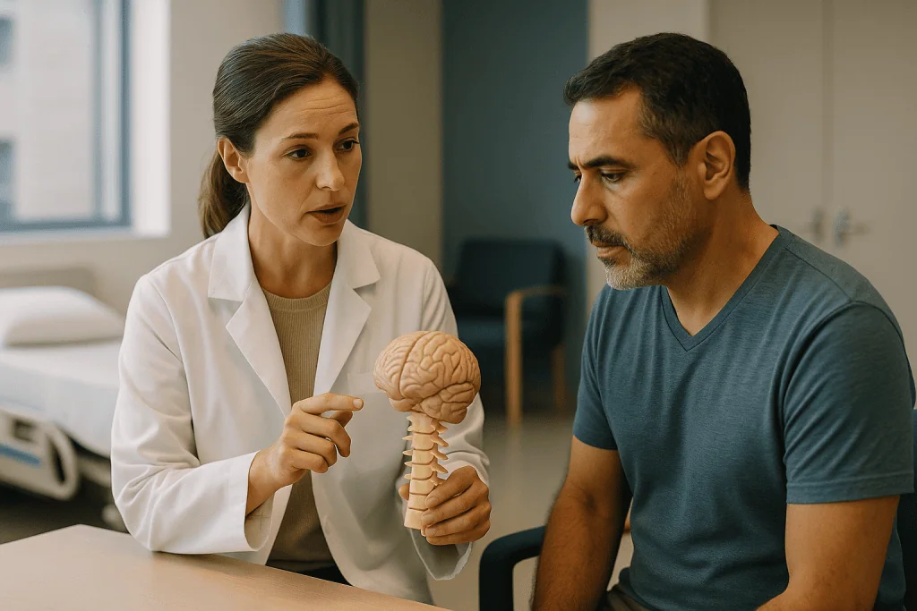
The first is a section of the occipital bone, the plate that forms the lower back of the skull. The surgeon removes a window of it just above the foramen magnum, an operation called a suboccipital craniectomy. “Craniectomy” simply means removing bone from the skull without putting it back; the muscles and skin close over the gap afterward and protect the area much as they do everywhere else on the head. The window is typically a few centimeters across, sized to the individual anatomy rather than to a fixed template.
The second piece is often the posterior arch of the first cervical vertebra, the ring-shaped bone at the very top of the spine that sits directly under the skull. Removing its back portion, a C1 laminectomy, extends the enlarged space downward so the descended tonsils have room below the skull as well as behind it. Whether C1 is included depends on how far the tonsils have dropped, according to the Mayo Clinic.
What is not routinely removed is the cerebellar tonsils themselves. Some surgeons do shrink or trim tonsil tissue in selected cases, but the core of the posterior fossa decompression procedure is subtraction of bone, not brain. This matters because people often picture the operation as cutting away part of the cerebellum, and that fear is worth putting down.
A useful analogy: the brain and its fluid are guests in a room that was built too small. The surgery knocks out a wall. It does not ask anyone to leave.
How does the posterior fossa decompression procedure enlarge the space for fluid?
Bone removal creates room. Whether that room reaches the fluid depends on the next layer down.
Beneath the skull lies the dura mater, a tough, leathery membrane that wraps the brain and spinal cord like a sleeve. In Chiari malformation the dura at the skull base is often thickened and tight, and it can hold the tonsils in a squeeze even after the bone above it is gone. Many surgeons therefore open the dura and sew in a patch, a step called duraplasty, so that the sleeve itself becomes wider.
The patch, or graft, may be taken from the patient’s own tissue, such as a strip of the thick fascia over the scalp muscles, or it may be a processed collagen or synthetic material. The choice varies between surgeons and is a reasonable question to ask. Once the graft is in place, the space beneath it, the cisterna magna, becomes a generous pocket of cerebrospinal fluid behind the cerebellum, restoring the reservoir that a normal posterior fossa provides.
Some surgeons go one step further and open the arachnoid, the thin, cobweb-like membrane just inside the dura, to release adhesions and confirm that fluid flows freely from the fourth ventricle, the small chamber in the brainstem where cerebrospinal fluid exits the brain. Others deliberately leave the arachnoid intact to lower the risk of fluid leak.
The endpoint the surgeon is looking for is visible in the operating room: cerebellar tonsils that are no longer wedged, and a pulsation of fluid that matches the heartbeat. Cleveland Clinic describes the purpose plainly as creating more room for the cerebellum and relieving pressure on the spinal cord, per its overview.
Duraplasty vs bone-only decompression: which approach do surgeons choose?
This is the live debate in Chiari surgery, and honest surgeons will tell you the evidence has not settled it.

Bone-only decompression removes the occipital bone and, usually, the C1 arch, but leaves the dura closed. Its appeal is straightforward: no opening in the membrane means a lower chance of cerebrospinal fluid leaking through the wound, less risk of infection or meningitis, and typically a shorter operation. Some surgeons check fluid flow with an ultrasound probe during the operation and open the dura only if the tonsils still look compressed.
Decompression with duraplasty adds the dural patch described above. Its appeal is a more reliable expansion of the fluid space, and it is the approach many surgeons prefer when a syrinx, a fluid-filled cavity inside the spinal cord, is present, or when the tonsils sit particularly low.
Systematic reviews indexed on PubMed have compared the two strategies repeatedly. The broad pattern is that duraplasty tends to show lower rates of needing a second operation, while bone-only decompression tends to show fewer fluid-related complications. Neither approach has been proven superior for symptom relief across all patients, and most studies are retrospective, meaning they look back at records rather than randomly assigning treatment. That is a lower grade of evidence than a trial.
The practical upshot: the choice is often made on the individual’s anatomy and the surgeon’s experience rather than on a rule. In children, bone-only approaches are used more often; in adults with a syrinx, duraplasty is more common. Asking which approach is planned, and why, is one of the most useful conversations a person can have before consenting.
Who is usually offered surgery, and who is usually asked to wait?
Surgery is offered for symptoms, not for a picture.
The people most consistently offered a posterior fossa decompression are those whose symptoms clearly match the anatomy: the characteristic cough- or strain-provoked headache at the back of the head, brainstem signs such as swallowing difficulty or sleep-disordered breathing, progressive weakness or numbness, or a syrinx that is enlarging or already causing spinal cord symptoms. In these situations the goal is to stop the process before nerve tissue is permanently damaged, a rationale laid out by the NIH’s NINDS.
People asked to wait fall into several groups. The largest is those whose Chiari was found incidentally, on a scan done for an unrelated reason, and who have no symptoms. For them, both NHS and Mayo Clinic guidance favors monitoring, sometimes with repeat MRI, because many will never develop problems. Operating on someone without symptoms exposes them to surgical risk with nothing measurable to relieve.
A second group has symptoms that do not fit the anatomy well, for instance daily tension-type or migraine-pattern headaches without the pressure-related trigger. Chiari is common enough on imaging that it can be an innocent bystander, and surgery cannot be expected to help a headache it did not cause. A neurologist’s assessment often precedes a surgical referral for exactly this reason.
A third group has a different problem masquerading as Chiari: tonsillar descent caused by low spinal fluid pressure, by a mass, or by hydrocephalus, an excess of fluid in the brain’s ventricles. Decompressing the skull base in those cases can make things worse, so the treating team screens for them first.
Children add another layer: growth can change the anatomy, and decisions weigh that.
What happens on the day of Chiari decompression surgery, step by step?
The operation is performed under general anesthesia, meaning the patient is fully asleep and unaware. It usually lasts a few hours, with the exact time depending on whether the dura is opened and on individual anatomy.
Positioning comes first and takes longer than people expect. The patient lies face down with the head fixed gently in a frame and the neck flexed forward, which opens the angle between skull and spine and gives the surgeon a clear line to the foramen magnum. Padding protects the eyes, chest, and pressure points.
A small strip of hair at the back of the head is clipped. The incision runs vertically in the midline, from roughly the bump at the back of the skull down to the upper neck, typically several centimeters long. The neck muscles are parted along their natural seam rather than cut across, which is one reason recovery focuses so heavily on neck stiffness.
Bone removal follows: the suboccipital window, then usually the back of C1. If duraplasty is planned, the surgeon opens the dura in a Y-shaped cut under the operating microscope, inspects the tonsils, and sews in the graft with fine sutures, sometimes adding a sealant to reduce leakage. Many teams use intraoperative ultrasound to confirm fluid movement before closing.
Closure is layered: muscle, fascia, skin. Some surgeons place a temporary drain. The patient wakes in a recovery area, and most spend the first night under close monitoring, often in a neurosurgical or high-dependency unit, before moving to a regular ward. The NHS notes a hospital stay of a few days is typical, per its Chiari malformation page.
Syringomyelia and Chiari surgery: why the fluid cavity in the spinal cord matters
Syringomyelia, a syrinx for short, is a cavity of cerebrospinal fluid that forms inside the spinal cord itself. It is the complication that most often turns a Chiari malformation from an observation into a surgical conversation.
The leading explanation ties it directly to the blocked flow at the skull base. Each heartbeat pushes a small pulse of fluid downward into the spine; when the tonsils plug the opening, that pressure wave cannot dissipate normally and instead drives fluid into the substance of the cord. Over months and years the cavity can widen, stretching nerve fibers from the inside. The Mayo Clinic lists syringomyelia among the recognized complications of Chiari type I.
Symptoms follow the cord’s wiring. Because the fibers carrying pain and temperature cross near the center of the cord, an early sign is often a loss of those sensations in the hands or across the shoulders in a cape-like pattern, sometimes noticed as painless burns. Hand weakness, stiffness in the legs, and changes in bladder function can follow.
Surgery does not drain the syrinx directly in most cases. Instead, the decompression restores normal flow at the top, and the cavity is expected to shrink on its own once the pressure pulse is relieved. Follow-up MRI, often at several months and then at intervals decided by the team, tracks whether that is happening. A syrinx that persists or grows despite adequate decompression prompts a look for residual blockage or scar tissue, and occasionally a small tube is placed to drain the cavity into the surrounding fluid space.
The presence of a syrinx is also the single factor that most often tips surgeons toward opening the dura rather than relying on bone removal alone.
How do the treatment options for Chiari malformation compare?
Laying the options side by side clarifies what each one is trying to do. None of these is universally right; the table describes the usual reasoning, not a rule.
| Option | What is done | Usually considered when | Main trade-off |
|---|---|---|---|
| Monitoring | Periodic clinical review, repeat MRI at intervals set by the team | No symptoms, or symptoms that do not match the anatomy; no syrinx | Avoids surgical risk; requires accepting uncertainty and follow-up |
| Symptom management | Headache and pain strategies guided by a neurologist; activity advice | Mild, stable symptoms; alongside monitoring | Does not change the anatomy or fluid flow |
| Bone-only decompression | Suboccipital craniectomy, usually with C1 laminectomy; dura left closed | Symptoms present, no or small syrinx; often preferred in children | Lower fluid-leak risk; possibly higher chance of needing revision |
| Decompression with duraplasty | Bone removal plus opening and patching the dura | Symptoms with syrinx, very low tonsils, or after failed bone-only surgery | More reliable expansion; higher fluid-leak and meningitis risk |
| Shunt procedures | Tube diverting fluid from ventricles or from a syrinx | Hydrocephalus present, or syrinx persisting after decompression | Hardware can block or infect; addresses fluid, not the crowding |
Two points deserve emphasis. First, hydrocephalus, when present, is generally treated before or instead of decompression, because excess ventricular fluid can itself push the tonsils down, and decompressing the skull base without addressing it can be hazardous. Second, monitoring is a genuine treatment plan, not a failure to act. The NHS is explicit that people without symptoms often need no treatment beyond regular review.
What are the risks of posterior fossa decompression surgery?
Every neurosurgical operation carries risk, and this one has a particular profile worth understanding in plain terms.
The most characteristic complication is a cerebrospinal fluid leak, in which fluid escapes through the dural repair and collects under the skin as a soft swelling, or drips from the wound. It is more common when the dura has been opened. Small collections often settle with time and positioning; persistent leaks may need a temporary lumbar drain, a thin tube in the lower back that lowers pressure while the repair seals, or a return to the operating room.
Meningitis, inflammation of the membranes around the brain, can be bacterial, which is a surgical emergency, or “aseptic,” an irritant reaction to blood or graft material that causes fever and headache without infection. Distinguishing the two usually requires testing the fluid. Both are recognized risks noted by the Mayo Clinic.
Other risks include wound infection, bleeding, injury to the cerebellum or brainstem with resulting balance or swallowing problems, and a rare complication called cerebellar slump, where the cerebellum sags into an overly generous opening, which is one argument against removing too much bone. Instability of the joint between skull and spine is uncommon but is watched for, especially in people with connective tissue conditions. Pseudomeningocele, a fluid pocket under the skin, overlaps with leak and is usually self-limiting.
Finally, there is the risk that surgery goes technically well and symptoms do not improve, or that they return years later because scar tissue narrows the space again. Reoperation is needed in a minority of people; published rates vary widely between series, so a specific figure quoted without a source should be treated with caution.
Chiari decompression surgery recovery: what the first days and weeks usually look like
The first thing most people report is not headache but neck pain. The muscles that were parted to reach the skull base protest for days, and turning the head feels stiff and guarded. This is expected and usually eases week by week.
In the hospital, the team watches for fever, wound swelling, fluid leak, worsening headache when upright, and any new weakness or swallowing difficulty. Walking begins early, often the day after surgery, both to prevent blood clots and because gentle movement helps the neck. Pain is managed with a plan set by the anesthesia and surgical teams; the specifics belong to them, and the general principle is to reach comfort while keeping the person alert enough to be assessed.
Discharge typically follows within a few days, according to the NHS. At home, the wound is kept clean and dry as instructed, heavy lifting and straining are avoided, and driving waits until the neck moves freely and the team clears it.
The NHS describes full recovery as taking a few weeks, though people vary considerably, and those with desk-based work often return sooner than those with physical jobs. Fatigue is common and underappreciated; an operation at the skull base under general anesthesia is a real physiological event, and the tiredness that follows is not a sign something is wrong.
Headache patterns may fluctuate before settling. Some people notice the pressure-type headache gone almost immediately; others experience a different, muscular headache from the incision that fades over weeks. A follow-up visit, often around the six-week mark, checks the wound and symptoms, and an MRI at several months assesses fluid flow and any syrinx.
Children generally recover faster than adults and are back at school within weeks, with sports resumed on the team’s schedule.
Does Chiari surgery relieve headaches, and what does the evidence actually show?
The honest answer is: often, for the right headache, and less reliably for others.
The headache most responsive to decompression is the one that led to the diagnosis in textbooks: a pain at the back of the head or upper neck that surges with coughing, sneezing, bending, laughing, or straining and fades within seconds to minutes afterward. That headache is mechanically produced by the pressure spike hitting a blocked skull base, and relieving the block removes its cause. The NIH’s NINDS describes surgery as the only treatment that can address the structural problem and notes it can reduce or stabilize symptoms in many people.
Headaches that behave like migraine, that are present daily regardless of posture or exertion, or that are accompanied by light sensitivity and nausea are less predictable, because they may not be driven by the Chiari at all. People with both patterns often find the cough headache resolves while the migraine continues, which is disappointing but not a surgical failure.
Symptoms from a syrinx follow a different rule. Pain and abnormal sensation frequently improve as the cavity shrinks, but strength and sensation that were already lost to cord damage may not return. This is why the timing argument for surgery centers on preventing further loss rather than reversing it.
Evidence quality deserves a word. Most outcome data come from single-center case series, and definitions of “improvement” differ from one paper to the next, so percentages quoted online are rarely comparable. Systematic reviews on PubMed consistently call for randomized comparisons that do not yet exist in adequate numbers. A surgeon who says “most people with your symptom pattern do well, but I cannot promise” is describing the literature accurately.
What people often get wrong about Chiari malformation surgery
Several ideas circulate widely enough that correcting them is a public service.
“The surgeon removes part of the brain.” The standard operation removes bone and expands a membrane. Tonsil tissue is trimmed only in selected cases, and the cerebellum’s function is not sacrificed.
“Chiari is a rare disease.” Before MRI it was thought to be, because it was found mainly at autopsy. Modern imaging finds tonsillar descent in a meaningful fraction of scans done for other reasons, and the Johns Hopkins overview notes many people never know they have it. Rarity was an artifact of the technology.
“If it’s on the MRI, it must be operated on.” The opposite is closer to the truth. Without symptoms that fit, most guidance favors monitoring, because the anatomy alone does not predict problems.
“The millimeters of descent tell you how sick you are.” Someone with 6 mm of descent and a large syrinx can be far more affected than someone with 15 mm and no fluid disturbance. Flow, crowding, and symptoms matter more than the ruler.
“Surgery fixes everything, or it failed.” Decompression addresses fluid dynamics. Migraine, fibromyalgia, anxiety, and neck arthritis can coexist and are unaffected. Separating the strands beforehand prevents a lot of grief afterward.
“Chiari runs in families, so my children need scanning.” A hereditary component exists in some families, but routine screening of asymptomatic relatives is not standard practice. Scanning is reserved for those with symptoms, a point on which the Mayo Clinic and other sources are aligned.
“Once decompressed, the skull is unprotected.” Thick neck muscles cover the window. Normal daily life, including most sports after clearance, does not require special protection.
How does Chiari type I differ from type II, and does the operation change?
The numbered types describe different conditions that happen to share a name, and the surgery for each has a different purpose.
Chiari type I, the subject of nearly everything above, involves descent of the cerebellar tonsils alone, in a person whose spine and brain otherwise formed normally. It is usually recognized in adolescence or adulthood, sometimes never. The decompression described in this article is the operation for type I.
Chiari type II involves descent of the cerebellar tonsils, part of the brainstem, and the fourth ventricle, and it occurs almost exclusively alongside myelomeningocele, the most serious form of spina bifida, in which the spinal cord and its coverings protrude through an opening in the back. It is present at birth and typically identified before or shortly after delivery. Hydrocephalus is very common in type II, according to the MedlinePlus summary from the National Library of Medicine.
For type II, the first priorities are closing the spinal defect and managing hydrocephalus, most often with a shunt. Posterior fossa decompression is reserved for infants and children who develop brainstem symptoms such as breathing pauses, swallowing trouble, or a weak cry despite a working shunt. It is a higher-stakes procedure in this group, and pediatric neurosurgical teams weigh it carefully.
Types III and IV are rare and severe, involving herniation of brain tissue through a skull defect or an underdeveloped cerebellum; decompression as described here does not apply.
Some clinicians also use the label “Chiari 0” for people with syringomyelia and a crowded skull base but tonsils above the 5 mm line, and “Chiari 1.5” when the brainstem also sits low. These terms signal that flow and crowding, rather than a single measurement, drive the surgical decision.
Questions to ask your care team before agreeing to decompression
A good consultation leaves a person understanding not just what will be done but why this operation, in this form, for this set of symptoms. These questions tend to open that conversation.
- Which of my symptoms do you expect this operation to help, and which do you think are unrelated to the Chiari?
- Do I have a syrinx, and if so, how large is it and where? How will you know later whether it is shrinking?
- Do you plan to open the dura? If yes, what graft material will you use, and why that one? If no, how will you decide during the operation whether the bone removal is enough?
- Will you remove part of C1? Do you intend to trim the tonsils?
- Have you checked for hydrocephalus, low-pressure causes of tonsillar descent, and instability between my skull and spine?
- What are the most common complications you see with this approach, and how are they managed?
- What would you expect if I chose monitoring instead, and what changes would prompt you to recommend surgery later?
- How long do you expect me to be in the hospital, and what should recovery look like at two weeks, six weeks, and three months?
- When will follow-up imaging happen, and what will you be looking for?
- Who do I contact, day or night, if something worries me after discharge?
Bringing a family member or friend to take notes helps, as does asking for the plan in writing. A second opinion is a normal part of decision-making for elective neurosurgery and is not an insult to anyone; surgeons expect it. The Johns Hopkins overview and the NINDS pages both stress that the timing and type of surgery are individualized, which is exactly why these questions have no universal answers.
When to call your doctor after posterior fossa decompression
Most recoveries are uneventful, but a short list of warning signs should prompt a same-day call to the surgical team, or emergency care if the team cannot be reached.
- Fever, stiff neck, sensitivity to light, or a severe headache that is new or rapidly worsening, which can signal meningitis and needs urgent assessment.
- Clear or straw-colored fluid leaking from the wound, a soft swelling growing under the incision, or a headache that is markedly worse when upright and relieved by lying flat, all of which suggest a cerebrospinal fluid leak.
- Redness spreading from the incision, pus, or the wound edges separating.
- New or worsening weakness, numbness, unsteadiness, difficulty swallowing, choking on liquids, hoarseness, or changes in breathing, including pauses in breathing during sleep noticed by others.
- Persistent vomiting, extreme drowsiness, confusion, or a seizure.
- Calf pain, swelling in one leg, chest pain, or sudden shortness of breath, which can indicate a blood clot.
Beyond the immediate recovery, anyone with a known Chiari malformation, operated or not, should report a return of the pressure-type headache, new hand numbness or clumsiness, or painless burns and cuts on the hands, because these can indicate a syrinx forming or enlarging. The Mayo Clinic lists ongoing follow-up as part of care after decompression for precisely this reason.
None of this list is meant to be a self-diagnosis tool. Its purpose is to lower the threshold for picking up the phone. Surgical teams would far rather hear about a symptom that turns out to be nothing than learn about a leak or infection a week late.
Frequently asked questions
What is Chiari malformation in plain language?
Chiari malformation is a condition in which the lowest part of the cerebellum, the tonsils, sits below the opening at the base of the skull and into the upper spinal canal, because the compartment holding the cerebellum is smaller than average. This crowding can press on the brainstem and block the normal flow of cerebrospinal fluid. Many people have the anatomy without symptoms; others develop pressure-type headaches, neck pain, numbness, or balance problems.
How long does Chiari decompression surgery recovery take?
Hospital stays are typically a few days, and the NHS describes full recovery as taking a few weeks, though individuals vary widely. Neck stiffness and fatigue are the dominant early complaints. Desk-based work is often resumed sooner than physical work, and driving waits until the neck moves comfortably and the team clears it. Follow-up imaging is usually arranged at several months to check fluid flow and any syrinx.
Is duraplasty vs bone-only decompression a settled question?
No. Systematic reviews suggest duraplasty is associated with fewer repeat operations while bone-only decompression carries fewer fluid-leak complications, but most data are retrospective and no approach has been shown superior for symptom relief across all patients. Surgeons often decide based on whether a syrinx is present, how low the tonsils sit, the patient’s age, and their own experience, which is why asking about the planned approach is worthwhile.
Does syringomyelia go away after Chiari surgery?
A syrinx often shrinks after decompression because restoring fluid flow at the skull base removes the pressure pulse that drives fluid into the spinal cord, but this is not guaranteed and happens over months rather than days. Pain and abnormal sensation frequently improve; strength or sensation already lost may not return. If a syrinx persists or grows despite adequate decompression, the team looks for residual blockage and may consider draining it directly.
Is the posterior fossa decompression procedure brain surgery?
It is neurosurgery performed at the junction of skull and spine, but the standard operation removes bone and expands a membrane rather than removing brain tissue. The cerebellum is exposed and inspected, and in selected cases the tonsils are trimmed, but the core of the procedure is subtraction of bone and, often, patching of the dura. Risks to the cerebellum and brainstem exist and are part of the consent discussion.
Can Chiari malformation be treated without surgery?
Yes, for many people. Those without symptoms, or with symptoms that do not match the anatomy, are usually monitored with periodic review and sometimes repeat MRI rather than operated on. Symptom management guided by a neurologist can address headache and pain. No medication or exercise changes the anatomy or fluid flow, so surgery remains the only option that addresses the structural problem when symptoms warrant it.
What does 'cerebellar tonsils 8 mm below the foramen magnum' mean on an MRI?
It means the lowest tips of the cerebellum extend about 8 mm below the level of the large opening at the base of the skull, which exceeds the roughly 5 mm threshold radiologists commonly use to describe Chiari type I. The measurement identifies the anatomy but does not by itself indicate severity or the need for surgery; symptoms, fluid flow, and the presence of a syrinx matter more.
Will the back of my skull be unprotected after the bone is removed?
The bone window is covered by the thick muscles at the back of the neck and by the scalp, which protect the area much as soft tissue protects other parts of the head. Normal daily activity does not require special protection, and most people return to sports once the surgical team clears them. Some surgeons deliberately limit the size of the opening to reduce the small risk of the cerebellum sagging into it.
Can Chiari symptoms come back years after decompression?
They can, though this affects a minority of people. Scar tissue can narrow the enlarged space over time, or a graft can adhere to underlying structures and restrict fluid flow again. A return of the pressure-type headache, new hand numbness, or painless injuries to the hands should be reported, because they can indicate recurrence or a syrinx. Long-term follow-up with the treating team is recommended for this reason.
Should my children be scanned if I have Chiari malformation?
Routine MRI screening of relatives without symptoms is not standard practice, even though a hereditary tendency exists in some families. Scanning is generally reserved for children who develop symptoms suggestive of Chiari, such as pressure-type headaches at the back of the head, swallowing difficulty, or unexplained scoliosis. A pediatrician or neurologist can advise whether a child’s symptoms justify imaging.
References
- Chiari malformation (NHS)
- Chiari Malformation (NIH National Institute of Neurological Disorders and Stroke)
- Chiari Malformation (Cleveland Clinic)
- Chiari Malformation (MedlinePlus)
This article is for general information only and is not a substitute for professional medical advice. Please consult a qualified doctor about your individual situation.

More from the Blog

Broca’s, Wernicke’s and Global Aphasia: How the Type Changes the Therapy Approach
Therapy for aphasia is matched to the pattern of language loss. In Broca's aphasia, understanding is relatively preserved, so therapy drills spoken output, often…

Epilepsy Surgery and Neurostimulation: What Changes When Medication Alone Is Not Enough
Epilepsy surgery is usually considered when seizures continue despite two appropriately chosen and well-tolerated anti-seizure medicines, a situation neurologists call drug-resistant epilepsy. A specialist…

Which Hearing Devices and Vision Aids Help With Deafblindness, and How Are They Fitted?
Hearing devices for deafblindness include hearing aids, cochlear implants, bone-conduction devices, remote-microphone systems and vibrating alerting devices; vision aids include prescribed magnifiers, electronic video…

CBT-I or Sleep Medication for Insomnia: How the Two Approaches Compare Over Time
For chronic insomnia, major guidelines recommend cognitive behavioral therapy for insomnia (CBT-I) as the first treatment because its benefits usually persist after the course…

Insomnia and Daily Routine: Wake Times, Caffeine and Screens in a Personalized Sleep Plan
A personalized insomnia daily routine sleep plan is built around three anchors that research supports most strongly: a fixed wake time kept seven days…

When to Call Your Surgeon After Chiari Malformation Surgery: Fluid Leakage, Fever and Redness
After Chiari malformation surgery, call your surgical team the same day if you notice clear or watery fluid leaking from the incision, a fever,…