

Split-Thickness vs Full-Thickness Skin Grafts: How Surgeons Choose the Graft Type
Key Takeaways
- A split-thickness graft carries the epidermis and part of the dermis; a full-thickness graft carries the entire dermis, and that difference drives nearly every trade-off between them.
- New blood vessels begin growing into a graft within about 36 hours and the graft typically connects to the local blood supply over roughly 5 to 7 days, which is why the first week demands stillness.
- Thinner grafts survive more readily on imperfect wound beds, while full-thickness grafts need excellent blood supply but resist later shrinkage far better.
- Full-thickness grafts are favored on the nose, eyelids and ears because secondary contraction there can distort a feature or pull a lid away from the eye.
- A split-thickness donor site regrows its own surface over about one to three weeks and is often more uncomfortable than the grafted wound during that time.
- There is no single reliable success percentage for skin grafts; bed quality, freedom from fluid and shear, infection control and smoking status shape the outcome far more than graft type alone.
Surgeons choose between a split-thickness and a full-thickness skin graft mainly on wound size, location, and how much the graft may shrink. Split-thickness grafts take epidermis plus part of the dermis, cover large areas, and survive more readily on imperfect wound beds. Full-thickness grafts take the entire dermis, match facial skin better and contract less, but need small, well-vascularized wounds. The treating team weighs both for each patient.
The bandage comes off in the dermatology clinic, and the mirror shows a coin-sized wound on the side of the nose where a skin cancer used to be. The surgeon says the word “graft,” then two more words that sound almost identical: split-thickness, full-thickness. The patient nods, drives home, and types split thickness vs full thickness skin graft into a search bar before dinner.
That moment repeats itself in clinics every day, after cancer removal, after burns, after a scalp wound that will not close on its own. The choice sounds technical. In practice it rests on a handful of concrete questions a surgeon asks about the wound in front of them: How big is it? How good is the blood supply underneath? Will a shrinking scar pull on an eyelid or a lip? How much matching skin can be spared elsewhere?
This explainer walks through those questions in plain language, so the conversation in the consulting room feels less like a coin toss and more like a decision you can follow.
What a split thickness vs full thickness skin graft actually is
A skin graft is a piece of healthy skin moved from one part of the body (the donor site) to cover a wound elsewhere (the recipient bed), where it must grow a new blood supply to survive. Skin has two main layers: the epidermis, the thin outer barrier, and the dermis beneath it, the thicker layer that holds collagen, hair follicles, sweat glands and blood vessels. The difference between the two graft types is simply how much of that dermis travels with the graft.
A split-thickness skin graft takes the whole epidermis and only part of the dermis. The surgeon shaves it off with a dermatome, a precision instrument that works a little like an adjustable plane, leaving the deeper dermis behind so the donor area can regrow its own surface. According to the National Library of Medicine’s StatPearls review of skin grafting, split-thickness grafts are further described as thin, intermediate or thick depending on how deeply the blade is set.
A full-thickness skin graft takes the epidermis and the entire dermis, down to the fat, which is trimmed away. Because nothing is left behind to regrow skin, the donor wound must be closed with stitches, so these grafts are harvested from places with loose spare skin, such as behind the ear, the upper eyelid, the neck or the groin.
Neither type is “better” in the abstract. A thin graft is a survivor: it demands less from the wound bed and covers a lot of ground. A thick graft is a perfectionist: it looks and behaves more like the skin it replaces but needs ideal conditions. Nearly every decision a surgeon makes here flows from that single trade-off.
How skin graft take works: the first week under the dressing
“Take” is the word surgeons use for a graft that has successfully become living, attached skin. It does not happen in the operating room; it happens quietly over roughly a week, while the graft sits under a firm dressing and the patient is asked not to disturb it.
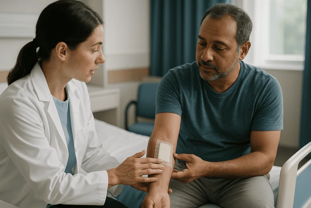
The StatPearls review describes three overlapping phases. In the first day or two the graft has no blood supply of its own and survives by imbibition, soaking up nutrient-rich fluid from the wound bed the way a sponge holds water. Next comes inosculation, in which tiny blood vessels at the base of the graft line up with vessels in the bed and begin to connect. Then true revascularization follows, as new capillaries grow up into the graft. MedlinePlus notes that new blood vessels begin growing from the recipient area into the transplanted skin within about 36 hours, and NHS guidance describes the graft typically connecting to the local blood supply within about 5 to 7 days.
Three things can interrupt that sequence, and they explain most graft failures: fluid or blood collecting under the graft and lifting it off the bed, infection, and shearing, where the graft slides even a millimeter and the fragile new connections tear. This is why dressings are bulky and why surgeons ask patients to keep a grafted leg elevated or avoid touching a facial graft.
Thickness matters here. A thinner graft has less tissue to feed during the imbibition phase, so it tolerates a less-than-perfect bed. A full-thickness graft is asking the bed to nourish far more tissue on faith, which is why it is reserved for wounds with excellent blood supply.
When to use a split thickness skin graft: the surgeon's checklist
Ask a surgeon when they reach for a split-thickness graft and the answer usually starts with size. Large wounds, whether from burns, trauma, or removal of a big skin cancer on the scalp or leg, cannot be closed with a full-thickness graft because no donor site could spare that much skin and still be stitched shut. A split-thickness graft can be harvested from a broad, flat area such as the thigh, and the donor site heals on its own.
The second item on the checklist is the quality of the wound bed. Split-thickness grafts, particularly thinner ones, are the choice when the base is healthy granulation tissue, the pink bumpy tissue that forms in healing wounds, but not pristine. The StatPearls review lists contaminated or less vascular beds as settings where a thinner graft is favored precisely because it needs less nourishment to survive.
Third comes the option of meshing. The graft can be passed through a device that cuts a pattern of slits so it stretches like a net; StatPearls describes typical expansion ratios from 1:1.5 up to 1:3. Meshing also lets fluid drain through the graft rather than pooling beneath it. The trade-off is a permanent lattice pattern in the healed skin, which is acceptable on a leg but rarely on a cheek.
Finally, surgeons think about what the area does. A split-thickness graft is well suited to a scalp after cancer removal, the trunk, or the lower leg, where coverage and reliable healing matter more than a perfect color match. In the facial aesthetics setting, it appears mostly on the scalp, the temple, or as a temporary cover while a larger reconstruction is planned.
When surgeons choose a full-thickness skin graft, especially on the face
A full-thickness graft is chosen when the wound is small enough to be covered from a spare-skin donor site, the bed is richly vascular, and the appearance or function of the result matters a great deal. That combination describes much of facial reconstruction after skin cancer surgery.
Consider the nasal tip, the lower eyelid, the inner corner of the eye, or the rim of the ear. These are areas where even a few millimeters of scar shrinkage can pull a lid down, notch a nostril, or distort a contour that everyone notices in conversation. Because a full-thickness graft carries the entire dermis, it resists that later shrinkage far better than a split graft, a point returned to below. Its surface also keeps more of the natural texture, pore pattern and pigment of donor skin, so a graft taken from behind the ear or the upper eyelid can settle into a similar tone to the surrounding face.
Surgeons also prefer full-thickness grafts where the skin will need to stretch and move, such as the fingertip or the palm side of a finger, and in children whose skin is still growing, since a graft that contracts can restrict movement later.
The prerequisites are strict. MedlinePlus points out that full-thickness grafts are used for smaller wounds on highly visible parts of the body and that they need a longer recovery than split grafts. The bed must be free of infection and dead tissue and must have a good blood supply; exposed bare bone, tendon without its covering, or heavily irradiated tissue are generally poor beds for any graft and may push the team toward a flap instead.
Split thickness vs full thickness skin graft: side-by-side comparison
The table below gathers the differences most patients ask about. Every entry is a general tendency drawn from the StatPearls review and patient-facing guidance from MedlinePlus and the NHS; individual wounds vary, and the treating team’s assessment of a specific wound outranks any table.
| Feature | Split-thickness graft | Full-thickness graft |
|---|---|---|
| Layers included | Epidermis plus part of the dermis | Epidermis plus the entire dermis |
| Typical wound size | Large areas, can be meshed to stretch further | Small to moderate wounds |
| Wound bed needed | Tolerates a less ideal bed | Requires an excellent blood supply |
| Donor site | Thigh, buttock, back, scalp; heals on its own | Behind ear, eyelid, neck, groin, inner arm; closed with stitches |
| Later shrinkage (contraction) | More over time | Less over time |
| Color and texture match | Often paler, shinier; mesh pattern if meshed | Closer to surrounding skin |
| Durability | Thinner, more fragile surface | Thicker, more durable |
| Hair, sweat and oil glands | Few carried over | Many carried over |
| Common uses in the face and head | Scalp, temple, large defects, temporary cover | Nose, eyelid, ear, cheek after cancer removal |
Two rows deserve emphasis. Contraction and bed quality pull in opposite directions: the graft that shrinks least is the one that is hardest to keep alive. Reconstructive surgery is largely the art of resolving that tension for a particular wound on a particular person. When the two graft types both seem workable, surgeons often ask which failure would be worse, a graft that partly fails and needs a second procedure or a graft that survives but distorts a feature, and choose accordingly.
What are the advantages of a split thickness skin graft?
The split-thickness graft is the workhorse of reconstructive surgery, and its advantages are practical rather than glamorous. The first is reach. A single strip from the thigh can cover a wound many times the size of anything a full-thickness donor site could yield, and meshing extends that further. For someone with a large burn or a wide excision on the scalp, it is often the only realistic graft option.
The second is resilience. Because the graft is thin, the wound bed needs to deliver less nourishment during the days before new vessels grow in. The StatPearls review notes that thinner grafts have a better chance of surviving on beds that are less vascular or mildly contaminated, which matters in older adults, in people with diabetes or circulatory disease, and in wounds that have been slow to heal.
The third is the donor site. Since part of the dermis stays behind, the donor area regrows its own epidermis from the skin cells lining hair follicles and glands. StatPearls describes this re-epithelialization typically occurring over about one to three weeks depending on graft thickness, and the same site can sometimes be reharvested later. No stitched wound is created there.
Fourth is speed and simplicity. Harvesting is quick, and recovery from the harvest itself is usually straightforward; MedlinePlus notes that people generally recover more quickly after split-thickness grafting than after full-thickness grafting.
The honest counterweight is appearance and behavior over time. Split grafts tend to heal paler or darker than their surroundings, with a shinier surface and, if meshed, a lasting pattern. They also shrink more as they mature. On a shin that is a fair trade. On a lower eyelid it usually is not, which is exactly why the two graft types coexist.
Graft contraction: the shrinkage trade-off behind most decisions
Contraction is the tendency of grafted skin to shrink, and understanding it explains why a surgeon will insist on a full-thickness graft near an eye but accept a split graft on a leg.
There are two kinds. Primary contraction happens on the operating table the moment the graft is cut free, as elastic fibers in the dermis recoil. StatPearls notes that full-thickness grafts show more primary contraction because they contain more dermis and therefore more elastin. Surgeons simply account for it by cutting the graft slightly larger than the wound.
Secondary contraction is the one that matters to patients. It unfolds over weeks to months as the healed wound bed pulls inward, driven by specialized cells called myofibroblasts that behave a little like tiny muscle fibers in the scar. Here the relationship flips: the more dermis a graft carries, the more it resists this later pull. A thin split graft offers little resistance and may shrink noticeably; a full-thickness graft holds its ground far better.
Why does this matter so much on the face? Facial features are held in position by delicate balances. A scar that tightens by even a small margin below the lower eyelid can pull the lid away from the eye, a problem called ectropion that causes watering and irritation. Around the nostril, contraction can narrow the airway or lift the rim. Near the mouth it can distort a smile. In each case the surgeon is not choosing the graft that looks best on day one but the one that will still look right after the scar has finished maturing, which MedlinePlus and NHS guidance describe as a process that can extend over many months.
The same logic applies to joints and to children, where a contracting graft can become a contracture that limits movement.
Where the skin comes from: donor sites and skin graft donor site healing
Patients are often surprised that the donor site, not the graft, is what bothers them most in the first week. Knowing why helps.
For a split-thickness graft the donor site is a shallow, raw area, usually on the front or outer thigh, sometimes the buttock, back or scalp. Because it is essentially a large abrasion with millions of exposed nerve endings, it can be more tender than the grafted wound. It weeps fluid for several days and then re-epithelializes, meaning the surface regrows from the cells left in the deeper dermis. StatPearls places that healing window at roughly one to three weeks depending on how thick a graft was taken. The area typically heals paler or slightly darker than surrounding skin and may remain a faint patch permanently. Surgeons frequently choose a site that is easily covered by clothing for that reason.
For a full-thickness graft the donor site is a stitched incision, chosen where skin is loose and a fine scar can hide in a natural crease: behind the ear, in the fold of the upper eyelid, along the neck, in the groin crease, or on the inner upper arm. The scar is small and closes like any surgical cut. In facial work the choice of donor site is itself a matching exercise. Skin from behind the ear or above the collarbone has seen sun exposure and carries a tone closer to the face than skin from the groin.
Dressing choices for donor sites vary between teams, and instructions differ accordingly. The one universal message is to leave the dressing alone unless told otherwise and to report increasing pain, spreading redness or a bad smell, which can indicate infection.
Who is usually a candidate, and who is usually asked to wait
Skin grafting is offered to people whose wound cannot be closed directly and will not heal well or quickly enough on its own. In the facial aesthetics setting that most often means defects left after skin cancer removal; elsewhere it includes burns, traumatic skin loss, and chronic ulcers that have finally developed a healthy base.
Candidacy rests as much on the wound bed as on the person. A graft needs a clean, bleeding, living surface to bond to. Surgeons therefore look for pink granulation tissue without dead material, no active infection, and no bare bone, cartilage or tendon stripped of its covering, since those surfaces cannot feed a graft. If a wound is not ready, the usual answer is not “no” but “not yet”: the bed is cleaned, dressed and sometimes treated with negative pressure dressings until it is healthy enough.
People are often asked to wait or to prepare when factors that threaten graft survival can be improved first. Smoking narrows small blood vessels and is consistently linked to poorer wound healing in surgical guidance; teams generally ask patients to stop before and after surgery. Poorly controlled diabetes, heavy alcohol use, poor nutrition and swelling in the legs each slow healing. Certain medicines, including blood thinners and drugs that suppress the immune system, are reviewed by the team, but any change is the prescribing clinician’s call, never the patient’s alone.
Some wounds are better served by a different reconstruction altogether. Deep defects that need bulk, areas over exposed bone, and heavily irradiated tissue often call for a flap, which brings its own blood supply. When that is the recommendation, the team should be able to explain in plain language why a graft would be at higher risk in that particular spot.
Full thickness skin graft healing time and the weeks that follow
Recovery has a rhythm, and knowing it in advance takes much of the anxiety out of the first dressing change.
Week one is about stillness. The graft is covered by a padded dressing, often stitched or glued at the edges, sometimes with a foam or cotton bolster tied over it to press it gently onto the bed. For a graft on a leg, elevation is usually requested; for a facial graft, the request is simply not to touch, rub or sleep on it. NHS guidance describes the graft typically connecting to the blood supply within about 5 to 7 days, and many teams schedule the first look at around that point. A newly uncovered graft is often purple, mottled or swollen. That is expected, not a sign of failure.
Weeks two to four bring color change and settling. Stitches or clips are removed, the surface may flake or blister lightly, and moisturizing and gentle care usually begin under instruction. MedlinePlus advises avoiding activity that could stretch or injure the graft for about three to four weeks. Full-thickness grafts on the face frequently look thick, shiny or raised during this phase and then gradually flatten.
Months one to twelve are the maturation phase. Scars remodel, redness fades, and the graft’s texture softens. Sun protection matters throughout, because healing skin pigments unpredictably. Some grafts benefit from massage, silicone products or, later, minor revision procedures, all of which are the treating team’s decisions.
MedlinePlus notes that full-thickness grafts generally need a longer recovery than split-thickness grafts and that hospital stays, when needed, are shorter for split grafts. None of these windows is a promise; they are typical ranges, and an individual timeline depends on the wound, the site and general health.
Risks of skin grafting and the alternatives surgeons weigh
Every reconstruction carries risk, and a fair discussion names them without drama.
The specific risk of grafting is partial or complete graft loss, most often from bleeding or fluid under the graft, infection, or movement in the first days. A partly failed graft may heal from the edges on its own or need re-grafting. Other recognized risks listed in patient guidance from MedlinePlus and the NHS include infection at either site, bleeding, delayed healing, uneven color or texture, altered sensation, and scarring that is raised, tight or contracted. Facial grafts carry the added concern of distortion of a feature if contraction is greater than expected. General surgical risks such as reactions to anesthesia apply as well.
Alternatives exist, and a good team will explain why one was chosen over another.
- Healing by secondary intention means letting a wound close on its own from the edges and base. On concave areas such as the inner corner of the eye or the temple this can give a surprisingly good result with no donor site.
- Primary closure, simply stitching the edges together, works when there is enough loose skin.
- A local flap moves neighboring skin, still attached to its own blood supply, into the defect. Flaps bring bulk, resist contraction and match color almost perfectly, but create longer incision lines and require more dissection.
- Skin substitutes and dermal templates are manufactured or processed materials used to build a bed before or instead of grafting in some complex wounds; their role is specialized and decided case by case.
No option is universally superior. The surgeon’s job is to fit the reconstruction to the wound, the face and the person, and the patient’s job is to ask enough questions to understand the reasoning.
What is the success rate of a split thickness skin graft? What the evidence actually shows
This is one of the most searched questions about grafting, and the honest answer is that no single number describes it well.
Published series report graft take differently. Some count a graft as successful only if it survives entirely; others count partial take as success if the wound goes on to heal. Some report at one week, others at three months. The wounds themselves vary enormously, from a clean excision on a healthy scalp to a chronic leg ulcer in someone with poor circulation. A percentage lifted out of that context tells a patient almost nothing about their own wound.
What the evidence does support, consistently, is the list of factors that shift the odds. The StatPearls review and patient guidance from MedlinePlus agree on the fundamentals: a well-prepared bed with a good blood supply, control of bleeding and fluid beneath the graft, prevention of shear, and absence of infection are the strongest predictors of take. Thinner grafts survive more readily on marginal beds than thicker ones. Smoking, uncontrolled diabetes, poor nutrition and swelling reduce the chance of a clean take. MedlinePlus states plainly that most skin grafts are successful but that some fail to heal and may need to be repeated.
For a patient, the useful reframing is this: rather than asking for a universal success rate, ask the surgeon what they consider the specific risks for this wound, on this site, given this health history, and what the plan would be if the graft only partly takes. That conversation yields a far more realistic picture than any figure, and it keeps the decision where it belongs, with the treating team and the person in front of them.
What people often get wrong about split-thickness and full-thickness grafts
Some myths surface again and again in clinic conversations. Correcting them makes the real decision easier to understand.
“Full-thickness is the premium option, so it must always be better.” Thicker is not automatically better. A full-thickness graft placed on a marginal bed is more likely to fail than a thin one, and a failed graft is worse than a slightly paler one. Surgeons choose thickness to match the bed and the location, not to signal quality.
“The graft is skin from a donor, like an organ.” Almost all grafts described in this article are autografts, meaning the patient’s own skin. Skin from another person or from animal sources is occasionally used as a temporary biological dressing on large burns, but it is not a permanent replacement and is not what a facial reconstruction involves.
“If the graft looks purple at the first dressing change, it has died.” Early grafts are frequently dark, mottled or swollen as new vessels connect. Color settles over weeks. Only the team can judge whether an area is failing, and they will look at the edges, the smell and the underlying bed rather than the color alone.
“Once it has taken, the result is final.” Grafts continue to change for many months. Redness fades, contraction can develop, and pigment shifts, which is why sun protection and follow-up matter long after the wound has closed.
“The donor site is the easy part.” A split-thickness donor site is often the most uncomfortable wound in the first week, because it is a broad raw surface with exposed nerve endings.
“Grafts always leave a visible patch.” Some do, particularly meshed split grafts on the body. Well-matched full-thickness grafts on the face frequently blend closely once mature, though nothing about a scar can be promised in advance.
Questions to ask your care team before a skin graft
A short list of good questions can turn a rushed consent conversation into a genuine discussion. Bring these, and add your own.
- Which graft type are you recommending for my wound, and what about the size, location or blood supply led you there?
- What would happen if we chose the other type, or a flap, or let the wound heal on its own instead?
- Where will the donor skin come from, what will that site look like once healed, and can I choose a spot that is easier to cover or that matches my face better?
- Will the graft be meshed? If so, what will the pattern look like once healed?
- How much shrinkage do you expect over the coming months, and how might that affect my eyelid, nostril, lip or ear?
- What should the graft look like at the first dressing change, and what would worry you?
- What are the specific risks for me, given my health, my medicines and whether I smoke, and is there anything I should change before surgery with my prescribing clinician’s guidance?
- What would the plan be if the graft only partly takes?
- How long should I protect the area from stretching, bumping and sun, and when can I return to work, exercise, driving and normal washing?
- How will the result change over the first year, and would any revision or scar treatment be considered later?
- Who do I call, at any hour, if something looks wrong?
The answers will differ from one surgeon and one wound to another, and that variation is appropriate. What should not vary is the willingness to explain. A team that can walk through its reasoning in plain language is showing you how the decision was made, which is the best reassurance a patient can have that it was made carefully.
When to call your doctor after a skin graft
Most grafts heal without drama, but a few signs should prompt a same-day call to the surgical team, and a small number warrant urgent care. Patient guidance from MedlinePlus and the NHS on surgical wound infection and graft aftercare is consistent on the following.
Call your care team promptly if you notice:
- Spreading redness, warmth or increasing swelling around the graft or the donor site, especially if it is getting worse rather than better day by day.
- Pain that increases after the first few days instead of easing, or pain out of proportion to what you were told to expect.
- Thick, cloudy, green or yellow discharge, or a foul smell from either dressing.
- Bleeding that soaks through the dressing or does not stop with gentle pressure.
- The dressing becoming loose, wet or dislodged before the planned change, or any accidental knock to the graft.
- Fever or chills, which can indicate infection spreading beyond the wound.
- Any part of the graft turning black or dry, or the edges lifting away from the wound.
- New difficulty closing an eyelid, watering of the eye, or a change in the shape of a nostril or lip after a facial graft.
Seek urgent or emergency care, rather than waiting for a clinic callback, for rapidly spreading redness with fever, confusion, a racing heartbeat, difficulty breathing, or a sudden severe increase in pain, because these can signal a serious infection or, rarely, sepsis. After surgery on a leg, new calf pain or swelling, or chest pain and breathlessness, also need immediate assessment.
When in doubt, call. Teams would far rather look at a graft that turns out to be fine than hear about a problem a week late. Every decision about dressings, medicines and further treatment rests with the clinicians who know your wound.
Frequently asked questions
When should a split thickness skin graft be used instead of a full thickness graft?
Surgeons generally choose a split-thickness graft for larger wounds, for wound beds with a less-than-ideal blood supply, and for areas where coverage and reliable healing matter more than an exact color match, such as the scalp, trunk or lower leg. A full-thickness graft is preferred for small, well-vascularized wounds on the face, hands or other visible or mobile areas where minimal shrinkage and a close match are the priority.
What are the advantages of a split thickness skin graft?
The main advantages are reach, resilience and a self-healing donor site. A split-thickness graft can cover large areas, can be meshed to stretch further, and survives more readily on marginal wound beds because it needs less nourishment before new vessels grow in. The donor area regrows its own skin over roughly one to three weeks without stitches. Its drawbacks are greater later shrinkage and a poorer color and texture match.
How long does it take for a split thickness skin graft to heal?
The graft typically connects to its new blood supply within about 5 to 7 days, after which the first dressing change usually happens. MedlinePlus advises protecting the area from stretching or injury for around three to four weeks. The donor site re-epithelializes over roughly one to three weeks. Color, texture and scar maturity continue to change for many months, and individual timelines depend on the wound and general health.
What is the full thickness skin graft healing time on the face?
A full-thickness facial graft also attaches over roughly the first week, but MedlinePlus notes that full-thickness grafts generally need a longer overall recovery than split grafts. Facial grafts often look thick, dark or raised for several weeks before flattening and lightening. Final appearance emerges over many months as the scar matures, and the team may discuss sun protection, massage or later revision during that period.
What is the success rate of a split thickness skin graft?
No single figure applies, because studies define and measure success differently and wounds vary enormously. MedlinePlus states that most skin grafts are successful but some fail to heal and may need repeating. The factors that most influence take are a well-prepared, well-vascularized bed, prevention of fluid collection and shearing under the graft, absence of infection, and general health factors such as smoking and diabetes control.
How does skin graft take actually happen?
A graft survives at first by imbibition, absorbing fluid from the wound bed like a sponge. Over the next days, vessels in the graft align with vessels in the bed, a step called inosculation, and new capillaries then grow into the graft. MedlinePlus notes that vessel growth begins within about 36 hours. Fluid under the graft, infection and movement are the three main things that interrupt this process.
How does skin graft donor site healing differ between the two types?
A split-thickness donor site is a shallow raw area, usually on the thigh, that regrows its own skin from cells left in the deeper dermis over about one to three weeks and often heals as a paler or darker patch. A full-thickness donor site is a stitched incision in an area of loose skin, such as behind the ear or in the groin crease, and heals like any surgical cut with a fine linear scar.
Why do skin grafts shrink, and which type shrinks more?
Grafts shrink through secondary contraction, driven by cells in the healing bed that pull the scar inward over weeks to months. The more dermis a graft contains, the better it resists this pull, so split-thickness grafts shrink more over time and full-thickness grafts shrink less. Full-thickness grafts do recoil more immediately after harvest, but surgeons simply cut them slightly larger to compensate.
Is a skin graft or a flap better for facial reconstruction?
Neither is universally better. A flap moves neighboring skin with its own blood supply, offering excellent color match, bulk and resistance to shrinkage, but requires longer incisions and more dissection. A graft is simpler and leaves fewer new lines on the face but depends on the wound bed for survival. The choice depends on the depth, location and blood supply of the defect and is made by the treating surgeon.
What should a healing skin graft look like, and when is color a warning sign?
In the first week or two a graft is often purple, mottled or swollen as new vessels form, and this is expected. The team becomes concerned when an area turns black and dry, when edges lift off the bed, or when there is spreading redness, discharge, a bad smell, fever or worsening pain. Because color alone is unreliable, any doubt should be checked by the surgical team rather than judged at home.
References
- MedlinePlus Medical Encyclopedia: Skin graft
- NHS: Plastic surgery – What happens
- NIH National Library of Medicine, StatPearls: Skin Grafting
- MedlinePlus: Surgical wound infection – treatment
This article is for general information only and is not a substitute for professional medical advice. Please consult a qualified doctor about your individual situation.

More from the Blog
When Can Acne Scar Treatment Begin? Why Active Acne Must Be Controlled First
Acne scar treatment usually begins only after active acne has been controlled for several months, because every new breakout can create a new scar…
Microdermabrasion Downtime: Why Most People Return to Normal Skincare Within Days
Microdermabrasion downtime is usually short: most people have mild redness, tightness, and a sunburn-like feel for about a day, then return to a gentle…
How Cryotherapy Freezes Skin Lesions: Liquid Nitrogen, Freeze Time and What You Feel
Cryotherapy treats skin lesions by applying liquid nitrogen, a liquid colder than minus 190 degrees Celsius, to a wart, actinic keratosis or similar growth…
Before Scar Treatment: Sun, Tanning and the Products to Pause in the Weeks Before
Preparing for scar treatment usually means protecting the area from tanning and unprotected sun for several weeks beforehand, pausing self-tanner, waxing and strong exfoliating…
Surgical Tattoo Removal vs Laser: When Cutting the Tattoo Out Makes More Sense
Surgical tattoo removal cuts the inked skin out in one or a few operations and always leaves a scar, while laser removal breaks pigment…
Laser Resurfacing Side Effects: Expected Crusting vs Pigment Change or Infection Signs
Expected laser resurfacing side effects include redness, swelling, oozing, crusting, itching and peeling, most intense in the first days and settling over one to…