

The Imaging Behind Robotic Knee Replacement: How Your Scans Shape the Implant Plan
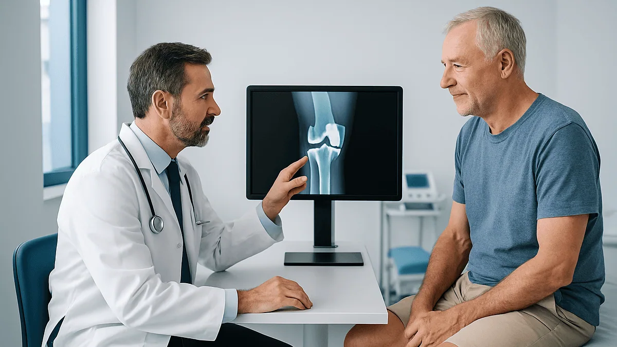
Key Takeaways
- Image-based robotic systems require a pre-operative CT of the hip, knee and ankle, while imageless robotic and conventional knee replacements plan from standing X-rays and mapped landmarks instead.
- The hip and ankle are scanned to locate the mechanical axis, the straight line from hip center to ankle center that determines how load passes through the new knee.
- CT is preferred over MRI for planning because it shows bone geometry and rotation precisely, whereas MRI excels at soft tissue, which is not what implant positioning depends on.
- The implants used in almost all robotic knee replacement are standard components in standard sizes; the scan guides their selection and position rather than producing a custom part.
- Comparative studies have not shown a clear accuracy advantage for image-based over imageless robotic systems, so the absence of a CT is not a marker of lower-quality care.
- Mainstream guidance places hospital stays at around one to three days and a return to most daily activities at roughly three to six weeks, with full recovery taking many months regardless of whether a robot was used.
Imaging before robotic knee replacement usually means a CT scan of the hip, knee and ankle, or detailed X-rays, that software turns into a three-dimensional model of your leg. The surgical team uses that model to plan implant size, position and alignment before the operation, then confirms the plan on your actual bone during surgery. Some robotic systems work from landmarks mapped in the operating room instead of a CT, so the exact imaging depends on the system your surgeon uses.
The envelope from the imaging department feels oddly thin for something that is supposed to decide the shape of your new knee. Inside is an appointment for a CT scan that, the letter says, will cover your hip, your knee and your ankle. You only have a problem with the knee. Why scan the rest?
That question is one of the most common ones patients bring to a pre-operative clinic, and it gets to the heart of what imaging before robotic knee replacement is for. The scan is not looking for a diagnosis; you already have one, usually advanced osteoarthritis, the wear-and-tear breakdown of joint cartilage. The scan is a measuring tool. It lets a computer build a model of your leg so a surgeon can decide where every cut and every implant surface should sit before anyone picks up an instrument.
This article walks through what those images are, why some systems need them and others do not, what else gets tested, and what the weeks afterward tend to look like, with the evidence stated plainly where it is thin.
How imaging before robotic knee replacement actually works
Start with what a knee replacement is. The worn ends of the thigh bone (femur) and shin bone (tibia) are trimmed and capped with metal components, with a plastic spacer between them. Mayo Clinic describes the goal simply: resurface the damaged joint so it moves with less pain. A robotic system does not perform that operation on its own. It is a tool held or guided by the surgeon that limits the cutting instrument to a planned zone and tracks the bones in real time.
For the tool to know where that zone is, it needs a map. Imaging before robotic knee replacement is how that map is drawn. In image-based systems, a CT scan is taken days or weeks before surgery. CT, or computed tomography, is a rotating X-ray that produces cross-sectional slices of the body. Software stacks those slices into a three-dimensional model of your femur, tibia and kneecap, accurate to the contours of your particular bone, including the bony spurs (osteophytes) that osteoarthritis grows along joint edges.
On that model the surgeon rehearses the operation: choosing the implant size that best covers the bone surface, setting the angle of each cut, and checking how the leg would line up from hip to ankle once the parts are in. Think of it as the tailor taking measurements before cutting cloth, except the measurements can be adjusted and re-checked repeatedly with nothing lost.
In imageless systems, no pre-operative CT is used. Instead, the surgeon touches a tracked pointer to dozens of bony landmarks during the operation and the software builds the model there and then. Both routes end at the same place: a plan the robot helps execute. Which route you take is decided by the system your hospital uses, not by anything about your knee.
Is a CT scan necessary before knee replacement surgery?
The honest answer is: it depends on the system, not on the surgery. Conventional knee replacement, which is still the most common way the operation is done, is planned from standing X-rays. Surgeons have been doing this for decades with durable results, and no major guideline body requires CT for a routine knee replacement.

A CT becomes necessary only when the surgeon is using an image-based robotic platform. Those systems are built around a pre-operative CT model, and they cannot generate a plan without one. In that case the scan is a part of the procedure in the same way a blood test is part of an anesthetic assessment.
Imageless robotic systems, and computer navigation systems that are not robotic at all, skip the CT and gather their landmark data in the operating room. If your surgeon uses one of these, you may only have X-rays beforehand, and that is not a shortcut. It is simply a different way of building the same map.
What about radiation? Mayo Clinic notes that CT uses more ionizing radiation than a plain X-ray. A pre-operative knee CT, however, is a limited study of the lower limb, usually without contrast dye, and is a one-time exposure in the setting of a major operation. For most adults the trade-off is considered reasonable, but it is a fair question to raise, particularly if you have had many scans recently or are pregnant. Contrast is almost never needed for this planning scan, which matters if you have kidney problems or a previous reaction to iodine-based dye, two issues Mayo Clinic flags for CT generally.
If you are not sure whether your scan is diagnostic or planning, ask. The purpose changes how the images are taken and read.
Why the CT scan before knee replacement covers your hip and ankle too
This is the part that puzzles the most people. The knee hurts; the scan includes two joints that do not. The reason is a single straight line.
Surgeons talk about the mechanical axis of the leg, which is the imaginary line from the center of the hip joint to the center of the ankle joint. In a well-aligned leg, that line passes through or close to the middle of the knee. In an arthritic knee that has worn more on one side, the leg bows inward or outward and the line drifts off center, loading one compartment of the joint far more than the other. An implant placed without regard to this line can wear unevenly or feel unstable.
To find that line you need the two ends of it. A CT that includes thin slices through the hip center and the ankle center lets software calculate the mechanical axis to a fraction of a degree. The knee itself is scanned in finer detail, while the hip and ankle are captured in a quick, lower-detail pass, which is also why the scan is faster than you might expect.
Standing long-leg X-rays do the same job in two dimensions, and many surgeons use them in addition to or instead of CT. The CT adds the third dimension: rotation. Bones can be twisted along their length as well as angled, and rotation is nearly impossible to judge on a flat X-ray. In robotic planning, rotational alignment of the femoral component is one of the variables the model exists to settle.
Nothing about this means your hip or ankle will be treated or even reported on in detail. They are reference points, like the pegs at either end of a chalk line.
From scan to 3D model: how robotic knee replacement planning uses your images
After the CT, a technician or engineer segments the scan, which means outlining the bone on each slice so software can separate femur from tibia from kneecap and discard the soft tissue. The output is a robotic knee replacement 3D model that the surgeon can rotate on a screen, cut through virtually and measure.
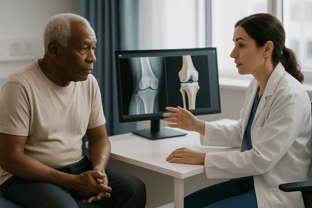
Three decisions are then made on that model, in roughly this order.
Size comes first. Knee implants come in a range of sizes for each component, and an implant that overhangs the bone edge can irritate soft tissue, while one that is too small leaves bone uncovered. The model lets the surgeon check fit around the whole circumference of the bone, not just the front view an X-ray gives.
Position comes next. Where the components sit, how far forward or back, and how they rotate relative to the bone, all influence how the kneecap tracks and how the ligaments tension through bending and straightening. The surgeon moves the virtual components until those relationships look right.
Alignment is the final check. The software draws the mechanical axis through the planned implants and reports the angle at the knee. Some surgeons aim for a perfectly straight line; others deliberately keep a little of the patient’s natural lean, a philosophy sometimes called kinematic alignment. The evidence on which approach produces better long-term function is still evolving, and there is no consensus that one is superior for every knee. Your surgeon’s choice is a clinical judgment, and it is a reasonable thing to ask about.
What the model cannot show is the soft tissue: ligaments, capsule and scar. That information is collected in the operating room, and it is why the plan made on the screen is a draft, not a contract.
Do you need an MRI before knee replacement?
Usually not, and it is worth understanding why, because many people arrive assuming an MRI is the gold standard for any knee problem.
MRI, or magnetic resonance imaging, uses a strong magnet and radio waves rather than X-rays, and it is superb at showing soft tissue: cartilage, menisci, ligaments and bone marrow swelling. Mayo Clinic describes it as the preferred test for many joint injuries. That is exactly why it is so useful earlier in the story, when a doctor is working out whether a painful knee is arthritis, a torn meniscus or something else.
By the time a joint replacement is being planned, that question has been answered. The cartilage is known to be worn because plain X-rays show the joint space narrowed and the bone surfaces touching. What the planning team needs now is bone geometry, and bone is where CT outperforms MRI. CT gives crisp edges, true-to-scale dimensions and reliable detail of the dense cortical shell that implants rest on.
A few robotic or patient-specific instrument systems were designed to work from MRI models, and a surgeon may order one for other reasons, such as suspected bone death (osteonecrosis) or an unusual deformity. If an MRI before knee replacement is requested, it is reasonable to ask what question it is answering.
There are also practical limits. MRI takes longer, is noisy, and is unsuitable for some people with certain implanted devices or metal fragments, points the Mayo Clinic MRI page spells out. For a procedure whose planning hinges on bone shape, the shorter, bone-focused CT or a set of standing X-rays is generally the better fit.
If you have had an MRI already, bring the report. It will not usually be repeated, but the surgeon may want to see it.
What other tests are done before knee replacement surgery?
The planning scan is one line in a longer checklist. The rest of the pre-operative assessment is about whether your body is ready for a major operation and an anesthetic, and it is similar whether or not a robot is involved.
Blood tests are near-universal. A full blood count checks for anemia, which matters because joint replacement involves some blood loss. Kidney function and electrolytes guide fluid and medication choices. A blood sugar measure, often including a longer-term average marker, is checked because poorly controlled diabetes raises infection and wound-healing risk. Blood grouping is done in case a transfusion is needed.
Heart assessment usually includes an electrocardiogram, a tracing of the heart’s electrical rhythm. People with known heart or lung disease may be referred for further tests or a specialist opinion.
Infection screening is routine in most services. Swabs of the nose and sometimes the skin look for bacteria such as Staphylococcus aureus, including resistant strains, so that a decolonization routine can be arranged before surgery if needed. A urine test may be included. Dental health is frequently asked about, since untreated dental infection is a recognized source of bacteria that can seed a new joint.
A medication review is where decisions about blood thinners, diabetes medicines and some arthritis treatments are made. These are individual decisions for the prescribing clinician and the anesthetic team; do not stop or change anything on your own.
Finally, a physical examination of the knee records how far it bends and straightens and how stable the ligaments feel. The NHS also describes a pre-operative assessment visit as the moment to discuss what to expect afterward and to arrange help at home. Treat it as a planning meeting, not an exam to pass.
Image-based vs imageless robotic knee replacement: how they compare
Patients often ask which type of robotic system is better. The research so far does not show a clear winner in alignment accuracy or early outcomes between the two approaches, and comparative studies are still relatively small and short in follow-up. What differs is the workflow, and that is what affects you.
| Feature | Image-based robotic | Imageless robotic | Conventional (non-robotic) |
|---|---|---|---|
| Pre-operative imaging | CT of hip, knee and ankle, usually without contrast | Standing X-rays; no CT required | Standing X-rays |
| When the 3D model is built | Before surgery, from the CT | During surgery, from mapped bone landmarks | No 3D model; plan drawn on X-rays |
| Implant size chosen | On the model beforehand, confirmed in theater | In theater after mapping | In theater with trial components |
| Extra radiation | One limited CT | None beyond X-rays | None beyond X-rays |
| Added steps in theater | Registration of bone to the CT model | Landmark mapping, often longer | Standard jigs and guides |
| Who decides which you have | The surgical team, based on the equipment and training available |
Two caveats belong next to this table. First, the words "robotic" and "conventional" describe how bone is cut, not what is implanted. The metal and plastic parts are often identical across all three columns. Second, surgeon experience with a given method shapes results at least as much as the method itself, and this is one reason why head-to-head studies struggle to isolate the technology.
If you are offered an image-based system, expect the CT. If you are offered an imageless system or a conventional operation, do not read the absence of a CT as a lesser standard of care. The evidence does not support that reading.
What happens in the operating room once the plan exists
The scan and model are the preparation. On the day, a few extra steps turn them into something the robot can act on.
Once you are anesthetized and the knee is opened, the surgeon fixes small tracking arrays to the femur and tibia. These are posts with reflective markers that an infrared camera watches, so the system knows where each bone is in space, even as the leg is bent and moved. They require a couple of small additional pin sites in the bone, which heal like the main incision.
Next comes registration, the process of matching the real bone to the model. In an image-based system the surgeon touches a tracked probe to a series of points on the exposed bone, and the software checks those points against the CT surface until the match is within a tight tolerance. In an imageless system this same probing is how the model is created in the first place. If registration does not reach the required accuracy, the surgeon repeats it or converts to a conventional technique. That fallback is planned for, not a failure.
Then the plan is tested against reality. The surgeon moves the knee through its range while the system measures the gap between femur and tibia with the ligaments under tension. If one side is tighter than the other, the planned cuts or implant position can be adjusted on screen, in millimeters and degrees, before anything is removed. This soft-tissue balancing is where the surgeon’s judgment matters most, because no scan can measure a ligament.
Only after that does cutting begin. Depending on the system, the robotic arm either holds the saw within the planned boundary or positions a cutting guide for the surgeon. The implants are trialed, the final components fixed in place, and the knee is closed. The robot has shaped the precision; the surgeon has made every decision.
Who robotic knee replacement is usually for, and who is asked to wait
The criteria for a robotic knee replacement are, in practice, the criteria for any knee replacement. Mayo Clinic lists the usual trigger as severe knee pain or stiffness that limits everyday activities such as walking, climbing stairs or getting up from a chair, when non-surgical options have been tried. Those options include weight management, physiotherapy, activity modification, walking aids, oral anti-inflammatory medicines and joint injections. Their mechanisms and timelines vary, and whether they have been given a fair trial is a judgment for your treating team.
Imaging confirms that the pain has an anatomical source: joint-space narrowing, bone-on-bone contact or deformity visible on X-ray. Pain without those changes usually prompts a search for other causes before surgery is considered.
Who is asked to wait? Patients and search engines phrase this as what disqualifies you from knee replacement surgery, and the honest answer is that very few things are absolute, but several things prompt a pause.
- Active infection anywhere in the body, including skin, urinary tract or teeth, because bacteria can settle on a new implant.
- Poorly controlled diabetes, which raises wound and infection risk until blood sugar is steadier.
- Unstable heart or lung disease that makes the anesthetic itself a greater danger than the knee.
- Open skin wounds or poor circulation in the leg that could compromise healing.
- Smoking, which many services ask patients to stop before surgery because it impairs healing.
- A body weight at which anesthetic and surgical risk is judged too high for the moment; this is a conversation about safety, never a judgment of worth.
For the robotic element specifically, there are a few additional considerations: a patient who cannot have a CT, or whose bone is so distorted by previous fracture or hardware that registration would be unreliable, may be better served by a conventional approach. The surgeon makes that call after seeing the images.
How painful is robotic total knee replacement?
Set expectations honestly: knee replacement is a major operation on bone, and the days afterward are uncomfortable by any route. The robotic method changes how the bone is cut; it does not change the fact that bone is cut.
Where the method may make a difference is in the soft tissue. Because the robotic system confines the saw to a planned volume, there is a theoretical reduction in incidental injury to ligaments and capsule around the joint. Several studies have reported lower early pain scores and less need for pain relief in the first days and weeks after robotic knee replacement compared with conventional surgery. The quality of that evidence is mixed, follow-up is often short, and differences tend to narrow over the following months. It would be inaccurate to promise a less painful recovery; it is fair to say early comfort may be modestly better and that the long-term picture appears similar.
What reliably reduces pain is the anesthetic and analgesic plan, and that is independent of the robot. Most services use a combination: a spinal or general anesthetic, local anesthetic injected around the joint during surgery, sometimes a nerve block that numbs the front of the thigh, and a schedule of oral medicines afterward. The choice and timing of each belongs to the anesthetic and surgical team and is tailored to your health history and other medicines.
Ice, elevation and early movement do real work too. Getting up on the day of surgery or the next morning, which the NHS describes as the norm, stiffens the knee less than lying still, and a knee that bends early tends to hurt less later.
Pain that is getting worse rather than better after the first week, or that comes with fever, redness or calf swelling, is not normal recovery pain. The final section covers what to do.
What the days and weeks after surgery usually look like
The scan shaped the plan; the timeline afterward looks much like any knee replacement. These are typical ranges from mainstream guidance, not promises, and your own progress will be set by your team.
The first day or two are spent in hospital, where a physiotherapist helps you stand, take steps with a frame or crutches and begin bending exercises. According to the NHS, most people go home after around one to three days, provided they can walk safely and manage stairs if needed.
In the first two weeks at home, the priorities are swelling control, wound care and daily exercises. Ice and elevation help the swelling; the wound is kept clean and dry until the clinic or surgeon confirms it has sealed. Walking short distances several times a day is encouraged. Clot prevention, with compression stockings, movement and often a prescribed blood-thinning medicine, continues as directed by the surgical team.
Between roughly three and six weeks, Mayo Clinic notes that most people can resume most daily activities, including light housework and short outings. The NHS suggests that walking aids can usually be set aside at around six weeks, and that returning to work depends heavily on the job: sooner for desk work, later for physically demanding roles.
Driving is a common question. The usual advice is to wait until you can bend the knee enough to sit comfortably, control the pedals and perform an emergency stop, and until you are no longer taking medicines that impair reaction. Your surgeon will confirm the timing, and insurers may have their own rules.
Full recovery is longer than many expect. The NHS notes it can take up to a year for the knee to settle fully, with strength and swelling improving over months. The robot does not shorten this biology; good physiotherapy and patience shape it more than any scan did.
Risks, limits and alternatives: what the imaging cannot fix
A precise map does not remove the risks of the journey. Knee replacement by any method carries recognized complications, and MedlinePlus and Mayo Clinic list the main ones: infection in the wound or deep around the implant, blood clots in the leg or lung, bleeding, injury to nerves or blood vessels, stiffness, persistent pain, and, over the long term, loosening or wear of the components that may require revision surgery. Anesthetic risks apply as well.
Robotic surgery adds a handful of its own. The tracking pins create extra small holes in the bone, and rare fractures at those sites have been reported. Registration can fail, requiring a switch to conventional technique mid-operation. The additional steps can lengthen surgery, particularly early in a surgeon’s experience with the system. None of these are common, but they are real and your consent discussion should include them.
Imaging has its own limits. A CT shows bone but not the ligaments that decide how a knee feels. A model built from a scan taken lying down may not perfectly reflect how the leg aligns under load when standing. And a flawless plan executed precisely can still produce an unhappy knee if the expectations attached to it were unrealistic; a replaced knee generally bends less than a healthy natural one and may feel different for a long time.
Alternatives exist at every stage. Before surgery: structured exercise, weight management, bracing, walking aids, and medicines or injections whose role is for the treating clinician to judge. Surgical alternatives include a partial knee replacement if only one compartment is worn, a realignment operation (osteotomy) in some younger patients, and conventional or computer-navigated total knee replacement. None is right for everyone, and a good consultation lays them side by side rather than steering toward one.
What people often get wrong about imaging before robotic knee replacement
"The scan will tell me how bad my knee is." The planning CT is a measuring study. The decision that your arthritis warrants surgery has already been made from X-rays, examination and your account of daily life. If you are hoping the CT will settle whether you need the operation, that is a different conversation to have with your surgeon.
"A robot does the operation." Every decision, from implant size to how much ligament tension is acceptable, is the surgeon’s. The system constrains the saw and tracks the bone. It does not plan, cut or close on its own.
"No CT means a lower standard." Imageless robotic systems and conventional surgery do not use a pre-operative CT, and the current evidence does not show them to be less accurate in alignment. The CT is a requirement of a particular system, not a marker of quality.
"The implant is custom-made from my scan." In almost all robotic knee replacement, the implants are standard components in standard sizes. The scan helps choose and position them. Truly custom implants exist but are a separate, uncommon approach.
"MRI is better because it shows more." It shows more soft tissue, which is not what the planning needs. For bone geometry, CT or standing X-rays are the appropriate tools.
"Robotic means painless or faster recovery." Early pain may be modestly lower in some studies; the overall recovery timeline, including the months it takes swelling and strength to settle, is similar. Marketing phrases like "minimally invasive" or "quicker return" should be read against the evidence, which is cautious.
"A perfect plan guarantees a perfect knee." Alignment is one variable among many. Ligament balance, rehabilitation effort, overall health and the realistic limits of an artificial joint all shape the result. A good scan improves the odds of good positioning; it does not promise an outcome, and no honest clinician will say otherwise.
Questions to ask your care team
Consultations are short and the information is dense. Writing questions down beforehand, and bringing someone to take notes, changes what you come away with. These are the ones most relevant to imaging and planning.
- Which type of system do you use, image-based or imageless, and what imaging will I need because of that?
- Will the CT include my hip and ankle, and will it need contrast dye? I have kidney issues or a previous reaction to contrast, if either applies to you.
- How much extra radiation does the planning scan involve compared with the X-rays I have already had?
- What alignment philosophy do you use, and why do you prefer it for my knee?
- Will my implant be standard or custom, and what happens if the size planned on the scan does not match on the day?
- What is your plan if registration fails or the system is unavailable? Have you converted to conventional surgery before?
- How many robotic knee replacements have you performed, and how does your experience with this system compare with your experience in conventional surgery?
- What are the risks specific to the robotic approach, including the tracking pin sites?
- Which of my current medicines need to be reviewed before surgery, and who will tell me what to do about each one?
- Would a partial knee replacement, an osteotomy or a conventional total knee replacement be reasonable for me, and why or why not?
- What does the first two weeks at home realistically look like, and what help should I arrange?
- Who do I call, at any hour, if something worries me after I go home?
A team that welcomes these questions is telling you something about how they will handle the harder conversations later. Ask them plainly; you are entitled to plain answers.
When to call your doctor
Discomfort, bruising and swelling that slowly improve are expected after knee replacement. Certain changes are not, and they need prompt attention rather than a wait-and-see approach. Contact your surgical team or seek urgent care if you notice any of the following in the weeks after surgery.
- Calf pain, tenderness, warmth or swelling in either leg, which can signal a deep vein thrombosis, a clot in a deep leg vein.
- Sudden shortness of breath, chest pain or coughing up blood, which can signal a pulmonary embolism, a clot that has traveled to the lung. Treat this as an emergency.
- Fever or shaking chills, particularly alongside a knee that is becoming hotter, redder or more swollen.
- Wound changes: spreading redness, increasing fluid or pus, a foul smell, or edges that have opened.
- Pain that is escalating after the first week rather than easing, or pain that no longer responds to the plan you were given.
- Numbness, tingling or weakness in the foot that is new or worsening, or a foot that turns pale or cold.
- A fall onto the operated leg, a new deformity, or a sudden inability to bear weight.
- Bleeding that soaks through dressings, or black stools, which can indicate bleeding related to blood-thinning medicine.
These signs are drawn from mainstream guidance on knee replacement recovery and apply whether or not a robot was used. If you are unsure whether something is normal, ring. Surgical teams would far rather answer an unnecessary call than miss an infection or a clot that was caught late. Any decision about further tests or treatment rests with the clinicians caring for you.
Frequently asked questions
Is a CT scan necessary before knee replacement surgery?
Only if your surgeon uses an image-based robotic system, which builds its plan from a CT model. Conventional knee replacement and imageless robotic systems plan from standing X-rays and landmarks mapped during the operation, and no major guideline requires CT for a routine knee replacement. The scan is a requirement of the technology, not a measure of how thorough the care is.
Why does the CT scan before knee replacement include my hip and ankle?
To find the mechanical axis, the line from the center of the hip to the center of the ankle that shows how weight travels through your leg. The knee is scanned in detail while the hip and ankle are captured quickly as reference points. Nothing about those joints is being treated; they are the two pegs that let software measure your alignment to a fraction of a degree.
Do I need an MRI before knee replacement?
Usually not. MRI shows cartilage, menisci and ligaments, which is valuable when the diagnosis is uncertain, but by the time a replacement is planned the diagnosis is settled and the team needs bone shape, which CT or X-rays show better. An MRI may be ordered for a specific question such as suspected bone death or an unusual deformity. If one is requested, ask what it is meant to answer.
What is the difference between image-based and imageless robotic knee replacement?
Image-based systems create a three-dimensional model from a CT taken before surgery; imageless systems build the model in the operating room by touching a tracked probe to bony landmarks. Both guide the surgeon’s cuts and track the bones in real time. Current comparative studies have not shown a consistent accuracy advantage for either approach, and the implants used are typically the same.
How is a robotic knee replacement 3D model used during the operation?
The model is matched to your actual bone through registration, where the surgeon touches points on the exposed bone and the software confirms they align with the scan. The knee is then moved through its range to measure ligament tension, and the planned cuts can be adjusted on screen before anything is removed. Only after that does the robotic system help guide the saw within the planned boundary.
What tests are done before knee replacement surgery besides imaging?
Blood tests for anemia, kidney function, blood sugar and blood group; an electrocardiogram to check heart rhythm; swabs for bacteria that could infect the implant; and a medication review covering blood thinners and diabetes medicines. A physical examination records how far the knee moves. The NHS describes this pre-operative assessment as the time to plan help at home as well.
How painful is robotic total knee replacement compared with conventional surgery?
Both involve cutting bone, so the early days are uncomfortable either way. Some studies report modestly lower pain scores in the first weeks after robotic surgery, possibly from less disturbance to surrounding soft tissue, but the evidence is mixed and differences tend to fade over months. The anesthetic and pain-relief plan, chosen by your team, has a larger influence on comfort than the cutting method.
What disqualifies you from knee replacement surgery?
Few things are absolute, but active infection anywhere in the body, poorly controlled diabetes, unstable heart or lung disease, open skin wounds on the leg and smoking commonly lead teams to ask patients to wait until the issue is addressed. For the robotic element specifically, bone distorted by old fractures or hardware, or an inability to have a CT, may favor a conventional approach. The surgeon makes that judgment.
Does the planning CT mean my implant is custom-made?
In almost all cases, no. The implants are standard components available in a range of sizes, and the CT helps the surgeon choose which size and where to position it. Truly custom, patient-specific implants exist but are a separate and uncommon approach. If you want to know which applies to you, ask directly whether the components are off-the-shelf or made for your anatomy.
How long after robotic knee replacement can I walk without aids?
Most people stand and take steps within a day of surgery, and the NHS suggests walking aids can usually be set aside at around six weeks, with Mayo Clinic placing a return to most daily activities at roughly three to six weeks. These are typical ranges, not promises, and the robotic method does not shorten the underlying healing. Your physiotherapist and surgeon will set the pace for you.
References
This article is for general information only and is not a substitute for professional medical advice. Please consult a qualified doctor about your individual situation.

More from the Blog

What a Slipped Disc Feels Like, and How to Tell If You Have One
A slipped (herniated) disc most often feels like a deep ache in the lower back or neck paired with sharp, burning or electric pain…

Falls Prevention After 65: The Strength and Balance Program That Cuts Fractures
Falls prevention after 65 works best with a structured strength and balance program: progressive balance exercises, leg-strengthening and regular walking, done about three times…

What Kind of Anesthesia Is Used for Hip Replacement? Spinal, General and the Recovery Room
Most hip replacements are done under spinal anesthesia, a numbing injection in the lower back that blocks feeling from the waist down, usually paired…

Hand Surgery Recovery Milestones: Splint Removal, Grip Strength and Returning to Work
Hand surgery recovery time depends on what was repaired. The skin usually heals within about two weeks, a splint often comes off somewhere between…

Can Clubfoot Come Back After Treatment? Relapse, Bracing Lapses and What Happens Next
Yes, clubfoot can come back after treatment. Relapse most often appears in the first few years of life, and the strongest known predictor is…

Caring for a Baby in a Spica Cast After Hip Dysplasia Surgery: Feeding, Diapers and Sleep
Spica cast care for a baby after hip dysplasia surgery centers on four routines: keep the cast dry and intact, change diapers often with…