

The PET Scan: How It Works and Why It Is Ordered: Explained Calmly
Key Takeaways
- The scan itself takes only 20 to 40 minutes; the two-hour appointment exists because the tracer needs about an hour of quiet rest to distribute through your body.
- The most common tracer is a radioactive sugar look-alike with a half-life of roughly 110 minutes, and it is essentially gone from your body within a day, mostly through urine.
- Skip strenuous exercise for 24 hours before your scan, worked muscles absorb the sugar tracer and can create bright spots that obscure or mimic disease.
- A PET scan can miss cancer clusters smaller than about 7 to 8 millimeters and certain slow-metabolizing tumors, so a clear result means no detectable active disease, not certainty.
- Bright spots are not automatically cancer: infection, healing tissue, arthritis, and even recent vaccination sites consume extra glucose and can light up on the images.
- For a few hours after the scan, avoid prolonged close contact with pregnant women and small children while the small amount of remaining tracer decays.
A PET (positron emission tomography) scan is an imaging test that uses a tiny amount of a radioactive tracer, usually a sugar-like compound, to show how tissues are functioning rather than just how they look. Doctors most often order it to find, stage, or monitor cancer, and sometimes to evaluate the heart or brain. The scan itself usually takes 20 to 40 minutes, after roughly an hour of quiet preparation.
The strangest part of a PET scan is not the machine. It is the hour before it, when a technologist places a small IV in your arm, dims the lights, and asks you to sit as still as a library. No phone scrolling, no pacing, ideally not even much talking. That quiet hour has a purpose: the tracer circulating through your bloodstream is quietly mapping which of your cells are working hardest.
Most imaging tests take a photograph of your anatomy. This one records a live broadcast of your metabolism. That difference is why an oncologist may order a PET scan after a CT already found something, and why the results can change a treatment plan in a single afternoon.
If the word “radioactive” on the consent form made your stomach tighten, you are in good company. Here is what actually happens, what the scan can and cannot see, and why the whole process is calmer than it sounds.
What is a PET scan, exactly?
Positron emission tomography builds a three-dimensional picture of chemical activity inside the body. Before the scan, you receive a small injection of a radiotracer, most commonly a compound built to resemble glucose, the sugar every cell burns for energy. Hungry, fast-dividing cells pull in more of it. Sluggish or dying tissue pulls in less.
As the tracer settles into tissues, its radioactive atoms decay and release positrons. Each positron collides almost instantly with an electron, and the two annihilate each other, sending out a pair of gamma rays in opposite directions. The doughnut-shaped scanner detects those paired rays, and a computer traces each pair back to its point of origin. Millions of these tiny flashes, plotted together, become a color-coded map of metabolic activity: bright where cells are burning fuel fast, dim where they are not.
According to MedlinePlus, this functional view is the whole point. A tumor, an area of inflammation, or a patch of heart muscle starved of blood flow often behaves abnormally before it looks abnormal on other scans. PET catches the behavior.
One clarification worth making early: PET rarely travels alone anymore. Nearly every modern PET scan is actually a PET/CT, meaning a CT scan runs in the same machine during the same appointment. The CT supplies the anatomical street map; the PET supplies the traffic report. Radiologists read them as one fused image.
How does the radioactive tracer actually work?
Cancer cells are famously greedy. Many divide faster than normal tissue and consume glucose at several times the usual rate: a quirk biologists have known about for nearly a century. The most widely used PET tracer exploits this appetite. Because it is chemically dressed up as sugar, metabolically active cells absorb it eagerly, then get stuck with it; the molecule enters the cell but cannot be fully processed, so it accumulates exactly where activity is highest.
The radioactive atom attached to that sugar analog has a half-life of roughly 110 minutes. In practical terms, half the radioactivity is gone in under two hours, and the vast majority has decayed or been flushed out in urine within a day. This is why the tracer must be produced fresh, often the same morning, and why appointment times at PET centers are strict: the material is literally disappearing on the shelf.
Not every tracer is sugar-based. Specialized compounds can highlight amyloid protein in the brain, blood flow in heart muscle, or specific receptors on certain tumor cells. Your care team chooses the tracer based on the question being asked, which is one reason two people can both “get a PET scan” and have quite different experiences.
A few tissues glow brightly on every sugar-based scan no matter what: the brain, which burns glucose constantly, and the bladder, where the tracer collects on its way out. Radiologists expect this background and read around it.
Why would a doctor order a PET scan?
Cancer care accounts for the great majority of PET scans, and the test earns its keep at several distinct moments in that journey.
- Characterizing a finding. A CT or X-ray spots a nodule. Is it metabolically active, which raises concern, or quiet, which is reassuring? PET helps sort the two, though it cannot make the distinction with certainty on its own.
- Staging. Before treatment begins, doctors need to know whether a known cancer has traveled. Because PET surveys the whole body in one pass, it can reveal distant deposits that targeted scans would miss.
- Checking whether treatment is working. A tumor that stops taking up tracer partway through therapy is often responding, sometimes before it shrinks visibly. That early signal can spare someone months of an ineffective regimen.
- Watching for recurrence. Scar tissue from surgery or radiation can look identical to tumor on a CT. Metabolic imaging helps distinguish dead scar from active disease.
Beyond oncology, cardiologists use PET to assess blood flow through heart muscle and to identify tissue that is damaged but still salvageable. Neurologists use it to investigate certain dementias and to pinpoint seizure origins before epilepsy surgery. The Mayo Clinic lists all three arenas, cancer, heart disease, and brain disorders, as core uses.
What PET is not: a general screening test. No major medical body recommends whole-body PET scans for healthy people without symptoms, because false alarms would vastly outnumber real finds.
How long does a PET scan take?
Plan on about two hours door to door, sometimes closer to three. The scan itself is the short part; the choreography around it takes longer, and every step exists for a reason.
First comes check-in and a brief review: recent illness, medications, blood sugar if you have diabetes, and any chance of pregnancy. Then the tracer injection through a small IV, no more uncomfortable than a routine blood draw. What follows is the quiet stretch that surprises most first-timers: roughly 45 to 60 minutes of resting while the tracer distributes through your body.
Stillness during this window genuinely matters. Muscles absorb glucose when they work, so fidgeting, walking the hallway, or even vigorous chewing can pull tracer into muscle tissue and muddy the images. Some centers ask you not to read or talk much, since jaw and eye muscles are metabolically busier than you would think.
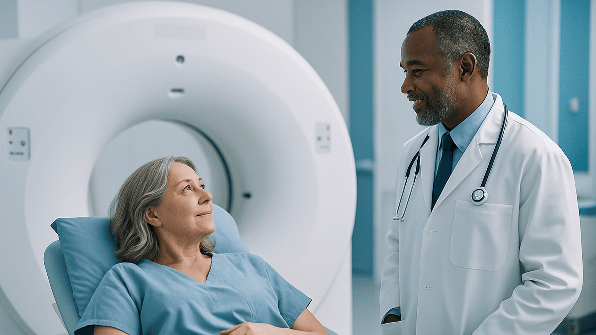
The scan itself typically runs 20 to 40 minutes, per Cleveland Clinic. You lie on a padded table that glides slowly through the scanner’s opening. The machine is quieter than an MRI, no jackhammer clatter, mostly soft whirring, and the ring is shorter and more open than many people expect, which claustrophobic patients often find a relief.
Afterward there is no recovery period. Most people walk out, drive themselves home, and go about their day, with one standing instruction: drink plenty of water to help your kidneys flush the remaining tracer.
What happens on scan day, step by step
Knowing the sequence in advance takes most of the anxiety out of it. A typical appointment unfolds like this:
- Arrival and screening. Staff confirm you have fasted as instructed and ask about diabetes, kidney function, pregnancy, and breastfeeding. If you have diabetes, they may check your blood sugar, since a high level competes with the tracer and can degrade image quality.
- IV and injection. The tracer arrives in a shielded container, dramatic looking, but the dose inside is small. The injection itself takes seconds and does not cause a flush, a taste, or any sensation for most people.
- The uptake hour. You rest in a recliner in a dim, quiet room, usually alone, for 45 to 60 minutes. Warm blankets are common; being cold makes a specialized tissue called brown fat light up and can complicate the read.
- A bathroom stop. Emptying your bladder just before scanning improves images of the pelvis, where the tracer pools.
- The scan. You lie still on the table, usually arms overhead or at your sides depending on the target area, for 20 to 40 minutes. Breathing normally is fine; a technologist watches from the next room and can hear you the whole time.
- Done. No sedation to wear off in most cases, no bandage beyond a small dot over the IV site.
Results are not instant. A radiologist or nuclear medicine physician analyzes the images and sends a report to the doctor who ordered the scan, usually within a few days. Ask before you leave how and when you will hear back: it saves days of inbox-refreshing.
Do you wear clothes during a PET scan?
Usually, yes, often your own. Policies vary by imaging center, but the deciding factor is metal, not modesty. Zippers, underwire, snaps, belt buckles, jewelry, and clothing with metallic threads can distort the CT portion of a PET/CT, so anything metal near the area being scanned has to go.
Many centers ask patients to change into a gown or scrubs simply because it is faster than auditing everyone’s wardrobe. Others let you stay dressed if you arrive in metal-free clothing, think sweatpants, a sports bra without underwire, and a plain T-shirt. If keeping your own clothes on matters to you, call ahead and ask what qualifies; most centers are happy to explain.
A few related practicalities people wish they had known:
- Leave jewelry, watches, and piercings at home when possible, rather than trusting a locker.
- Dress warmly or ask for blankets. Shivering activates brown fat, which absorbs the tracer and can create confusing bright spots, particularly around the neck and shoulders.
- Eyeglasses, hearing aids, and dentures can typically stay on until the scan itself, and staff will tell you if and when to remove them.
- If you use an insulin pump or continuous glucose monitor, mention it during scheduling so the team can plan around it.
You are never left exposed. Positioning is done with drapes and blankets, and you can ask for a same-gender technologist at most facilities if that would make you more comfortable. This is a routine outpatient test, and the staff run it dozens of times a week.
How should you prepare, and why each rule exists
PET prep instructions can read like arbitrary rules. They are not. Each one protects the accuracy of a scan that is fundamentally a picture of your metabolism, which means your recent behavior literally shows up in the images.
Fasting, usually four to six hours. Eating raises blood glucose and triggers insulin, which shoves sugar into muscles throughout your body. If that happens right before your injection, muscle everywhere soaks up tracer and the tumor-to-background contrast collapses. Water is not just allowed but encouraged; hydration helps clear excess tracer from the bloodstream.
No strenuous exercise for about 24 hours beforehand. A hard workout leaves muscles hungrily absorbing glucose for a full day afterward. A radiologist can usually tell who went for a long run the evening before: their leg muscles glow. That glow can obscure or mimic disease.
Blood sugar management if you have diabetes. Elevated glucose competes directly with the sugar-based tracer for entry into cells, dimming the signal. Centers typically want blood sugar below a specific threshold at injection time and will coordinate the timing of your usual regimen with your care team. Never adjust anything on your own; call the imaging center for tailored instructions.
Tell staff about pregnancy or breastfeeding. Radiation exposure is weighed differently in pregnancy, and the tracer passes into breast milk, so temporary adjustments to feeding are usually advised. The NHS covers both scenarios in its patient guidance.
Caffeine and gum are often on the no list too. Caffeine nudges metabolism; chewing works the jaw muscles. Small things, but this scan sees small things.
Is the radiation from a PET scan dangerous?
Honest answer: the dose is real, it is larger than an X-ray, and for the situations in which PET is ordered, mainstream medical consensus holds that the diagnostic benefit clearly outweighs the small theoretical risk.
Some context makes the numbers less abstract. Everyone on Earth absorbs natural background radiation, from soil, cosmic rays, even the food we eat, averaging roughly 3 millisieverts per year in the United States. A PET/CT delivers a dose in the range of several years’ worth of that background exposure, with the exact figure depending on the tracer amount and how the CT portion is configured. Low-dose CT protocols, now standard at many centers, trim the total meaningfully.
Two features of the tracer keep exposure contained. Its half-life of about 110 minutes means radioactivity fades quickly on its own, and your kidneys actively excrete it, which is why you are told to drink water and urinate frequently afterward. Within roughly a day, essentially none remains.
Sensible precautions for the first several hours after the scan: keep some distance from pregnant women, and avoid prolonged close contact, long cuddles, lap-sitting, with infants and small children. Brief, ordinary interaction is fine. If you are breastfeeding, follow your center’s specific guidance on pumping and timing.
The more useful frame than “is it dangerous” is “is it justified.” PET is not ordered casually or repeated on a whim. When a doctor requests one, it is because the answer it provides, is this cancer spreading, is this treatment working, carries far more weight for your health than the exposure does. If the justification is not obvious to you, asking “what will this scan change about my care?” is a fair and welcome question.
Will a PET scan show all cancer?
No, and any answer that skips past this deserves your skepticism. PET is a powerful test with well-documented blind spots, and understanding them helps you interpret both reassuring and worrying results.
Size matters. Clusters of cancer cells smaller than roughly 7 to 8 millimeters, about the width of a pencil eraser, may not produce enough signal to stand out. A “clean” PET scan means no detectable metabolically active disease, which is genuinely good news, but it is not the same as zero cancer cells anywhere.
Some cancers do not take the bait. The standard tracer works because most tumors devour sugar. A minority do not. Certain prostate cancers, some kidney tumors, particular slow-growing lymphomas, and a subset of lung cancers metabolize glucose sluggishly and can appear deceptively quiet. For some of these, specialized tracers now exist; for others, MRI or CT remains the better tool.
Location matters too. The brain burns glucose constantly, so a bright spot there may be lost in the ambient glow of normal tissue. The bladder and kidneys concentrate the tracer on its way out, complicating reads in the pelvis.
False positives are the flip side. Infection, healing surgical sites, arthritis flares, recent vaccination sites, and ordinary inflammation all consume extra glucose and can light up convincingly. A bright spot is a question, not a verdict: it typically prompts correlation with other imaging or a biopsy before anyone changes your treatment.
This is why Johns Hopkins Medicine and others describe PET as one instrument in an orchestra. It answers a specific question, where is metabolism abnormal, and physicians assemble the full picture from PET, anatomy scans, labs, and tissue samples together.
Is a PET scan better than an MRI?
Neither wins; they answer different questions. MRI produces exquisitely detailed pictures of soft-tissue anatomy: the shape, size, and borders of structures. PET reports on cellular behavior. Asking which is better is like asking whether a floor plan beats a utility bill: it depends entirely on whether you need to know what the house looks like or how much energy it is using.
| PET (usually PET/CT) | MRI | CT alone | |
|---|---|---|---|
| What it shows | Metabolic activity, how hard cells are working | Detailed soft-tissue anatomy | Anatomy, especially bone, lung, and blood vessels |
| Typical scan time | 20–40 minutes (plus ~1 hour uptake) | 30–90 minutes | Seconds to minutes |
| Ionizing radiation | Yes (tracer plus CT component) | None | Yes |
| Excels at | Whole-body cancer staging, treatment response, distinguishing scar from active tumor | Brain, spine, joints, pelvic organs, liver detail | Fast surveys, lung nodules, emergencies |
| Struggles with | Very small lesions, low-metabolism tumors, areas of high background activity | Cannot show whether tissue is metabolically active; slower; challenging with some implants | Cannot distinguish scar from living tumor |
In practice, doctors often use them in sequence: MRI to map a brain tumor’s exact borders before surgery, PET to check whether disease has spread elsewhere, CT to guide a biopsy needle. Hybrid PET/MRI machines exist at some academic centers, marrying metabolic data with MRI’s soft-tissue detail, particularly useful in brain and pelvic imaging, though they remain less widely available than PET/CT.
If your doctor orders one and you expected the other, ask what specific question the chosen scan answers. There is nearly always a concrete reason.
What do the results actually mean?
PET reports are written in a vocabulary of brightness. Radiologists describe areas of increased tracer accumulation, “uptake,” “avidity,” or informally “hot spots”, and quantify them with a number called the standardized uptake value, or SUV. Roughly speaking, SUV compares how much tracer a spot absorbed against what an even distribution throughout the body would predict. Higher numbers mean hungrier tissue.
Resist the urge to treat SUV as a cancer score. Normal organs have baseline values; the liver, for instance, always shows moderate uptake. Inflammation can post numbers that overlap with malignancy. And SUV thresholds vary by tumor type, scanner, and protocol, which is why comparing your number to something you found online is a recipe for needless dread. What matters far more is the pattern, where the uptake is, what the fused CT shows at that spot, and how the value compares to your own previous scans if you have had any.
Radiologists also weigh the change over time. A lesion whose uptake drops sharply between scans during treatment is often responding; one that brightens or multiplies suggests the opposite. That trend line frequently carries more clinical meaning than any single image.
Two practical notes. First, if you read your report through a patient portal before your doctor calls, expect unfamiliar language and possible “incidental findings”, minor observations that are common and usually benign. Write down your questions rather than searching each phrase at midnight. Second, ask your care team to walk you through the images if seeing them helps you; most physicians are glad to, and many patients find that the picture makes the plan easier to understand.
PET/CT hybrids: why two scans in one machine
Early PET machines had a frustrating flaw: they could tell doctors that something metabolically abnormal existed, but only vaguely where. A bright blob might sit in a lymph node, or the bowel loop beside it, or the muscle behind both. Radiologists had to mentally overlay a separate CT, taken on a different day with the patient in a slightly different position: an imprecise art.
Hybrid PET/CT scanners, which became standard through the 2000s, solved this by bolting the two technologies into one gantry. You pass through both during a single appointment without moving from the table, and software fuses the datasets pixel by pixel. The result is a map where every metabolic hot spot has an exact anatomical address.
The CT component serves a second, less obvious purpose: correcting the PET data itself. Gamma rays from deep tissue get partially absorbed on their way out of the body, and the CT’s density map lets the computer compensate, making the metabolic measurements more accurate.
For patients, the hybrid design changes little about the experience beyond a slightly longer table ride. It does mean the radiation dose includes both components, though many centers use a low-dose CT protocol when the CT is only needed for localization rather than diagnosis.
PET/MRI is the newer hybrid, swapping the CT for an MRI. It cuts radiation and adds superb soft-tissue contrast, which shines in brain, head-and-neck, and pelvic imaging. The trade-offs are longer scan times, higher cost, and limited availability, for most whole-body cancer questions, PET/CT remains the workhorse, and there is no evidence patients are shortchanged by it.
Beyond cancer: what PET reveals about the heart and brain
Oncology may dominate the schedule at most PET centers, but two other organs benefit from metabolic imaging in ways nothing else quite matches.
The heart. Cardiac PET can measure blood flow through heart muscle with impressive precision, helping cardiologists judge how significant a coronary artery narrowing really is. Its most distinctive role, though, is the viability question. After a heart attack, some muscle is scarred beyond recovery while neighboring tissue is stunned, alive, but hibernating on reduced blood flow. On PET, hibernating muscle still takes up the sugar tracer; dead scar does not. That distinction helps determine whether a procedure to restore blood flow is likely to help, information that can shape a major treatment decision, as the Mayo Clinic notes among PET’s cardiac uses.
The brain. The brain consumes about 20 percent of the body’s glucose despite being 2 percent of its weight, and diseases alter that consumption in recognizable patterns. In certain dementia evaluations, characteristic regions of reduced metabolism can support one diagnosis over another when the clinical picture is ambiguous. Specialized tracers can also visualize amyloid protein deposits associated with Alzheimer’s disease. In epilepsy, PET performed between seizures can reveal the underactive patch of cortex where seizures originate, critical intelligence when surgery is being considered.
These applications share PET’s core logic: function changes before structure does. A brain region can look anatomically normal on MRI while its metabolism has already shifted. Catching that shift is sometimes the earliest objective evidence available, used, always, alongside a full clinical evaluation rather than in place of one.
When to call your doctor, before and after the scan
PET scans are safe outpatient procedures, and complications are genuinely rare. Still, there are moments when picking up the phone is the right move.
Before your appointment, call the imaging center if:
- You have diabetes and are unsure how to time medications or food around the fasting window, never improvise this on your own.
- You are pregnant, might be pregnant, or are breastfeeding.
- You have significant claustrophobia. Centers can often help with positioning, music, or a mild relaxation plan arranged through your doctor in advance, but only if they know ahead of time.
- You develop a fever, infection, or received a vaccination in the days before the scan. Inflammation lights up on PET, and your team may prefer to reschedule rather than risk a confusing image.
- You cannot lie flat or still for 30 minutes because of pain. Solvable, but only with warning.
After the scan, contact your doctor if:
- The IV site becomes increasingly red, swollen, warm, or painful over the following days, signs of possible infection rather than normal bruising.
- You experience anything that feels like an allergic reaction, such as hives or trouble breathing. True tracer reactions are exceptionally rare, but they warrant immediate attention.
- A week has passed with no word on results. Reports occasionally stall in transit between facilities, and a polite nudge is entirely appropriate.
And a broader point: if you are having a PET scan because of unexplained symptoms, persistent pain, unintended weight loss, a lump, night sweats, keep your doctor updated on any changes while you wait for results. New or worsening symptoms are always worth a call, whatever the scan eventually shows.
Frequently asked questions
How long does a PET scan take?
The scanning portion takes about 20 to 40 minutes, but plan on roughly two hours total. After the tracer injection, you rest quietly for 45 to 60 minutes while it distributes through your body: this uptake period is essential for accurate images. Add check-in, screening questions, and a bathroom stop before the scan, and some appointments stretch closer to three hours. There is no recovery time afterward; most people leave immediately and resume their day.
Do you wear clothes during a PET scan?
Usually yes, though many centers provide a gown or scrubs for simplicity. The real issue is metal: zippers, underwire, snaps, and jewelry can distort the CT portion of the scan, so anything metallic near the scanned area must come off. Arriving in metal-free clothing, sweatpants, a plain shirt, a non-underwire bra, often lets you stay dressed. Call the imaging center ahead of time if keeping your own clothes matters to you.
Will a PET scan show all cancer?
No. PET can miss cancer clusters smaller than roughly 7 to 8 millimeters, and some tumor types, including certain prostate, kidney, and slow-growing cancers, absorb little of the standard sugar-based tracer and may not stand out. High background activity in the brain and bladder also complicates detection there. A clear PET scan is genuinely good news, but doctors interpret it alongside other imaging, lab work, and sometimes biopsy rather than treating it as final proof.
Is a PET scan better than an MRI?
Neither is better; they answer different questions. MRI shows detailed anatomy, the size, shape, and borders of structures, without radiation. PET shows metabolic function: which tissues are unusually active. Doctors often use both, such as an MRI to map a tumor precisely and a PET to check whether disease has spread elsewhere in the body. The right test depends on the clinical question, which is worth asking your doctor to explain if the choice seems puzzling.
Is the radioactive tracer safe?
For the situations in which PET is ordered, the medical consensus is that benefits clearly outweigh the small radiation exposure. The tracer dose is small, its half-life is about 110 minutes, and your kidneys flush most of it within hours; essentially none remains after a day. Allergic reactions are exceptionally rare. Drinking water afterward speeds clearance. Pregnant and breastfeeding patients need individualized guidance, so mention either situation when scheduling.
Can I drive myself home after a PET scan?
Almost always, yes. Standard PET scans involve no sedation, so there is nothing to wear off: you can drive, return to work, and eat normally as soon as you leave. The main instruction is to drink plenty of water through the day to help flush the remaining tracer. The exception is if you arranged medication for claustrophobia or anxiety beforehand; in that case, you will need someone else to drive, so plan ahead.
Can I be around children after a PET scan?
Yes, with brief common-sense precautions. For several hours after the scan, avoid prolonged close contact, extended cuddling, sleeping beside, or carrying an infant for long stretches, and keep a little extra distance from pregnant women. Ordinary interaction across a room or a quick hug is fine. The tracer’s radioactivity fades quickly on its own and is largely excreted in urine, so by the next day no special measures are needed at all.
Why do I have to fast before a PET scan?
Because the most common tracer is a sugar look-alike, and eating sabotages it. Food raises blood glucose and triggers insulin, which pushes sugar into muscles throughout your body; if that happens near injection time, muscle everywhere soaks up tracer and abnormal areas lose their contrast. Most centers ask for four to six hours without food. Water is encouraged. If you have diabetes, call the center for specific instructions rather than adjusting anything yourself.
What does it mean if something lights up on a PET scan?
It means that tissue is consuming more of the sugar tracer than its surroundings: a question, not a verdict. Cancer often causes bright spots, but so do infections, healing surgical sites, arthritis, inflammatory conditions, and even recent vaccination sites. Some organs, like the brain and bladder, glow normally on every scan. Radiologists weigh the location, intensity, pattern, and the fused CT image together, and a concerning spot typically leads to further testing before any conclusion.
Can I have a PET scan if I have diabetes or claustrophobia?
Yes to both, with planning. For diabetes, the center will coordinate timing of food and your usual regimen so blood sugar is in range at injection time, since high glucose competes with the tracer and blurs images. For claustrophobia, it helps to know the PET ring is shorter and more open than an MRI tunnel and much quieter; centers can also offer blankets, music, or a doctor-arranged relaxation plan. Mention either concern when scheduling.
References
This article is for general information only and is not a substitute for professional medical advice. Please consult a qualified doctor about your individual situation.

More from the Blog
Preventive Care Checklist: The Screenings and Vaccines Recommended by Age
Preventive care is the set of screenings, vaccines and counseling that find or prevent disease before symptoms appear. For most adults that means blood…

Whole Genome Sequencing and Polygenic Risk Scores: What Consumer Genomics Can Predict
Whole genome sequencing reads nearly all 3 billion letters of a person's DNA and can reliably identify rare single-gene variants linked to conditions such…
Protein in Urine: What a Urinalysis Finding Means for Your Kidneys
Protein in urine, called proteinuria, means the kidneys' filters are letting protein such as albumin leak through. A single positive result on a urinalysis…
Lynch Syndrome: The Inherited Cancer Risk Worth Knowing About
Lynch syndrome is an inherited condition, caused by a change in one of the DNA mismatch repair genes, that raises lifetime risk of colorectal,…
Genetic Testing: What It Can Detect and How Long Results Take
Genetic testing can detect single-gene conditions such as cystic fibrosis, sickle cell disease and Huntington disease, chromosome differences such as Down syndrome, inherited cancer…
Pap Smear vs HPV Test: What Each Detects and the New Screening Intervals
A Pap smear examines cervical cells under a microscope for abnormal changes, while an HPV test looks for the genetic material of high-risk human…