

Ureteroscopy vs Shock Wave Lithotripsy for Ureteral Stones: How the Choice Is Made

Key Takeaways
- Neither procedure is universally superior; urologists weigh stone size, density on CT, position in the ureter, anatomy, medicines, pregnancy and personal preference before recommending one.
- Shock wave lithotripsy breaks the stone from outside the body but cannot remove it, so fragments must pass on their own over days to weeks and a repeat session is sometimes needed.
- Ureteroscopy lets the surgeon see, break and remove the stone under direct vision, which is why it more often clears a ureteral stone in a single procedure.
- A temporary stent after ureteroscopy is a planned precaution rather than a sign of trouble, and its urgency, flank ache and pink urine usually fade once it is removed.
- Pregnancy, bleeding disorders, blood-thinning medicines, an aneurysm near the stone and infected obstruction are common reasons shock wave lithotripsy is set aside in favor of the scope.
- Fever or chills after either procedure can signal infection behind a blocked kidney and warrants same-day contact with the care team, or emergency care if severe.
Ureteroscopy and shock wave lithotripsy both treat ureteral stones, and neither is better for everyone. Urologists weigh stone size, location, hardness, how far the stone sits from the skin, pregnancy, bleeding risk, anatomy and patient preference. Ureteroscopy tends to clear a stone in fewer sessions but usually involves a temporary stent; shock wave lithotripsy is less invasive but more often needs repeating. The treating team makes the final call.
The CT report comes back a little after two in the morning. A seven millimeter stone, lodged in the left ureter, kidney swollen behind it. The pain is finally easing, and the urologist pulls up a chair with two options: shock waves from outside, or a scope from inside. Most people in that bed have never heard the phrase ureteroscopy vs shock wave lithotripsy, and now they are being asked to weigh in on it.
What follows is a real decision, not a coin toss. Both treatments are established, both are outpatient, and both come with trade-offs that matter more for some people than for others. The urologist has a mental checklist running: size, position, density, anatomy, medicines, pregnancy, and what the person in front of them can tolerate.
This article opens up that checklist. It explains what each procedure actually involves, who tends to be steered toward which, where the evidence is strong and where it is honestly unsettled, and what the weeks afterward usually look like.
Ureteroscopy vs shock wave lithotripsy: why there are two ways to treat one stuck stone
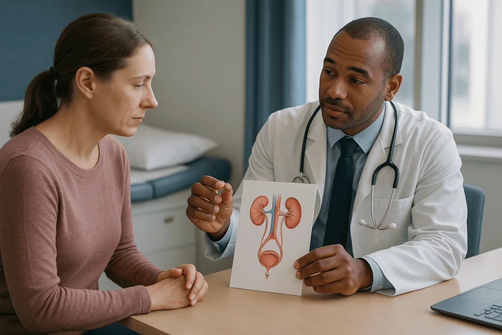
Nobody plans to learn a comparison of urological procedures in an emergency bay, yet that is exactly where many people first hear these two words. The ureter is the narrow muscular tube, roughly the width of a drinking straw, that carries urine from each kidney to the bladder. When a stone leaves the kidney and lodges there, urine backs up, the kidney swells, and the result is the wave-like pain most people describe as the worst they have known.
Two very different technologies grew up to solve that single problem. Shock wave lithotripsy attacks the stone from outside the body, sending focused pressure pulses through skin and tissue until the stone crumbles. Ureteroscopy goes in from the inside, following the urinary tract upward with a thin telescope so the surgeon can see the stone and break or remove it directly. Neither approach involves cutting through skin, both are usually done as same-day procedures, and both appear as accepted options for ureteral stones in mainstream guidance summarized by the NIDDK and the NHS.
That is precisely why the decision feels confusing. If one were clearly superior, there would be no conversation. Instead, the urologist is balancing trade-offs: fewer repeat sessions against a temporary stent, a lighter procedure against a lower chance of finishing in one go, and the physics of where the stone sits against what you personally can tolerate. Stone density, position in the ureter, body shape, medicines, pregnancy and your own priorities all move the needle. The rest of this article walks through those trade-offs one at a time, so the conversation in the clinic feels less like a quiz and more like a shared decision with someone who has already done the reading.
What actually happens during shock wave lithotripsy
The name is more intimidating than the experience. Lithotripsy simply means stone-crushing, and the shock waves involved are pulses of acoustic energy, not electricity. You lie on a padded table, usually on your back, with a soft water-filled cushion pressed against the flank on the side of the stone. Water carries sound energy efficiently, which is why that unglamorous cushion matters.
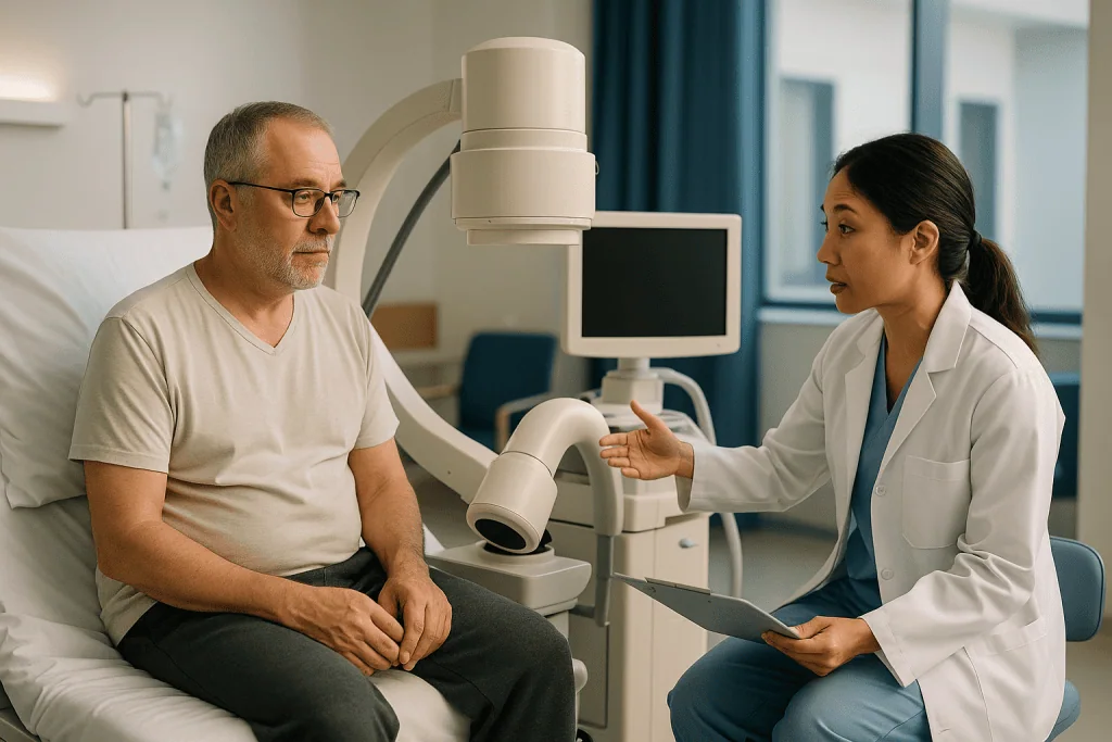
Before anything starts, the team locates the stone with X-ray or ultrasound and lines up the machine so that thousands of tiny wave fronts converge on one point a few millimeters wide. Tissue in the path of the waves receives a gentle, diffuse pulse; only the stone at the focal point receives enough concentrated energy to fracture. Sedation or a light general anesthetic keeps you still and comfortable, because even changes in breathing can nudge the stone out of focus.
A typical session delivers a set number of shocks over about 45 minutes to an hour, according to MedlinePlus. You may hear a rhythmic clicking and feel a tapping sensation against the skin. When imaging shows the stone has fragmented into sand-like pieces, the session ends. Most people go home the same day and pass the fragments in their urine over the following days to weeks, a timeline MedlinePlus describes as sometimes taking several weeks before every piece has cleared.
The technique works best, MedlinePlus notes, for stones smaller than about 2 centimeters that can be seen clearly on imaging. Very dense stones, stones deep behind a thick layer of tissue, and stones shadowed by bone can be harder for the waves to reach, which is one of the first things a urologist checks on the CT scan before offering this route.
What actually happens during ureteroscopy and laser lithotripsy
Ureteroscopy is the inside route. A ureteroscope is a thin flexible or semi-rigid telescope, narrower than a pencil, with a light and camera at its tip and a working channel for tiny instruments. Under general anesthesia, the surgeon passes it through the urethra, across the bladder, and up into the ureter until the stone appears on the video screen. No incision is made anywhere.
Once the stone is in view, a laser fiber roughly the thickness of a human hair is threaded through the scope. Laser lithotripsy uses short bursts of light energy, absorbed by the stone, to break it into fragments or fine dust. Larger pieces can be captured with a wire basket and pulled out; dust is left to wash away in the urine. Because the surgeon watches in real time, very hard stones and stones tucked in awkward positions can still be treated, which explains why this approach has expanded as scopes and lasers have shrunk.
At the end, a ureteral stent is often placed. A stent is a soft, hollow plastic tube that runs from the kidney to the bladder and keeps the ureter open while swelling settles. Cleveland Clinic describes it as a temporary measure, removed after a period the surgeon sets. Some surgeons skip the stent for straightforward cases; others use it routinely. Practice genuinely varies here, and it is worth asking what your surgeon plans and why.
Ureteroscopy is usually a same-day procedure too. You wake with a mild burning sensation on urinating and, if a stent is in place, an awareness of it that most people describe as pressure or urgency rather than sharp pain. Walking, eating and drinking normally that evening is typical rather than exceptional.
What size stone needs ureteroscopy? Size, location and hardness
Size is the first filter, but rarely the last. Many small stones never need a procedure at all. Mayo Clinic notes that most small stones will not require invasive treatment and can pass with fluids, pain relief and time, and the NIDDK gives the same reassurance: small stones frequently pass on their own, so the urologist may simply monitor.
Above that range, the physics start to favor one tool or the other. Shock wave lithotripsy is described by MedlinePlus as most effective for stones under about 2 centimeters, and in practice urologists reserve it for stones that are not too dense, sit within reach of the wave focus, and are not blocking an infected kidney. Ureteroscopy has no fixed upper size limit for ureteral stones because the surgeon can break the stone progressively under direct vision; very large stones simply take longer or need a second stage.
Location matters as much as size. A stone high in the ureter near the kidney behaves differently from one down near the bladder. Lower ureteral stones are often easy to reach with a semi-rigid scope, while upper stones can be approached either way. Stones visible on plain X-ray are easier to target with shock waves than stones that show up only on CT.
Hardness is the quiet third variable. CT scans report stone density in Hounsfield units, a measure of how much X-ray the stone absorbs. Very dense stones, and certain chemical types such as cystine, tend to resist shock waves and are more likely to be steered toward the laser. So when people ask what size stone needs ureteroscopy, the honest reply is that size opens the conversation, and density, position and anatomy usually settle it.
Who is usually offered each option, and who is asked to wait
The person offered watchful waiting usually looks like this: a small stone, pain controlled with ordinary measures, no fever, normal kidney function, and two working kidneys. In that setting the NHS and NIDDK both describe a period of observation with fluids and follow-up imaging as a reasonable first plan. Some clinicians add a medicine from the alpha-blocker class, which relaxes smooth muscle in the lower ureter and may help a stone travel; whether it is appropriate, and for how long, is a decision for the prescribing clinician.
Others are asked not to wait. Fever or chills with a blocking stone suggests infected urine trapped above the obstruction, which can become dangerous quickly; that situation usually calls for urgent drainage rather than definitive stone treatment. A single functioning kidney, kidneys blocked on both sides, deteriorating kidney blood tests, or pain that cannot be controlled are also reasons to move promptly toward a procedure.
Within the procedural group, certain features push firmly toward ureteroscopy. Pregnancy rules out shock wave lithotripsy, as MedlinePlus notes. Bleeding disorders and blood-thinning medicines raise the risk of bleeding inside the kidney from shock waves, so the endoscopic route is often preferred, although stopping or adjusting any anticoagulant is strictly a matter for the prescribing team. An aneurysm of the aorta or renal artery near the target, certain skeletal abnormalities, and a very long skin-to-stone distance also count against shock waves.
Fewer features point exclusively toward lithotripsy. A strong wish to avoid general anesthesia, instruments inside the urinary tract, and a stent is a legitimate one, and urologists take it seriously when the stone is suitable. Preference is data, not noise, in this decision.
Shock wave lithotripsy vs laser ureteroscopy: side by side
Seen together, the two approaches are less rivals than complementary tools, each strong where the other is weak. The table below summarizes the comparison a urologist is running in their head when the CT images come up.
| Feature | Shock wave lithotripsy | Ureteroscopy with laser |
|---|---|---|
| Route | From outside the body, through the skin | Through the urethra and bladder, no incision |
| Anesthesia | Sedation or light general | General anesthesia |
| How the stone is treated | Fragmented; pieces pass on their own | Fragmented or dusted under direct vision; pieces can be removed |
| Best-suited stones | Smaller, less dense, visible on imaging, within wave reach | Any size in the ureter, hard or soft, wherever the scope can reach |
| Chance of needing another session | Higher | Lower |
| Stent afterward | Uncommon | Common, temporary |
| Typical discomfort | Flank bruising, colic as fragments pass | Burning, stent irritation until removal |
| Usually avoided when | Pregnant, bleeding risk, aneurysm near target, infected obstruction, very dense stone | Ureter too narrow to admit the scope safely, active untreated infection |
Two rows deserve emphasis. The row on needing another session is where systematic reviews consistently separate the procedures, and it is the reason many urologists lean toward ureteroscopy for ureteral stones when both are feasible. The stent row is where patients most often push back, and rightly so; the stent is the part of ureteroscopy people remember. A shock wave lithotripsy vs laser decision, in other words, is often a choice between accepting a higher chance of coming back and accepting a few weeks of stent symptoms.
No table captures the person. Someone whose work cannot absorb a second unplanned absence weighs the rows differently from someone who dreads anesthesia. Both readings are valid, which is exactly why the choice is shared rather than dictated.
How painful is shock wave lithotripsy, and how does ureteroscopy compare?
How painful is shock wave lithotripsy? Less than most people fear during the session, and more variable afterward. The shocks are delivered under sedation or anesthesia, so the tapping many describe is felt as firm, rhythmic pressure rather than a strike. Cleveland Clinic and MedlinePlus both describe the procedure as generally well tolerated with medication. The real discomfort tends to arrive later: a bruised feeling over the flank where the waves entered, and cramping colic as fragments travel down the ureter. That colic can be sharp, because each fragment behaves like a miniature stone.
Ureteroscopy is painless during the procedure because you are under general anesthesia. Afterward the picture is different again. Burning on urination for a day or two is common, as is a small amount of blood in the urine. If a stent was left in place, stent symptoms dominate: urgency, frequency, a dull flank ache when the bladder empties, and occasional spasms. Your team will advise on managing these, and they resolve once the stent is removed.
Comparing the two honestly, the discomfort after lithotripsy is spread across the weeks of fragment passage, while the discomfort after ureteroscopy is concentrated in the stent period. Neither is trivial; both are temporary. People who have experienced both often say the stent was the harder part of ureteroscopy and the fragment colic the harder part of lithotripsy. Your own tolerance for a steady, nagging irritation versus unpredictable sharp episodes is therefore a genuine medical variable, and a good urologist will ask about it rather than assume.
Why one treatment may take more than one session
A single shock wave session does not always finish the job, and this is the trade-off most worth understanding. Shock waves break the stone but cannot remove it; the fragments must travel out on their own. Some stones fracture into fine sand that passes easily. Others split into a few stubborn chunks that stall in the ureter, or barely break at all. The NHS notes plainly that more than one session may be needed, and urologists typically arrange a follow-up X-ray or ultrasound to check.
Several factors predict a harder time: high density on CT, certain stone chemistries, a long distance from skin to stone, and a position where the waves struggle to reach. A shower of fragments can also line up in the lower ureter, a situation clinicians call steinstrasse, German for stone street, which sometimes requires ureteroscopy to clear. That is why some people who start with lithotripsy end up having a scope anyway.
Ureteroscopy more often clears the stone in one visit because the surgeon sees the fragments and removes or dusts them; that single-procedure advantage is the consistent finding of the comparative reviews summarized by the NIDDK and NHS. It is not infallible. Very large stones, a tight ureter that will not admit the scope safely, or a stone that has embedded in the wall can lead to a staged approach: a stent first to gently widen the ureter, then a second procedure. Your surgeon should be able to estimate, for your stone specifically, how likely a second step is with each route. Ask for that estimate rather than a general figure; the general figure describes an average stone, and yours is not average to you.
Ureteroscopy stent recovery: what the tube does and how long it stays
The stent generates more questions than the operation. It stays in for whatever period the surgeon judges necessary for swelling to settle and residual fragments to pass, commonly days to a few weeks according to Cleveland Clinic. Removal is quick, done in the clinic with a small scope or by pulling a retrieval string if one was left in place.
What does ureteroscopy stent recovery feel like? Most people notice the stent constantly at first. Its curled ends sit in the kidney and bladder; the bladder end rubs and triggers a sense of needing to urinate even when the bladder is nearly empty. A dull flank ache when you void is common, because a stent lets a little urine flow backward toward the kidney under pressure. Pink or rusty urine can come and go, often worse after activity. These are expected effects, not complications, and they fade once the tube is out.
Practical adjustments help more than most expect. Staying well hydrated dilutes the urine and eases irritation. Gentle activity is fine; vigorous exercise tends to increase bleeding and discomfort. Your team may suggest medicines that relax the bladder or ureter, and if so they will explain what to take and for how long; that decision rests with them.
One point is non-negotiable: a stent must not be forgotten. A tube left in far beyond its intended time can encrust with mineral deposits and become very difficult to remove. If your removal appointment is unclear, ask. If the string breaks or the stent shifts, call. Knowing your removal date is as much a part of recovery as any instruction on the discharge sheet.
Risks and complications of each procedure, in plain terms
Both procedures are considered safe in experienced hands, and both carry real risks that deserve plain language. Shock wave lithotripsy commonly causes blood in the urine and bruising of the skin and the kidney itself. Less commonly, bleeding can collect in or around the kidney as a hematoma, which usually settles with observation but occasionally needs treatment. Fragments can obstruct the ureter and cause renewed colic or, if infected urine is trapped, a serious infection. Cleveland Clinic and MedlinePlus list these as the main concerns, alongside rare injury to nearby organs.
Ureteroscopy carries the risks of any procedure under general anesthesia plus those specific to working inside a narrow tube. The ureter can be scraped or, rarely, perforated; small injuries heal with a stent in place, while a significant tear may require a longer stent or further surgery. Infection can be introduced or pushed upward, which is why urine is often tested beforehand and antibiotics are frequently given around the time of surgery. Long-term scarring that narrows the ureter, called a stricture, is uncommon but does happen. Stent-related discomfort remains by far the most frequent complaint.
Neither procedure has been shown to reduce kidney function in the long term for the typical patient, though whether repeated shock wave treatments raise blood pressure over years has been studied without a settled answer. When the evidence is unsettled, a good urologist will say so. What the decision usually comes down to is the shape of the risk: lithotripsy concentrates it in bleeding and incomplete clearance, ureteroscopy in instrumentation and stent effects. Which shape you would rather live with is a fair question to put on the table.
What the days and weeks after treatment usually look like
After shock wave lithotripsy, the first day is usually spent at home, drowsy from sedation, with soreness over the flank and pink urine. Over the following days the urine may carry visible grit; some people strain it through a filter so fragments can be analyzed. Colic can flare unpredictably as pieces move, then quiet again. Cleveland Clinic describes most people returning to normal activities within a few days, with fragments continuing to pass for weeks in some cases. A follow-up imaging test tells the team whether the stone is gone or a second session is needed.
After ureteroscopy, the first 24 hours bring burning on urination, blood-tinged urine and stent awareness. Most people are walking the same day and back to desk work within a few days, per Cleveland Clinic, though strenuous activity is usually deferred until the stent is out. The stent removal visit is the real milestone; many describe relief within hours. If the stone was sent for analysis, results typically return over the following weeks and shape any prevention advice.
Both pathways share the same longer view. People who have formed one stone are more likely to form another, the NIDDK notes, which is why the weeks after treatment are also when fluid intake, dietary review, and sometimes blood and urine testing come into focus. Drinking enough to keep urine pale is the single measure every source agrees on. Treating the stone that is there is the urgent part; understanding why it formed is the part that pays off years later, and it starts with that small jar the lab asks you to fill.
What people often get wrong about ureteroscopy vs shock wave lithotripsy
The most persistent myth is that lithotripsy is the easy option and ureteroscopy the serious one. Both are outpatient procedures; both involve anesthesia of some kind; both have recovery periods. The lighter footprint of shock waves is real, but it is paid for in a higher chance of a return visit, and a second session is not free of discomfort or risk.
A second misconception runs the other way: that the laser vaporizes the stone so nothing is left to pass. Laser lithotripsy leaves dust and small fragments, and some will still travel out in the urine over the following days. The difference is that the surgeon can remove larger pieces and confirm what remains, not that nothing remains.
Many people believe a stent is a sign something went wrong. Usually it is the opposite: a planned precaution to protect a ureter irritated by the stone and the scope. Similarly, blood in the urine after either procedure is expected for a period, not automatically a complication.
Another common belief is that shock waves permanently damage the kidney. Bruising of the kidney does occur, and possible long-term effects on blood pressure have been studied, but the evidence has not shown lasting loss of kidney function for the typical patient, and the question remains open rather than settled in either direction.
Finally, the notion that there is one right answer for a given stone size ignores density, location, anatomy, medicines, pregnancy and preference. When someone tells you a seven millimeter stone always gets one treatment or the other, they are describing a habit, not a guideline. Guidelines list both as options for exactly that reason.
Questions to ask your care team before choosing
Walking into the consultation with specific questions changes the conversation. General questions get general answers; specific ones make the urologist show you their reasoning for your stone. Bring the CT report if you have it, and consider asking:
- How big is my stone, where exactly is it in the ureter, and how dense does it look on the scan?
- Given those features, which option do you recommend for me, and what makes you lean that way?
- For each option, how likely is it that I will need a second procedure, based on stones like mine?
- Will I have a stent, for how long, and how and when will it be removed?
- What kind of anesthesia is planned, and what risks are specific to me?
- Is waiting still a safe option for me, and what would change that?
- Should any of my regular medicines be paused, and who will give me that instruction?
- What symptoms afterward should make me call you, and what number do I call?
- Will the stone be sent for analysis, and how will the result change what I do next?
Two of these matter more than the others. The question about a second procedure forces a personalized estimate rather than a textbook figure. The question about medicines matters because blood thinners, some diabetes medicines and others may need adjustment around anesthesia, and that instruction must come from the prescribing clinician, never from a leaflet or an article like this one. If the answers leave you unsure, it is reasonable to ask for time to think, or for the plan to be written down. A good team will welcome both, and will not be offended by a second opinion.
When to call your doctor
Both procedures are followed by a set of symptoms that are expected and a smaller set that are not. Knowing the difference is the single most useful piece of aftercare knowledge. Expected: mild to moderate flank soreness, pink or rusty urine that comes and goes, burning when urinating for a day or two, urgency and frequency with a stent in place, and intermittent cramping as fragments pass.
Call your doctor or the urology team the same day if you notice any of the following:
- Fever, chills or shaking, which can signal infection above a blocked ureter and needs urgent assessment
- Pain that is severe, escalating, or not relieved by the measures you were given
- Inability to pass urine, or passing only tiny amounts despite drinking normally
- Urine that is heavily bloody, thick like tomato juice, or contains large clots rather than a pink tinge
- Persistent vomiting that prevents you from keeping fluids down
- A stent string that has come out, or a stent you can see or feel emerging
- Faintness, rapid heartbeat or confusion
Seek emergency care immediately, rather than waiting for a callback, for high fever with flank pain, signs of sepsis such as confusion or very rapid breathing, or severe pain with vomiting and no urine output. Infection trapped behind an obstructed kidney can deteriorate within hours, and the NHS and Mayo Clinic both flag fever with stone symptoms as a reason to seek help without delay.
One quieter red flag: a stent removal appointment that never materializes. If you were told a stent would come out and weeks have passed without a date, contact the team. Every decision about what happens next, from imaging to a second procedure, sits with them, but the phone call sits with you.
Frequently asked questions
Which is better, ureteroscopy or lithotripsy?
Neither is better for every person or every stone. Ureteroscopy more often leaves someone stone-free after a single procedure but usually involves general anesthesia and a temporary stent. Shock wave lithotripsy is less invasive and often avoids a stent, but fragments must pass on their own and a repeat session is more likely. The right choice depends on stone size, density, location, your health and your preferences, decided together with your urologist.
Which is better for kidney stones, shock wave lithotripsy or laser removal?
Laser ureteroscopy tends to be favored for dense stones, stones the shock waves cannot reach well, and situations where finishing in one visit matters most. Shock wave lithotripsy suits smaller, softer stones visible on imaging in people who prefer to avoid instruments and a stent. For stones still inside the kidney rather than the ureter, location within the kidney also matters, and your urologist will weigh that separately.
How painful is shock wave lithotripsy?
During the session, sedation or anesthesia keeps you comfortable, and most people describe a firm tapping sensation rather than pain. Afterward, expect bruised-feeling soreness over the flank and, in the following days, cramping as fragments pass down the ureter. That fragment colic can be sharp but is temporary. Your care team will explain how to manage discomfort and when a symptom warrants a call.
What size kidney stone requires ureteroscopy?
There is no single cutoff. Many small stones pass on their own, and mid-sized stones can be treated either way. Ureteroscopy becomes the more likely recommendation as stones grow larger, appear denser on CT, sit where shock waves struggle to reach, or when pregnancy, bleeding risk or infection rule out lithotripsy. Size starts the discussion; density, location and your health usually settle it.
Do I always need a stent after ureteroscopy?
Not always. Some surgeons leave the ureter without a stent after a straightforward, uncomplicated procedure, while others place one routinely to protect the ureter while swelling settles. A stent is more likely when the ureter was tight, the procedure was long, fragments remain, or the ureter was scraped. Ask your surgeon what they plan for your case and why.
How long does ureteroscopy stent recovery take?
Stent symptoms such as urgency, a flank ache when urinating and pink urine usually last for as long as the stent is in, typically days to a few weeks depending on the surgeon’s plan. Most people feel markedly better within hours of removal. Staying hydrated and avoiding strenuous exercise helps in the meantime, and knowing your exact removal date is essential.
Can I have shock wave lithotripsy if I take a blood thinner?
Blood-thinning medicines raise the risk of bleeding inside or around the kidney from shock waves, so urologists often prefer ureteroscopy in that situation or discuss with your prescribing clinician whether the medicine can be safely paused. Never stop or adjust a blood thinner on your own; the decision depends on why you take it and must come from the team managing it.
Why does my urologist want a CT scan before deciding?
A CT scan shows the stone’s exact size, its position in the ureter, its density measured in Hounsfield units, and how far it sits from the skin. Those four details predict how well shock waves would work and whether the scope is the better route. The scan also reveals kidney swelling and anatomy that could change the plan, such as a narrowed ureter.
Can I choose the procedure myself?
Your preference is a genuine part of the decision, especially when both procedures are medically reasonable for your stone. What you cannot do is override a safety issue; pregnancy, infection behind a blockage, or bleeding risk may rule one option out. Tell your urologist what matters most to you, such as avoiding a stent or minimizing repeat visits, and let them explain how that fits.
Will treating this stone stop new stones from forming?
No. Both procedures remove the stone that is causing trouble but do nothing to change the chemistry that formed it. People who have had one stone are more likely to form another, which is why stone analysis, fluid intake, dietary review and sometimes blood and urine tests follow treatment. Prevention is a separate conversation your team will usually start once you have recovered.
References
This article is for general information only and is not a substitute for professional medical advice. Please consult a qualified doctor about your individual situation.

More from the Blog

Urinary Infections in Women and Men: Why the Causes Differ and How Care Is Adjusted
Urinary infections differ between women and men mainly because of anatomy. A woman's short urethra opens close to the bowel, so gut bacteria reach…

After Contrast Imaging: The Changes That Mean Your Kidneys Need a Check Sooner
Most people who receive contrast dye for a CT scan or MRI have no lasting effect on their kidneys, but anyone with reduced kidney…

How Renal Osteodystrophy Is Treated: Balancing Calcium, Phosphate and Parathyroid Hormone
Renal osteodystrophy treatment aims to correct the mineral imbalance that damaged kidneys create rather than to treat the bone directly. Care teams limit dietary…

Overactive Bladder Treatment in Order: Bladder Training, Medication and Advanced Therapies
Overactive bladder treatment usually moves through three steps. First come bladder training, pelvic floor exercises and fluid changes, tried for at least six weeks.…

Recovery After Phimosis Surgery: Wound Care, Loose Clothing and When Swimming Is Allowed
Recovery after phimosis surgery, most often a circumcision or a foreskin-widening procedure, typically takes about 10 days for the wound to knit and several…

Are You Stone-Free After Laser Lithotripsy? What Follow-Up Imaging Checks and Why It Matters
Being stone free after laser lithotripsy means no fragments remain in the kidney or ureter on follow-up imaging, not just that the surgeon saw…