

What a Root Canal X-Ray Shows: Infection at the Root Tip and How It Shapes the Plan

Key Takeaways
- Infection at the root tip appears on a root canal X-ray as a dark halo (radiolucency) where bone has dissolved, often with loss of the thin white socket outline.
- A fresh, acute abscess can leave the film looking normal because bone has not yet had time to dissolve; the X-ray records history rather than the present moment.
- A working-length X-ray taken mid-procedure confirms exactly where each canal ends so the filling reaches the tip without extending into bone.
- According to the NHS, root canal treatment is usually successful and in about 9 out of 10 cases a treated tooth can survive for up to 10 years.
- Cleveland Clinic gives a typical root canal duration of 30 to 60 minutes, with complex molars taking up to around 90 minutes.
- On a failed root canal X-ray the usual clues are a short fill, a missed canal, an overfill, a separated instrument or a J-shaped shadow suggesting a root fracture.
A root canal X-ray shows the tooth's inner canals, the bone around the root tip, and any dark area (a radiolucency) where infection has dissolved bone at the apex. Dentists use it to confirm the pulp is dead or infected, measure canal length, check that the filling reaches the tip, and later judge whether bone is regrowing. It cannot show pain or a living nerve directly.
The dentist turns the monitor toward you and points at a tooth that looks, to your eye, exactly like its neighbors. Then she traces a small gray smudge at the bottom of the root with the tip of her pen. “That,” she says, “is why it hurts when you bite.” You nod, though the smudge looks like nothing at all.
That smudge is the whole story of a root canal x ray. Most of what matters happens in bone you cannot see or feel, at the very tip of the root, millimeters from the nerve that has been keeping you awake. The image is a shadow map: dense things show white, hollow or dissolved things show dark, and infection leaves a dark footprint.
Understanding that map turns a frightening appointment into a series of answerable questions. What is the dentist looking at? Why does she take another picture halfway through? And months later, how will she know whether the bone has healed? This explainer walks through each of those images and what they mean for the plan.
What a root canal x ray is actually looking for
Start with the anatomy the picture is trying to capture. Every tooth has a hollow core called the pulp, a soft bundle of nerve and blood vessels that runs from the crown down each root in narrow tunnels called canals. The canals open at the root tip, or apex, where the vessels connect to the bone. A root canal x ray is a small, focused image, usually a periapical film, a view designed to show the whole root and a margin of bone around the apex.
On that film, the dentist is checking four things at once. First, how deep decay or a crack has traveled and whether it reaches the pulp. Second, the shape of the canals: how many there are, how curved they run, and whether they have narrowed with age. Third, the bone at the apex, because bone that is being dissolved by infection looks darker than healthy bone. Fourth, anything left over from earlier dentistry, such as a deep filling, a post, or an old root filling that stops short of the tip.
What the image cannot do matters just as much. An X-ray shows density, not biology. It cannot tell whether a nerve is alive, only whether the bone around it has changed. It cannot show pain. Early inflammation of the pulp, the stage where a filling might still be enough, often leaves the picture looking normal. That is why a dentist pairs the film with tapping, temperature tests, and a careful history before recommending anything.
According to MedlinePlus, dental X-rays use very low levels of radiation and are considered safe for most people when clinically justified; the point is to take the images that change the plan and no more.
How does infection at the root tip show up on a root canal x ray?
Think of bone as a dense sponge. On film, dense material blocks X-rays and appears white or pale gray; the technical word is radiopaque. Where bone has been eaten away, X-rays pass through easily and the area appears dark; that is a radiolucency. When bacteria from a dead pulp leak out of the apex, the body responds with inflammation that slowly resorbs the surrounding bone. The result is a rounded dark halo hugging the root tip, often called a periapical radiolucency or, loosely, an abscess on X-ray.

Several details help the dentist interpret that halo. Its edge may be fuzzy, suggesting active bone loss, or crisp and rimmed with a thin white line, suggesting a longer-standing process the body has walled off. Its size hints at duration; bone does not vanish overnight. The fine white line that normally outlines the root socket, the lamina dura, may be lost near the tip. The gap between root and bone, the periodontal ligament space, may look widened.
There is an important catch. A tooth that flared into a hot, swollen abscess two days ago may show almost nothing, because bone has not yet had time to dissolve. Mayo Clinic notes that dentists diagnose an abscess through examination and tapping the tooth as well as imaging. Conversely, a dark shadow can persist for a long while in a tooth that no longer hurts. The X-ray records history, not the present moment.
So when the dentist says the film shows infection, what she means is that bone has changed in a pattern that almost always follows a dead or dying pulp. That single finding is what moves the conversation from “let’s watch it” to “the pulp needs to come out.”
What actually happens during a root canal, step by step
The procedure sounds dramatic and is, in practice, methodical. Its goal is to remove the infected or dead pulp, clean and shape the empty canals, and seal them so bacteria cannot return. The NHS describes it as a sequence of numbing, isolating, cleaning, filling and restoring.
The dentist first numbs the tooth with local anesthetic. A small rubber sheet, called a dental dam, is stretched around the tooth to keep saliva and bacteria out and to protect your airway from tiny instruments. A drill opens the top of the tooth into the pulp chamber. From there, very thin flexible files, some barely thicker than a hair, are worked down each canal to remove pulp tissue and scrape the walls. Disinfecting solutions are flushed through repeatedly; the flushing does much of the real cleaning, because files cannot reach every branch.
Once the canals are clean and dry, they are filled with a rubbery material called gutta-percha, packed snugly to the tip and sealed with a cement. The opening in the crown is closed with a temporary or permanent filling. Because a tooth that has lost its pulp becomes more brittle, back teeth are often later fitted with a crown, a cap that wraps the whole tooth, to prevent fracture under chewing forces.
Some cases are completed in one visit. Others are left with a medicated dressing for a period and finished at a second appointment, especially when the infection is active or the anatomy is complex. Your dentist decides that based on what the canals look like on the day, not on a fixed rule. An endodontist, a dentist with additional training in root canal treatment, may be involved when the case is unusual.
Why X-rays are taken before, during and after treatment
Patients are sometimes surprised to be X-rayed three or four times over a single course of treatment. Each image answers a different question, and skipping one would mean guessing.
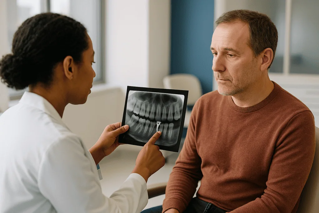
The pre-treatment film establishes the diagnosis and the map: how many roots, how curved, how long, and how much bone has been lost. It also reveals surprises that change the plan before the drill touches the tooth, such as a canal that has calcified shut or a root that splits into two branches near the tip.
During treatment, the dentist needs to know exactly where the canal ends. Filing short leaves infected tissue behind; filing long pushes debris and material into the bone. A file is placed in the canal and an image is taken to confirm its position relative to the apex, a step called a working-length film. Many clinicians also use an electronic apex locator, a device that measures where the canal meets the ligament, and use the X-ray to confirm it. Before the final fill, a second image may check that the gutta-percha cone fits to the correct depth.
The post-treatment film is the baseline. It documents that every canal is filled to the tip without gaps or overextension, and it is the picture future films will be compared against to judge healing.
Then there is the review film, taken at an interval your dentist sets. Bone rebuilds slowly, so the aim is to see the dark halo shrinking and the white outline of the socket returning. According to the NHS, root canal treatment is usually successful, and in about 9 out of 10 cases a treated tooth can survive for up to 10 years. Those review films are how your own tooth’s trajectory is tracked, rather than assumed.
Who is usually offered a root canal, and who is asked to wait
The clearest candidates are teeth whose pulp is irreversibly damaged. On the X-ray that means deep decay or a crack that reaches the pulp chamber, a periapical radiolucency, or both. Cleveland Clinic lists the typical triggers as untreated decay, repeated dental work on the same tooth, a chipped or cracked tooth, or trauma that has killed the nerve even without visible damage. A tooth that is dead but painless still qualifies, because bacteria keep leaking into bone whether or not you feel it.
Some people are asked to wait, and waiting has different meanings. If the pulp appears inflamed but possibly recoverable, a dentist may place a sedative filling and reassess over a period rather than commit to a root canal; the X-ray at that stage often looks normal and the decision rests on symptoms and vitality tests. If the tooth is acutely swollen, the dentist may first drain the abscess and calm the infection before beginning the definitive cleaning. The NHS notes that antibiotics are not a treatment for the tooth itself and are reserved for particular situations, such as spreading infection, with the decision resting with the clinician.
Others are steered away from a root canal altogether. When the X-ray shows a vertical root fracture, a root that has been shortened by extensive bone loss around the whole tooth, or too little sound tooth left above the gum to hold a crown, the honest conversation turns to extraction and replacement. Root canal treatment saves a tooth only if the tooth can be rebuilt afterward.
Medical history rarely rules anyone out, but people with bleeding disorders, recent heart valve surgery, or conditions affecting healing should tell the dentist so the plan can be coordinated with their physician. None of these decisions belongs to the X-ray alone; the image informs a judgment the treating team makes with you.
Root canal infection x ray vs. tooth abscess x ray vs. healthy tooth: a comparison
People searching for a tooth abscess on x ray often expect a single dramatic image. In reality the appearances form a spectrum, and the same dark spot can mean different things depending on when it is seen. The table below summarizes the common patterns and how each tends to steer the plan. It is a guide to the conversation, not a diagnostic key.
| What the film shows | Typical appearance | What it usually indicates | How it tends to shape the plan |
|---|---|---|---|
| Healthy tooth | Even bone density around the root, intact thin white socket line, uniform ligament space | No bone change; pulp may still be inflamed or healthy | Diagnosis relies on symptoms and vitality tests; watchful waiting or a filling may be enough |
| Deep decay approaching the pulp | Dark area within the crown reaching toward the pulp chamber | Pulp at risk or already involved | Removal of decay first; root canal if the pulp is exposed or tests show it is dying |
| Chronic root tip infection | Rounded dark halo at the apex, lost socket outline, sometimes a crisp border | Long-standing leakage from a dead pulp | Root canal treatment, or extraction if the tooth cannot be restored |
| Acute abscess | Often little or no visible change; possibly a slightly widened ligament space | Recent, active infection before bone loss is visible | Drainage and pain control first; definitive treatment once settled |
| Previously treated tooth with new dark area | Existing root filling with a halo at the tip, a short fill, or an untreated extra canal | Persistent or recurrent infection | Retreatment, surgical treatment of the tip, or extraction, depending on cause |
Two points deserve emphasis. A normal-looking film does not clear the tooth, and a dark halo does not by itself mean the tooth is beyond saving. Read together with the examination, the image narrows the options; it never chooses among them alone.
What does a failed root canal look like on X-ray?
The phrase “failed root canal” covers several different pictures, and a failed root canal x ray usually points to one of a handful of causes. The most common is a dark area at the root tip that has persisted or grown since the treatment film, meaning bacteria still have a route into the bone. Behind that shadow, the film often reveals why.
A short fill is the classic culprit: the white line of gutta-percha stops well before the apex, leaving uncleaned canal beyond it. The opposite problem, an overfill, shows material extruded past the tip into bone, where it can irritate tissue. A missed canal is subtler. Upper molars, for example, frequently have a second canal in the front-cheek root that is easy to overlook; on film the treated root may look filled while an adjacent untreated canal harbors infection. A separated instrument appears as a bright, sharply defined fragment lodged in the canal. A perforation, where a drill or post has exited the side of the root, shows as material or a dark area beside the root rather than at its tip.
Vertical root fractures are the hardest to see directly, because the crack runs in the same plane as the X-ray beam. Their signature is indirect: a narrow, J-shaped dark zone running up one side of the root, or bone loss isolated to a single root of a multi-rooted tooth. Leaking under an old crown produces recurrent decay at the margin, which shows as a dark notch where crown meets root.
Timing changes interpretation. Bone regrows slowly, so a halo that is smaller than before is a healing tooth, not a failing one. Your dentist compares films side by side and combines them with symptoms before deciding whether retreatment, a small surgical procedure at the root tip, or extraction is the right next step.
What is the hardest tooth to get a root canal on?
Ask endodontists and most will point to the upper first molar, and the reasons are visible on the X-ray. That tooth has three roots and often four canals, with the fourth, a second canal in the front-cheek root, so fine and hidden that it is a frequent cause of retreatment. Lower molars run a close second: their two roots typically carry three or four canals that curve sharply toward the back of the mouth, and files must bend without breaking to follow them.
Difficulty also comes from the imaging itself. Upper molar root tips sit just below the sinus floor, and the cheekbone can overlap them on a two-dimensional film, blurring the very region the dentist needs to see. Lower molars sit above a nerve canal in the jaw that appears as a dark band, complicating interpretation and demanding precision about working length.
Age adds another layer. Over decades the pulp lays down extra dentin, and canals that were wide tunnels in a teenager become hairline threads in an older adult. On film these calcified canals may be nearly invisible, and locating them takes patience and magnification. Teeth that have already been treated once, that carry a post, or that have unusual anatomy such as an extra root fall into the same demanding category.
When a standard film cannot resolve the anatomy, a cone beam CT scan, a low-dose three-dimensional X-ray of the jaw, may be considered. It reveals missed canals, curved roots and the true extent of bone loss in a way flat images cannot, at the cost of more radiation than a single film. Whether it is justified is a case-by-case judgment your dentist or endodontist makes, weighing the information gained against the exposure. Front teeth, with their single straight canals, are the far end of the same spectrum and are often the most straightforward.
When is it too late to avoid a root canal?
The honest answer has two halves, because “too late” can mean too late to avoid treatment, or too late for treatment to work.
The first threshold is crossed when inflammation in the pulp becomes irreversible. Early on, a pulp irritated by decay can recover if the decay is removed and the tooth is sealed. Past a certain point the pulp cannot heal, and the only paths forward are to remove it, through root canal treatment, or remove the tooth. No X-ray marks that line precisely. The clues are clinical: pain that lingers long after a cold drink, pain that wakes you at night, or a tooth that no longer responds to temperature at all. Once a periapical radiolucency appears on film, the pulp has almost always already died, and the window for a simple filling has closed. Mayo Clinic notes that an abscess will not resolve on its own, even if the pain settles as the nerve dies.
The second threshold concerns the tooth’s future. Root canal treatment is worthwhile when the tooth can be rebuilt and kept. If the X-ray shows a vertical fracture, bone loss so extensive that the root has lost most of its support, or decay so deep beneath the gum that no crown could grip what remains, the treating team may advise that extraction and a replacement are the more predictable route. That is not a failure of timing so much as a recognition of what the tooth can bear.
Between those thresholds sits most of dentistry, and the plan is a shared decision. Alternatives to root canal treatment, in neutral terms, are extraction followed by a bridge, an implant, a denture, or leaving the gap. Each has trade-offs in bone preservation, neighboring teeth and maintenance that your dentist should lay out plainly before you choose.
How long does a root canal take, and how many visits?
Shorter than most people fear. According to Cleveland Clinic, a root canal usually takes about 30 to 60 minutes, and more complex teeth, typically molars with several canals, can take up to around 90 minutes. The X-ray taken beforehand is one of the best predictors: a single straight canal in a front tooth is a very different appointment from four curved canals in a lower molar.
Whether the work is finished in one visit or two depends on what the dentist finds inside. When the canals can be cleaned thoroughly and dried, and there is no active drainage, many clinicians complete the filling the same day. When the tooth is acutely infected, when pus is still draining, or when the anatomy is unusually complex, the NHS notes that more than one appointment may be needed, with a medicated dressing placed between visits. Neither approach is inherently better; the choice follows the condition of the tooth on the day.
Then there is the restoration, which is a separate step. A back tooth that has been treated usually needs a crown to protect it from splitting under chewing forces, and that involves its own appointment or two. The total course, from first film to final crown, therefore runs across several weeks even when the root canal itself was quick.
Chairside, the experience is mostly stillness. With effective local anesthesia the procedure is not typically painful; discomfort, when it occurs, tends to come from keeping the mouth open and from a rubber dam that feels strange at first. If the tooth is very inflamed, achieving numbness can take longer, and telling the dentist immediately if you feel anything allows more anesthetic to be given. Pain during treatment is a reason to speak up, not to endure.
X ray after root canal: what the following days and weeks usually look like
The first evening, the tooth will feel odd. Numbness fades over a few hours, and what follows is usually tenderness rather than the deep, pulsing ache that brought you in. The NHS describes the treated tooth as feeling sensitive or tender for a few days, particularly when biting, which reflects irritation of the ligament around the root rather than ongoing infection. Cleveland Clinic advises avoiding chewing on the treated side until the final restoration is in place, since a tooth with a temporary filling is vulnerable to cracking.
Your dentist may suggest an over-the-counter pain reliever and will advise on which is appropriate for you and how to use it; that guidance, not general advice, is what you should follow. Brushing and flossing continue as normal. Swelling that appears or worsens, rather than easing, is not part of the expected course and is covered in the red-flag section below.
Within a few weeks, the tooth should be comfortable to bite on. The permanent filling or crown is then placed, and the tooth returns to ordinary service.
The X-ray after root canal is the quiet, longer chapter. Bone regrows far more slowly than gum heals, so the dark halo seen before treatment does not disappear in days. At a review appointment, scheduled at an interval your dentist judges appropriate, a film is compared with the treatment baseline. Healing looks like a shrinking radiolucency, a returning white socket line and a ligament space of normal width. A halo that is unchanged or larger after a suitable interval, or a tooth that remains sore to bite, prompts the conversation about whether something was missed. Review films are not optional extras; they are how the outcome is verified rather than assumed.
What people often get wrong about root canal X-rays
The first misconception is that a clear film means a healthy tooth. As covered above, early pulp inflammation and even a fresh abscess can leave bone looking normal, because bone has not yet had time to dissolve. The X-ray records what has already happened, and a dentist who orders vitality tests despite a normal image is being thorough, not indecisive.
The second is the mirror image: that a dark spot on the film means the tooth is lost or that the infection is spreading dangerously. Most periapical radiolucencies are chronic, walled-off and entirely treatable. Some are not infections at all; scars from previous healing and certain harmless anatomical shadows can mimic them, which is why interpretation rests with the clinician and not with a screenshot.
A third belief holds that a persistent shadow months after treatment proves failure. Bone heals slowly, and a shadow that is shrinking is a success in progress. The comparison between films over time matters more than any single image.
Fourth, there is the fear that repeated dental X-rays are hazardous. MedlinePlus notes that dental X-rays involve very low levels of radiation. The lead apron and thyroid collar, when used, and the targeted beam keep exposure small. The sensible question is not whether to have any X-rays but whether each one changes the plan, and a good dentist can tell you what each image is for. Pregnant patients should mention their pregnancy so that timing and shielding can be considered.
Fifth, that an abscess that has stopped hurting has resolved. Pain fades when the nerve dies; the bacteria remain. Mayo Clinic is explicit that a tooth abscess will not go away without treatment.
Finally, many people assume antibiotics can substitute for the procedure. They cannot reach inside a dead pulp where there is no blood supply. Cleaning the canal is the treatment; antibiotics, when a clinician prescribes them, address spreading infection around it.
Questions to ask your care team about your root canal x ray
Ask to see the film with the dentist beside you. Good clinicians welcome the request, and looking at the shadow together turns the plan from something done to you into something decided with you. The questions below are the ones that most reliably produce useful answers.
- Can you show me the area you are concerned about, and what would a healthy tooth look like in the same place?
- Is the pulp definitely beyond recovery, or is there a stage where you would reassess before committing to a root canal?
- How many roots and canals does this tooth appear to have, and does anything about the anatomy make it more complex?
- Do you expect to complete the treatment in one visit, and what would lead you to spread it over two?
- Will this tooth need a crown afterward, and how soon after the root canal should that happen?
- Would a three-dimensional scan add information that changes the plan, or is the standard film sufficient here?
- If this were a tooth that had already been treated, what on the film tells you whether retreatment or extraction is more appropriate?
- When will you take a review X-ray, and what will you be looking for as a sign of healing?
- What are the realistic alternatives to treating this tooth, and what are the trade-offs of each for the neighboring teeth and the bone?
- Is there anything in my medical history or medicines that should be coordinated with my physician before treatment?
Write down the answers, or ask for a printed summary. Root canal treatment unfolds over several appointments and sometimes several months of follow-up, and the reasoning behind each step is easier to trust when you can look back at it. If the answers leave you uncertain, asking for a referral to an endodontist for a second opinion is a normal part of care, not a criticism of your dentist.
When to call your doctor: red-flag signs after a root canal or with a dental infection
Most root canal recoveries are uneventful, with soreness that eases over a few days. A small number of situations need prompt attention, and knowing them in advance removes the guesswork.
Call your dentist the same day if pain is severe or worsening rather than easing after the first couple of days, if you notice new or increasing swelling of the gum, face or jaw, if the temporary filling falls out or the tooth cracks, if you develop a visible pimple-like bump on the gum that drains fluid, or if the tooth feels tall and hits first when you bite, which usually needs a simple adjustment. Numbness that has not worn off many hours after the appointment should also be reported.
Seek urgent medical care, at an emergency department or by calling emergency services, for signs that an infection may be spreading beyond the tooth. The NHS and Mayo Clinic highlight these as red flags: fever with facial swelling, swelling that makes it difficult to open your mouth or swallow, any difficulty breathing, swelling spreading toward the eye or under the jaw and neck, or feeling generally very unwell with a rapid heartbeat or confusion. These are uncommon, and they are emergencies precisely because they are treatable when acted on quickly.
For anyone with an untreated dental infection who has not yet had a root canal, the same rules apply. A toothache that has kept you awake, a tooth that no longer responds to hot or cold, or a bad taste from a draining gum all warrant a dental appointment soon rather than waiting to see whether the pain settles. Pain that fades on its own is often a nerve dying, not an infection resolving.
Every decision about whether to treat, retreat or remove a tooth rests with your treating team, who can see your films, your history and your mouth together. This article explains the pictures; it does not replace the person reading them.
Frequently asked questions
What does a failed root canal look like on X-ray?
It usually shows a dark area at the root tip that has persisted or grown since treatment, often alongside a cause: a filling that stops short of the apex, an untreated extra canal, material pushed past the tip, a bright instrument fragment, or a narrow J-shaped shadow suggesting a fracture. Because bone heals slowly, a shrinking shadow is healing, not failure; your dentist compares films over time.
Can a root canal infection show up on an X-ray before I feel pain?
Yes. Many teeth die quietly after old trauma or deep fillings, and the bone at the root tip slowly dissolves without symptoms. A routine film can reveal a periapical radiolucency in a tooth that has never hurt. That finding still matters, because bacteria continue to leak into bone, and it is typically what prompts a dentist to recommend treatment on a painless tooth.
What does a tooth abscess look like on an X-ray?
A chronic abscess appears as a rounded dark zone hugging the root tip, sometimes with a crisp white rim, and the fine white socket outline is often lost nearby. An acute abscess that flared days ago may show almost nothing, because bone loss takes time to become visible. Dentists therefore combine the image with tapping the tooth and temperature tests before deciding.
What is the hardest tooth to get a root canal on?
Upper first molars are widely regarded as the most demanding, with three roots and often a hidden fourth canal that is a common reason for retreatment. Lower molars with sharply curved canals and older teeth whose canals have narrowed with age are also difficult. Overlapping cheekbone on the X-ray adds to the challenge, which is when a three-dimensional scan may be considered.
When is it too late to avoid a root canal?
Once pulp inflammation becomes irreversible, marked clinically by lingering pain after cold, night pain, or a tooth that no longer responds to temperature, a filling alone will not suffice. When a dark halo has already appeared at the root tip, the pulp has almost always died. At that point the options are root canal treatment or extraction, and the choice rests with your treating team.
How long does a root canal take?
Cleveland Clinic gives a typical range of 30 to 60 minutes, with more complex teeth, usually molars with several canals, taking up to around 90 minutes. Some teeth are completed in one visit; the NHS notes that others need more than one appointment with a medicated dressing in between. Fitting a crown afterward is a separate step spread over additional visits.
Why do I need another x ray after root canal treatment?
The film taken immediately after treatment documents that every canal is filled to the tip and becomes the baseline. A later review film, at an interval your dentist sets, is compared against it to see whether the dark area is shrinking and the socket outline is returning. That comparison is how healing is verified rather than assumed, and it can reveal problems before symptoms return.
Are dental X-rays safe if I need several during a root canal?
MedlinePlus notes that dental X-rays use very low levels of radiation and are considered safe for most people when clinically justified. Each image in a root canal answers a specific question about diagnosis, canal length or the final fill. If you are pregnant or have concerns, tell your dentist so that timing and shielding can be considered; you are entitled to ask what each picture is for.
Can antibiotics clear an infection seen on a root canal X-ray instead of treatment?
No. A dead pulp has no blood supply, so antibiotics cannot reach the bacteria inside the canal; cleaning and sealing the canal is the actual treatment. Clinicians may prescribe antibiotics in specific situations such as spreading facial infection or fever, and that decision belongs to the prescribing dentist or doctor. Mayo Clinic notes that an abscess will not resolve without treatment of the tooth.
Does a dark spot on the X-ray always mean the tooth needs to come out?
No. Most periapical radiolucencies are chronic, contained and treatable with root canal therapy, and a treated tooth can be kept for many years. Extraction is generally discussed when the film also shows a vertical root fracture, extensive bone loss around the whole root, or too little sound tooth to support a crown. Some shadows are healed scars or normal anatomy, which is why interpretation rests with your clinician.
References
This article is for general information only and is not a substitute for professional medical advice. Please consult a qualified doctor about your individual situation.

More from the Blog

Daily Life With a Removable Appliance: Wear Schedule, Cleaning and Storing It Safely
A removable appliance stays healthiest when you rinse it every time it comes out, brush it daily with a soft brush and mild soap…
Eating After a Dental Filling: Numbness, Hard Foods and When the Tooth Feels Normal
With a composite (white) filling you can usually eat as soon as the numbness fades, typically within a few hours; with a silver amalgam…

Who Is a Candidate for Clear Aligners? Bite Types, Gum Health and Wear Commitment
Clear aligners candidates are usually adults and teenagers with healthy gums, no untreated tooth decay, and mild to moderate problems such as crowding, gaps…

How Orofacial Myology Retrains Tongue Thrust and Mouth Breathing Patterns
Orofacial myology, also called myofunctional therapy, retrains tongue thrust and mouth breathing through supervised exercises that teach the tongue to rest on the palate,…

When Fiber Application Is Used: Loose Teeth, Weak Teeth and Root Canal Restorations
Fiber application in dentistry means bonding a thin ribbon of glass or polyethylene fibers, set in tooth-colored resin, to teeth. Dentists use it to…

Questions to Ask Before Teeth Veneers: Enamel Removal, Repairs and Future Replacement
Before veneers, ask how much enamel will be removed (a thin layer, often around half a millimeter, that never grows back), whether porcelain or…