

What an Echocardiogram Shows in Pericardial Effusion and How It Guides Treatment
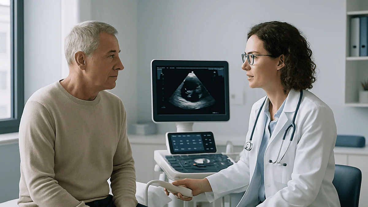
Key Takeaways
- Ultrasound sees fluid as black, so an echocardiogram can detect a pericardial effusion as a dark band between the heart muscle and the bright pericardial sac.
- The European Society of Cardiology grades effusions by fluid width at end-diastole: small under 10 mm, moderate 10 to 20 mm, large over 20 mm.
- Speed of accumulation matters more than size; a sudden small bleed can cause tamponade while a slowly accumulated large effusion may cause no pressure at all.
- Echo signs of tamponade include collapse of the right atrium or ventricle, a distended inferior vena cava that no longer narrows with breathing, and exaggerated breathing-related changes in valve inflow.
- Anti-inflammatory classes are first-line for effusion with pericarditis, but guidelines find no proven benefit from them when inflammation markers are normal.
- Drainage is the treatment for tamponade and for large symptomatic effusions, but fluid often returns if the underlying cause is not addressed.
An echocardiogram shows how much fluid surrounds the heart, where it sits, and whether it is pressing on the heart chambers. Measuring the fluid layer places an effusion into small, moderate or large groups, while signs such as chamber collapse or a swollen inferior vena cava point toward tamponade. Clinicians combine those findings with symptoms and blood tests to decide between monitoring, anti-inflammatory treatment or drainage.
The sonographer goes quiet for a moment, adjusts the probe below your ribs, and asks you to hold your breath. On the screen, a black crescent has appeared around the grey, beating muscle of your heart. You did not know there was a space there at all, let alone one that could fill with fluid.
That black crescent is what a pericardial effusion echocardiogram is designed to find and measure. The pericardium is a thin, two-layered sac around the heart, and a pericardial effusion means more fluid than usual has collected between those layers. Ultrasound sees fluid as darkness, so even a modest collection stands out against the tissue.
What happens next depends on details that are easy to miss in the moment: the width of that crescent in millimeters, whether it wraps all the way around, and how the right side of the heart behaves when you breathe. Those details, more than the word effusion itself, shape the plan your care team offers.
What a pericardial effusion echocardiogram actually shows
An echocardiogram is an ultrasound scan of the heart: a handheld probe sends sound waves through the chest wall and turns the returning echoes into moving pictures. Muscle and valve tissue reflect sound and look grey or white. Fluid lets sound pass straight through, so it appears black. That contrast is the whole trick behind the pericardial effusion echocardiogram, and it explains why ultrasound, rather than a plain X-ray, is the first test most clinicians reach for when they suspect fluid around the heart (MedlinePlus).
A standard scan takes images from several windows on the chest and from just below the breastbone. In each view, the operator looks for a dark band between the outer surface of the heart muscle and the bright line of the pericardial sac. A healthy pericardium normally holds only a few teaspoons of lubricating fluid, roughly 15 to 50 mL according to the European Society of Cardiology guideline on pericardial disease, and that thin film is usually invisible on ultrasound.
When more fluid gathers, the scan can answer four questions in one sitting. How thick is the fluid layer? Does it surround the whole heart or pool in one region? Is anything solid floating in it, such as clot or strands of inflammatory tissue? And, most urgently, is the fluid squeezing the heart enough to interfere with filling? The scan also shows how well the heart pumps, whether the valves work normally and whether the inferior vena cava, the large vein returning blood to the right side of the heart, is swollen.
The test itself is painless and involves no radiation. You lie on your left side, gel is applied, and the probe is pressed firmly against the skin. Most complete studies take between 20 and 60 minutes, though a focused bedside scan in an emergency setting may take only a few minutes (MedlinePlus).
How is pericardial effusion size grading done on echo?
Size is the first thing a report states, and it is measured, not guessed. The operator freezes the image at the end of diastole, the phase when the heart is fullest, and measures the width of the dark fluid band at its widest point. The European Society of Cardiology guideline groups effusions by that single measurement: small when the layer is under 10 mm, moderate between 10 and 20 mm, and large when it exceeds 20 mm (ESC guideline, PubMed).
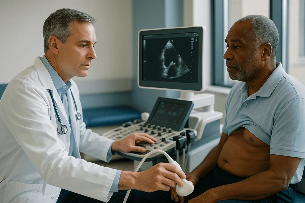
Those millimeters translate very roughly into volume. Ten millimeters of fluid all the way around the heart corresponds to a few hundred milliliters, while a band over 20 mm may represent half a liter or more. A useful comparison: a small effusion is about the thickness of a stack of two or three coins; a large one approaches the width of a thumb.
| Grade | Fluid width at end-diastole | What it usually means for next steps |
|---|---|---|
| Trivial or small | Under 10 mm | Often incidental; management follows the cause rather than the fluid |
| Moderate | 10 to 20 mm | Cause is investigated; repeat imaging is commonly arranged |
| Large | Over 20 mm | Closer follow-up; drainage considered if symptoms or pressure signs appear |
Grading has limits worth knowing. Fluid often pools unevenly, so a measurement behind the heart may differ from one in front. A report will normally state where the widest point was found. Size also says nothing about speed: a moderate effusion that appeared over two days behaves very differently from one of the same width that has been stable for a year. That is why size grading is the start of interpretation rather than its end (ESC guideline, PubMed).
Where the fluid sits, and why location changes the picture
Two effusions of identical width can carry different weight depending on where the fluid has settled. Most collections start behind the heart, in the low point of the sac when someone lies down, and spread forward as they grow. A circumferential effusion, meaning fluid visible all the way around, tends to be reported as more significant than fluid confined to one pocket, simply because it usually reflects a larger total volume (Cleveland Clinic).
Loculated effusions are the exception that worries clinicians. Loculated means the fluid is walled off into a compartment by scar tissue or adhesions, which is common after heart surgery, radiation or previous inflammation. A small loculated pocket pressing on one chamber can compromise filling out of proportion to its size, and it may hide in a corner of the sac that standard views do not reach. Reports often flag this with phrases such as regional or localized collection.
Echo also describes the character of the fluid. Clear, watery fluid looks uniformly black. Blood, pus or protein-rich fluid may show speckles, swirling echoes or strands, which the report may call fibrinous or complex. These features do not diagnose a cause on their own, but they raise or lower suspicion of infection, bleeding or malignancy and can prompt the team to sample the fluid.
Finally, the scan checks for things that mimic effusion. A layer of fat on the front of the heart, common with age and higher body weight, sits in the same place as anterior fluid but is usually slightly grey rather than black and does not move freely. A left-sided pleural effusion, fluid around the lung, can also appear behind the heart; its relationship to the descending aorta helps the operator tell the two apart (Cleveland Clinic).
What are the cardiac tamponade echo signs?
Cardiac tamponade is the situation everyone wants to rule out. It means fluid pressure inside the pericardial sac has risen high enough to stop the heart from filling properly, so less blood is pumped with each beat. The word is dramatic, but the echocardiogram tends to reveal it as a series of subtle, mechanical clues rather than one obvious picture (Mayo Clinic).

The first clue is usually collapse of the right atrium, the thin-walled chamber that receives blood from the body. Under external pressure its wall buckles inward briefly during each beat. As pressure climbs, the right ventricle, the pumping chamber beside it, also dents inward during its relaxation phase. In the most severe cases the whole heart swings within a large pool of fluid, a finding sometimes called a swinging heart (ESC guideline, PubMed).
The second clue is the inferior vena cava. When the heart cannot accept blood normally, this vein becomes distended and no longer narrows with a deep breath. The third clue is exaggerated breathing-related change in blood flow across the mitral and tricuspid valves, the doorways into the two ventricles, measured with Doppler ultrasound. In tamponade, breathing in boosts flow into the right ventricle while starving the left, a pattern echo can quantify beat by beat.
None of these signs is a diagnosis on its own. Tamponade is a clinical judgment that combines echo findings with a fast pulse, low blood pressure, shortness of breath and distended neck veins. A person may have several echo signs while still feeling reasonably well, particularly when fluid has built slowly and the sac has stretched to accommodate it. Conversely, a rapidly bleeding sac can cause tamponade with a small volume. Speed of accumulation, not size alone, determines danger (ESC guideline, PubMed).
Should I be worried about pericardial effusion?
The honest answer is that it depends on three things the echo report and your clinical picture together reveal: how big the collection is, how fast it appeared, and what is causing it. A small effusion discovered by chance during a scan for something else is a common finding, and in the absence of symptoms or an underlying disease it often requires nothing more than noting it in the record (Mayo Clinic).
Concern rises when the fluid is large, when it has grown between two scans, when any tamponade sign is present, or when the cause is one that tends to recur, such as cancer, kidney failure or an autoimmune condition. A large effusion with no identifiable cause deserves attention too. The European guideline notes that large chronic idiopathic effusions carry a real, if modest, risk of progressing to tamponade over time, which is why they are followed rather than dismissed (ESC guideline, PubMed).
Symptoms shift the calculation as well. Fluid that presses on the heart or nearby structures can cause breathlessness, especially lying flat, a feeling of chest fullness, difficulty swallowing, hoarseness or a persistent cough. Sharp chest pain that eases when sitting forward points toward pericarditis, inflammation of the sac, which often accompanies effusion (NHS).
What worry should not do is drive you to interpret the millimeters yourself. The same 15 mm band may be a stable feature in one person and a red flag in another with a recent viral illness and a rising heart rate. Your team weighs the scan alongside blood tests for inflammation, kidney and thyroid function, an electrocardiogram and sometimes further imaging before reaching a view. Asking them directly how they would grade your risk, and what would change their mind, is a reasonable request.
What causes fluid around the heart, and can echo tell?
Echo shows the fluid; it rarely names the cause. That distinction frustrates many patients, because the natural question after seeing the scan is why. The list of possible reasons is long, and in a meaningful share of cases no cause is ever confirmed, a situation clinicians call idiopathic. The European guideline reports that a known underlying disease accounts for up to 60 percent of effusions in developed countries, which leaves a sizable minority unexplained (ESC guideline, PubMed).
The common categories are inflammation, infection, systemic disease and injury. Viral or idiopathic pericarditis is the leading cause in many settings; the sac becomes inflamed and weeps fluid, much as a sprained joint swells. Autoimmune conditions such as lupus or rheumatoid arthritis can do the same. Kidney failure, underactive thyroid and heart failure raise fluid throughout the body, including the pericardium. Cancer, either spreading to the pericardium or arising nearby, can produce large and recurrent collections. Bleeding after heart surgery, a heart attack, catheter procedures or trauma fills the sac with blood rather than watery fluid. Certain medicines and chest radiation are occasional culprits (Mayo Clinic).
The scan does offer hints. Strands and speckling suggest inflammation, infection or blood. A thickened, bright pericardium supports pericarditis. Poor pumping function or leaky valves point toward heart failure as a contributor. Fluid that also appears around the lungs or in the abdomen suggests a body-wide process rather than a purely pericardial one.
Beyond those hints, the search for a cause relies on the story and on blood tests. When the cause remains hidden and the effusion is large or symptomatic, sampling the fluid during drainage allows laboratory analysis for cells, protein, bacteria and tumor markers. Whether that step is worthwhile in a given person is a judgment for the treating team, because the yield varies and drainage carries its own small risks.
Who is treated right away, and who is asked to wait
The clearest way to understand the decision tree is to picture three groups. The first group has tamponade or is heading toward it: echo shows chamber collapse and a distended vena cava, and the person has a fast pulse, low blood pressure or breathlessness at rest. For them, drainage is the treatment, and it is arranged urgently. Fluid removal is the only intervention that relieves the pressure; medicines cannot do it (ESC guideline, PubMed).
The second group has an effusion accompanied by signs of inflammation, typically chest pain, fever, raised inflammatory markers in the blood and characteristic changes on the electrocardiogram. Here the effusion is treated as part of pericarditis. Anti-inflammatory treatment is directed at the sac itself, and the fluid is expected to recede as inflammation settles. Drainage is reserved for cases where fluid is large, growing or causing symptoms despite treatment (NHS).
The third group, often the largest, has a small or moderate effusion without inflammation, without symptoms and without pressure signs. These people are usually asked to wait, though waiting is an active plan rather than neglect. The team looks for a cause with blood tests and a careful history, treats any condition found, and books repeat imaging. The European guideline suggests moderate effusions be rescanned roughly every six months and large ones every three to six months, with timing adjusted to the individual (ESC guideline, PubMed).
Who is asked to wait even when the effusion is large? Someone whose collection has been stable for years, who feels well and whose heart shows no compression may be monitored rather than drained, because needle procedures carry risk and fluid that is drained for no clear reason often returns. That balance, between the small chance of sudden deterioration and the small chance of procedural harm, is exactly the kind of judgment that belongs to the treating team after discussion with the patient.
What is the most common treatment for pericardial effusion?
People searching this question usually expect a single answer, and the most truthful one is that the most common treatment is treating whatever caused the fluid. When inflammation is the driver, anti-inflammatory medicine is the mainstay. When kidney failure, thyroid disease or heart failure is behind it, correcting that condition tends to shrink the effusion. When infection is confirmed, antibiotics or antituberculous therapy target the organism. Cancer-related effusions are managed alongside the cancer itself (Mayo Clinic).
For inflammatory pericarditis, the classes used are non-steroidal anti-inflammatory drugs and colchicine, a medicine originally derived from a plant that dampens the activity of certain white blood cells. The European guideline recommends these in combination as first-line treatment for acute pericarditis, and colchicine has been shown in randomized trials to reduce the chance of the inflammation returning (ESC guideline, PubMed). Corticosteroids are held back for people who cannot take the first-line classes or who have specific autoimmune causes, partly because they appear to raise recurrence risk when used early. Which class, for how long, and at what strength are decisions for the prescribing clinician; they depend on kidney function, stomach history, other medicines and how the person responds.
Where the effusion is present without any evidence of inflammation, the guideline is candid that anti-inflammatory drugs have no proven benefit, and it advises against using them simply because fluid is visible. That is one of the most common misunderstandings patients carry into the clinic.
Drainage, either by a needle procedure or a small surgical opening, is the treatment of choice for tamponade and for large effusions that cause symptoms or do not respond to medical management. It is common in hospital cardiology practice but is not the most common treatment across all effusions, because most effusions are small and never need it (Johns Hopkins Medicine).
How echo guided pericardiocentesis works
Pericardiocentesis is the procedure of removing fluid from the pericardial sac through a thin needle and catheter. Its history includes a reputation for danger, and echo guidance is largely what changed that. Before the procedure, the operator uses the ultrasound to find the point on the chest where the fluid pocket is deepest and closest to the skin, and to measure the distance and angle that will reach fluid while avoiding the lung, liver and heart muscle (Johns Hopkins Medicine).
You lie propped up, which lets fluid pool where it can be reached. The skin is cleaned and numbed with local anesthetic. Sedation may be offered, but most people remain awake and are asked to keep still and breathe steadily. The needle is advanced along the planned path, often under continuous ultrasound view. Once fluid returns, a soft wire is passed, the needle is exchanged for a flexible drainage catheter, and the needle is removed. Some operators inject a small amount of agitated saline to confirm on the screen that the catheter tip lies in the pericardial space rather than inside a heart chamber (Johns Hopkins Medicine).
Fluid is drained slowly rather than all at once, because a sudden pressure change can stress a heart that has adapted to compression. The catheter is commonly left in place for a period so that residual fluid can drain and the team can watch whether it re-accumulates. Samples go to the laboratory.
Risks are real but uncommon with imaging guidance: puncture of the heart muscle or a coronary vessel, bleeding, collapsed lung, infection, abnormal heart rhythm and, rarely, injury to the liver or bowel. Alternatives include a surgical pericardial window, a small opening made in the sac so fluid drains into the chest cavity, which is favored when fluid is loculated, clotted or keeps returning. The choice between approaches rests with the treating team (Cleveland Clinic).
Can mild pericardial effusion resolve on its own?
Yes, and it frequently does. Small effusions that accompany viral pericarditis typically disappear as the inflammation settles, usually over a few weeks. Fluid that appears after heart surgery or a heart attack often follows the same course. Effusions linked to fluid overload from kidney or heart problems tend to shrink once the underlying imbalance is corrected. In each case the fluid was a symptom of something else, and it departs when the cause is addressed (NHS).
Mild pericardial effusion treatment, in the sense of doing something to the fluid itself, is therefore rarely needed. What a small effusion does require is a sensible check for a cause. That usually means a conversation about recent illnesses, chest pain, weight change, fatigue or joint symptoms, plus blood tests looking at inflammation, kidney and thyroid function. If everything is normal and there are no symptoms, the European guideline suggests that small idiopathic effusions may not even need routine repeat imaging, though many clinicians will arrange a single follow-up scan for reassurance (ESC guideline, PubMed).
There are two cautions. First, mild refers to width on a scan, not to the seriousness of a cause. A small effusion in someone with known cancer, a recent chest procedure or a new autoimmune diagnosis still warrants investigation. Second, resolution is confirmed by imaging, not assumed. If a follow-up echo is arranged, keeping that appointment is the way to close the loop.
Some small effusions persist for years without changing and without causing trouble. Stability over time is itself reassuring, and a long-standing small effusion in a well person is generally regarded as a benign finding rather than a condition needing treatment (Mayo Clinic).
Is pericardial effusion a form of heart failure?
No. The two conditions are often confused because both involve fluid and both can cause breathlessness, but they describe different problems. Heart failure means the heart muscle itself pumps or relaxes inadequately, so blood backs up and fluid gathers in the lungs, legs and abdomen. A pericardial effusion is fluid in the sac around the heart; the muscle inside may be entirely healthy (Mayo Clinic).
That said, the two can travel together. Heart failure raises pressure in the veins and can lead to a small effusion as part of general fluid retention, so the echo report of someone with heart failure may mention a trivial effusion. In the other direction, a large effusion causing tamponade produces low output and congestion that look clinically like acute heart failure. The treatment, however, is drainage rather than diuretics, which is one reason getting the distinction right matters. Diuretic medicines, which remove fluid through the kidneys, can actually worsen tamponade by lowering the filling pressure the compressed heart depends on (ESC guideline, PubMed).
The echocardiogram is what separates them. In heart failure, the scan shows a weakened or stiff ventricle, often enlarged chambers and leaky valves, with little or no pericardial fluid. In effusion, the scan shows a normal-sized, normally contracting heart surrounded by a dark band. When both features appear, the report describes each separately.
Patients sometimes leave a consultation believing their heart is failing because they heard the phrase fluid around the heart. If your report says effusion and nothing about reduced ejection fraction, the pumping measure, then the finding concerns the sac, not the muscle. Asking your clinician to point to the ejection fraction on the report is a quick way to settle the question.
What the following days and weeks usually look like
The timeline branches according to which path you are on. For someone monitored without treatment, the coming weeks mostly involve waiting for blood results, treating any cause identified, and attending a repeat scan. That second echo is where the picture usually clarifies: fluid that has shrunk or stayed the same supports continued observation, while growth prompts a closer look.
For someone treated for pericarditis, chest pain typically improves within days of starting anti-inflammatory treatment, and the NHS notes that most people recover within a few weeks. The effusion lags behind symptoms and may take longer to disappear on imaging. Clinicians commonly recheck inflammatory blood markers before stopping treatment, and repeat imaging is arranged to confirm the fluid has resolved. Physical exertion is usually restricted while inflammation is active, particularly for athletes, because strenuous activity appears to raise the risk of recurrence (NHS; ESC guideline, PubMed).
Recurrence is the main disappointment on this path. The European guideline reports that pericarditis returns in roughly 15 to 30 percent of people after a first episode when colchicine is not used, and that colchicine lowers that figure substantially. Recurrent pericarditis is treated with the same classes for a longer duration, again at the discretion of the prescribing clinician.
After drainage, expect a hospital stay while the catheter remains in place, often a day or two but sometimes longer, with a bedside echo before it is removed to confirm the fluid has not returned. Soreness at the puncture site is common and settles. Follow-up imaging in the weeks afterward checks for re-accumulation, which is more likely when the cause is cancer or an untreated systemic disease (Johns Hopkins Medicine). Any timeline your team gives you will be a range, not a promise, because the cause dictates the course more than the procedure does.
What people often get wrong about pericardial effusion echocardiogram results
The first misunderstanding is that a bigger number means a more dangerous situation. Speed matters more than size. A sac that has stretched over months can hold a large volume calmly, while a sudden bleed of a small volume can cause tamponade within hours. This is why a repeat echo comparing two points in time often carries more information than a single measurement (ESC guideline, PubMed).
The second is that the echo tells you why the fluid is there. It usually does not. People sometimes decline further tests because the scan already found the fluid, but the scan cannot distinguish a viral cause from an autoimmune or malignant one. Blood work and history do that.
The third is that fluid around the heart means the heart is weak. As the previous section described, effusion and heart failure are separate findings. A normal ejection fraction on the same report is common and reassuring.
The fourth is that draining the fluid solves the problem. Drainage relieves pressure and provides a sample, but if the cause is not addressed the fluid frequently returns. Guidelines discourage draining asymptomatic effusions purely to remove them, because the procedure carries risk and the fluid tends to come back (Cleveland Clinic).
The fifth is that anti-inflammatory medicines shrink any effusion. They help when the sac is inflamed and have no demonstrated effect when it is not. Taking or requesting them for a non-inflammatory effusion exposes a person to side effects without benefit.
A final, quieter error is treating a small stable effusion as a lifelong worry. Long-standing small effusions in people who feel well are generally benign. Once a cause has been reasonably excluded and stability confirmed, most clinicians consider the matter closed unless symptoms change (Mayo Clinic).
Questions to ask your care team
A good consultation about an effusion should leave you able to explain your own report. The following questions tend to draw out the information that matters most. Bring the written report, and ask the clinician to point to the relevant lines as they answer.
- How wide was the fluid layer, where was it measured, and which size grade does that place me in?
- Does the fluid surround the whole heart, or is it pooled in one area? Was any of it loculated?
- Were there any signs that the fluid is pressing on the heart, such as chamber collapse or a distended vena cava?
- What was my ejection fraction, and is the heart muscle itself working normally?
- Do you think inflammation is driving this? Which blood tests support that view?
- What is your working theory about the cause, and what else are you testing for?
- If we are monitoring, when is the next echo, and what change on that scan would alter the plan?
- If medicine is recommended, what is it meant to do, roughly how long is it usually continued, and what should I watch for?
- If drainage is being considered, why now, what are the specific risks in my case, and what are the alternatives?
- Are there activities I should limit while this is being sorted out, and for how long?
- Which symptoms should make me call you, and which should send me to an emergency department?
Two habits make these questions more useful. Write the answers down, because effusion terminology is easy to muddle afterward. And ask what the clinician would want to know from you before the next appointment, whether that is a symptom diary, a record of your resting heart rate or simply how your breathing feels lying flat. Monitoring works best when it runs in both directions.
When to call your doctor
Most people with a pericardial effusion will not experience an emergency, but the consequences of missing tamponade are serious enough that the warning signs deserve a clear place in your memory. Seek emergency care immediately, by calling emergency services rather than driving yourself, if you develop sudden or rapidly worsening shortness of breath, particularly if you cannot lie flat; fainting or near-fainting; a racing or pounding heartbeat that does not settle with rest; cold, clammy or bluish skin; confusion or severe lightheadedness; or crushing chest pain. These can indicate that fluid is compressing the heart or that a separate cardiac event is under way (Mayo Clinic).
Call your care team the same day, without waiting for the next scheduled appointment, if breathlessness on exertion is clearly worse than it was; if chest pain returns or changes character after settling; if you develop fever, chills or night sweats; if your legs or abdomen swell noticeably; if swallowing becomes difficult or your voice turns hoarse; or if you notice a new persistent cough. After drainage, redness, discharge or increasing pain at the puncture site, or fever, should also prompt a call (Johns Hopkins Medicine).
Symptoms should be judged against your own baseline. Someone with known heart failure may already be breathless on stairs; what matters is a change from the usual. People taking anti-inflammatory treatment should also report black stools, vomiting blood, persistent stomach pain or unusual bruising, which can signal side effects that the prescribing clinician needs to know about.
If you are being monitored and simply feel uneasy, that is a reasonable reason to call as well. A brief conversation, sometimes followed by a quick bedside scan, resolves most worries and occasionally catches a change early. The team following your effusion expects to hear from you between appointments; that is part of what monitoring means.
Frequently asked questions
Should I be worried about a pericardial effusion found on an echocardiogram?
Usually the level of concern depends on size, speed of accumulation and cause rather than the finding itself. Small effusions discovered incidentally in people who feel well are common and often need only a check for a cause. Concern rises with large or growing collections, echo signs of pressure on the heart, symptoms such as breathlessness, or an underlying disease that tends to recur.
What is the most common treatment for pericardial effusion?
Treating the underlying cause is the most common approach. When inflammation is present, non-steroidal anti-inflammatory drugs and colchicine are first-line classes according to the European Society of Cardiology guideline. Effusions from kidney, thyroid or heart problems shrink as those are corrected. Drainage is reserved for tamponade or large symptomatic effusions and is not needed for most small collections.
Can a mild pericardial effusion go away on its own?
Yes, small effusions frequently resolve without specific treatment, particularly those linked to viral pericarditis, recent heart surgery or fluid overload. A sensible check for a cause is still recommended, and a follow-up scan confirms resolution. Some small effusions persist for years without changing, and a stable small effusion in a well person is generally considered benign.
Is pericardial effusion a form of heart failure?
No. Heart failure describes a heart muscle that pumps or relaxes poorly, while a pericardial effusion is fluid in the sac around the heart. The muscle inside may be perfectly healthy. The two can coexist, and severe effusion can mimic heart failure symptoms, but the echocardiogram distinguishes them, and the treatments differ.
How is pericardial effusion size grading measured?
The operator freezes the image at the end of diastole, when the heart is fullest, and measures the width of the fluid band at its widest point. The European Society of Cardiology grades under 10 mm as small, 10 to 20 mm as moderate and over 20 mm as large. Reports usually state where the widest point was found because fluid pools unevenly.
What are the cardiac tamponade echo signs I might see on my report?
Reports may mention right atrial or right ventricular collapse, a dilated inferior vena cava with little change on breathing, a swinging heart, or marked respiratory variation in mitral and tricuspid inflow. None of these alone confirms tamponade; clinicians combine them with pulse, blood pressure and symptoms. Ask your team what each phrase on your report means for you.
How does echo guided pericardiocentesis work and is it safe?
Ultrasound is used to choose the entry point where fluid is deepest and closest to the skin, then to guide a needle and soft catheter into the sac under local anesthetic. Fluid is drained gradually and the catheter may stay in place for a time. Imaging guidance has made complications uncommon, though heart puncture, bleeding, collapsed lung and infection remain possible risks.
Does mild pericardial effusion treatment involve medication?
Often it does not. If a small effusion is accompanied by pericarditis, anti-inflammatory treatment targets the inflammation rather than the fluid. If inflammation markers are normal and there are no symptoms, guidelines find no proven role for anti-inflammatory medicines, and management focuses on identifying and treating any cause. The prescribing clinician makes that judgment.
How often will I need a repeat echocardiogram?
The European Society of Cardiology guideline suggests moderate effusions be rescanned roughly every six months and large ones every three to six months, with small idiopathic effusions often needing little routine follow-up. Your own interval will depend on symptoms, cause and how the fluid changed between scans, so treat any schedule as a starting point set by your team.
Can I exercise with a pericardial effusion?
It depends on the cause and whether inflammation is active. During pericarditis, guidelines advise restricting strenuous activity until symptoms and inflammatory markers settle, because exertion appears to raise recurrence risk. With a stable, non-inflammatory small effusion and no symptoms, normal activity is often permitted. Ask your care team for specific limits and when to reassess them.
References
- Pericardial effusion: Cleveland Clinic
- Echocardiogram: MedlinePlus Medical Encyclopedia
- Pericarditis: NHS
This article is for general information only and is not a substitute for professional medical advice. Please consult a qualified doctor about your individual situation.

More from the Blog

How to Lower Blood Pressure Naturally: The Ten Changes With Trial Evidence, Ranked by Effect
The lifestyle changes with the strongest trial evidence for lowering blood pressure are a DASH-style eating pattern, cutting sodium, losing excess weight, regular aerobic…

Why Cardiorenal Syndrome Is Managed by Cardiology and Nephrology Together, Not in Turn
Cardiorenal syndrome is managed by cardiology and nephrology together because the heart and kidneys fail as a linked system, not one after the other.…

What an Electrophysiology (EP) Study Involves: Catheters, Rhythm Mapping and Test Pacing
During an electrophysiology (EP) study, a cardiologist threads thin, flexible catheters through a vein, usually in the groin, into the heart. Electrodes on the…

When Is Catheter Carotid Angiography Recommended After Ultrasound Shows Narrowing?
Catheter carotid angiography is usually reserved for situations where ultrasound and a second noninvasive scan, such as CT or MR angiography, disagree or cannot…

How the Extracardiac Fontan Procedure Reroutes Blood Flow: A Guide for Parents
The extracardiac Fontan procedure is the usual third stage of surgery for children born with one working heart pump. Surgeons attach a synthetic tube…

Preparing for Cardiothoracic Surgery: Chest Imaging, Stopping Smoking and the Pre-Op Talk
Preparing for cardiothoracic surgery usually means completing chest imaging and heart tests such as a chest X-ray, ECG and echocardiogram, stopping smoking as early…