

What Do Tests for Urethra Diseases Involve? Cystoscopy, Uroflowmetry and Ultrasound Explained

Key Takeaways
- Uroflowmetry measures how fast urine leaves the body but cannot tell a blockage from a weak bladder; that distinction needs a pressure-flow urodynamic study.
- Normal uroflowmetry values vary by age and sex, with peak flow in men declining with age, and a small voided volume makes the curve unreliable enough to repeat.
- A flexible cystoscopy is done awake with local anesthetic gel and, according to the Mayo Clinic, typically takes 5 to 15 minutes for the procedure itself.
- Standard ultrasound shows the bladder, kidneys and prostate well but rarely the urethra; a retrograde urethrogram is the test that maps a stricture's location and length.
- Mild burning and a trace of blood commonly last a day or two after cystoscopy per the NHS, while urinary tract infection is the most common true complication.
- An inability to pass urine, fever above 101.4 F (38.5 C), clots or pain lasting beyond two days after cystoscopy are reasons to contact the care team promptly.
Tests for urethra diseases usually combine a urine sample, uroflowmetry (urinating into a machine that measures flow rate), an ultrasound of the bladder and kidneys, and sometimes cystoscopy, in which a thin camera is passed along the urethra under local anesthetic gel. Contrast X-rays or urodynamic pressure studies may follow. Each test answers a different question, and the treating team decides which ones are needed.
The instruction on the appointment letter sounded almost comic: arrive with a comfortably full bladder, then wait. So he sits in the corridor, legs crossed, checking the clock, wondering how a clinic can turn the most private act of the day into a measurement. Down the hall, a woman with her third urinary infection this year is being told that the next step may involve a camera, and she has quietly decided to look it up before she agrees to anything.
Both of them are about to meet the standard urethra tests: cystoscopy, uroflowmetry, ultrasound and, occasionally, a contrast X-ray or a pressure study. None is glamorous. Together they answer a surprisingly precise set of questions about a tube that most people never think about until it stops behaving.
What follows is an honest walk through each test: what physically happens, what the numbers on the report mean, what tends to be uncomfortable, and where the limits of each method lie.
Which urethra tests come first: cystoscopy, uroflowmetry or a simple urine sample?
The urethra is the tube that carries urine from the bladder out of the body; in men it also carries semen. It can become inflamed (urethritis), narrowed by scar tissue (a urethral stricture), squeezed from outside by an enlarged prostate, or, less often, affected by a pouch in its wall called a diverticulum. Each problem produces overlapping symptoms, which is exactly why a stepwise approach exists.
The sequence matters more than any single test. A urologist will almost always begin with a conversation and an examination, then a urine sample checked with a dipstick and, where infection is possible, sent for culture. When urethritis is suspected, a urine or swab test for sexually transmitted infections is often part of that first visit. These simple steps rule out the most common and most treatable explanations before anyone reaches for equipment.
Only then do the machine-based urethra tests enter: uroflowmetry to measure how fast urine leaves the body, an ultrasound to see whether the bladder empties and the kidneys are under strain, and cystoscopy when a direct look at the lining is required. Contrast X-rays and urodynamic pressure studies sit further along the path, reserved for situations where the picture remains unclear or surgery is being planned.
If there is one idea worth holding onto, it is this: the least invasive tests are not the least informative. A flow curve and a bladder scan, read together, can steer the whole investigation. The camera comes later, and for many people it never comes at all. The order is set by the clinical question, not by how impressive the equipment looks.
What does uroflowmetry involve, and what is a normal uroflowmetry result?
Uroflowmetry is a test that measures the rate and pattern of urine flow. There are no needles and no instruments inside the body. You arrive with a comfortably full bladder, are shown to a private room, and urinate into a funnel connected to an electronic meter. The machine weighs the urine as it arrives and draws a curve of flow against time. MedlinePlus describes it as a quick, painless test that is best done when you feel a normal urge to go, not when you are bursting.
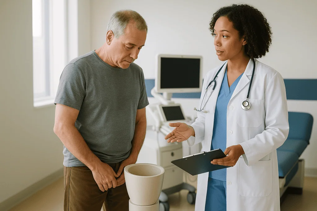
The printout typically reports several figures:
- Peak flow rate (Qmax): the fastest point in the stream, in milliliters per second.
- Average flow rate and total voided volume.
- Time to peak and total voiding time.
- Curve shape: a smooth bell is the usual pattern; a long, flat plateau is the classic signature of a fixed narrowing such as a stricture; a jagged, stop-start line often means straining.
So what is a normal uroflowmetry result? Honest answer: it depends on who is voiding. MedlinePlus notes that normal values vary with age and sex, and that flow in men declines as they get older while women change less. Standard urology references usually regard a peak flow above roughly 15 mL per second, produced from a voided volume of at least about 150 mL, as within the expected adult range, and a peak flow under about 10 mL per second as a prompt to look for obstruction or a weak bladder muscle. Your team applies age- and sex-adjusted ranges and will tell you where you sit.
Two caveats matter. A small voided volume makes the curve unreliable, so you may be asked to repeat the test. And a low flow cannot say whether the cause is a blockage or an underpowered bladder; that distinction needs the tests described later.
How does a cystoscopy work? Flexible versus rigid, step by step
Cystoscopy is an examination of the urethra and bladder using a cystoscope, a thin tube with a light and camera at its tip. Because the scope travels along the urethra on its way in, the urethra itself is inspected first, which is why this test is so useful for diagnosing strictures, inflammation and unusual growths.
There are two versions. A flexible cystoscopy uses a bendable scope roughly the width of a pencil and is done awake in an outpatient room. According to the NHS, a local anesthetic gel is applied to numb the urethra, and the procedure is usually over within minutes. You lie on your back, the opening of the urethra is cleaned, the gel is given a moment to work, and the scope is passed gently while sterile fluid flows through it to open up the passage so the lining can be seen. The image appears on a screen, and many clinicians will talk you through what they see.
A rigid cystoscopy uses a straight, wider scope and is generally performed under general or spinal anesthesia in an operating theater. Its advantage is the working channel: instruments can be passed to take a biopsy (a small tissue sample), remove a stone, or treat a narrowing. The Mayo Clinic explains that the choice between the two depends on the reason for the test and on whether treatment might be done at the same time.
In both cases the bladder is filled with fluid so its walls can be inspected fully. That filling produces the strong urge to urinate that people remember afterward. Once the examination is complete, the scope is withdrawn and you are usually allowed to empty your bladder straight away.
What should you not do before a cystoscopy?
Preparation is mostly about not sabotaging the test, and the list of things to avoid is short but real.

- Do not stop or change any prescribed medicine on your own. Blood thinners and antiplatelet drugs are the usual concern before a rigid cystoscopy or biopsy, but the decision to pause, bridge or continue belongs to the prescribing clinician and the urology team together.
- Do not hide symptoms of infection. Burning, cloudy urine or fever in the days before the appointment should be reported. The Mayo Clinic notes that a urine sample is often checked beforehand, and an active infection generally means the cystoscopy is postponed and treated first.
- Do not arrive having emptied your bladder completely if you have been asked for a sample. Many clinics want urine on arrival for a dipstick or culture.
- Do not eat or drink against instructions if you are having general or spinal anesthesia. Flexible cystoscopy under local gel usually has no fasting rule, but follow your letter, not a forum.
- Do not plan to drive yourself home after sedation or a general anesthetic. After a local-only flexible procedure, most people can travel normally.
- Do not forget allergies. Local anesthetic gel, antibiotics and latex are all worth mentioning.
Some teams give a preventive antibiotic around the time of the procedure; the Mayo Clinic describes this as a case-by-case decision based on infection risk, and it is never something to arrange yourself.
One more thing not to do: skip your questions. The few minutes before the scope goes in are the right time to ask what the clinician expects to see, whether a biopsy is likely, and how the results will reach you.
How long does a cystoscopy take, and does it hurt?
People ask about time because they want to know how long they have to tolerate the sensation, so both questions deserve straight answers.
The Mayo Clinic states that a simple outpatient cystoscopy can take 5 to 15 minutes, and that when sedation or general anesthesia is used the procedure itself usually takes 15 to 30 minutes. The whole visit is longer: changing, positioning, waiting for the anesthetic gel to work, and, after a general anesthetic, time in recovery until you are awake and have passed urine.
Does it hurt? Most people describe a flexible cystoscopy as uncomfortable rather than painful. The sensations come in three phases. The gel itself can sting briefly. As the scope passes the external sphincter, the muscular ring that holds urine in, men in particular feel a pressure or a short sharp moment; slow breathing out and consciously relaxing the pelvic floor genuinely helps, because a clenched sphincter narrows the path. Then the bladder fills and the urge to urinate becomes strong, sometimes to the point of feeling you cannot hold on. That is expected, and it eases the instant the scope comes out and you are allowed to void.
A rigid cystoscopy is not felt at the time because of the anesthetic, but afterward there is typically more burning and a little more blood, especially if tissue was sampled.
Anxiety amplifies every one of these sensations. Ask to watch the screen if that distracts you, or ask not to. Ask the clinician to narrate. Either approach is reasonable, and clinics are used to both.
Where do ultrasound and contrast X-rays fit among urethra tests?
Ultrasound uses sound waves to build a picture of internal organs; it involves no radiation and no instruments inside the body. In the urinary investigation it plays two supporting roles. The first is the post-void residual, the amount of urine left behind after you have emptied your bladder. A handheld probe on the lower abdomen estimates this within seconds, and the NIH’s National Institute of Diabetes and Digestive and Kidney Diseases lists it among the core urodynamic measurements. A consistently large residual suggests either a blockage or a bladder that is not contracting well.
The second role is upstream. A kidney ultrasound checks for hydronephrosis, the swelling of a kidney when urine cannot drain freely, which is the main structural danger of a long-standing obstruction. In men, an abdominal ultrasound can also estimate prostate size.
What ultrasound does poorly is the urethra itself. The tube is narrow and mostly collapsed, so standard scanning rarely shows a stricture directly. When the team needs a map of the urethra, they turn to contrast imaging:
- A retrograde urethrogram is an X-ray taken while a small amount of contrast dye is gently introduced at the opening of the urethra. It shows the location and length of a narrowing, information a surgeon needs before deciding how to treat it.
- A voiding cystourethrogram fills the bladder with contrast through a thin catheter and takes images while you urinate, revealing how the urethra opens under real pressure and whether urine flows back toward the kidneys.
Because these use X-rays, they are usually deferred or replaced in pregnancy. MRI is occasionally used for complex cases such as diverticula, but it is not a routine urethra test.
Urodynamic studies: when uroflowmetry alone cannot settle the question
A low flow rate poses a fork in the road. Either something is narrowing the outlet, or the bladder muscle is not pushing hard enough. Uroflowmetry cannot tell these apart, and treating the wrong one is a waste of everyone’s time. Urodynamic testing exists to resolve that fork.
Urodynamics is a group of tests that measure how well the bladder stores and releases urine. The NIDDK describes the main components: cystometry, in which a thin catheter fills the bladder slowly with fluid while sensors record pressure and you report when you first feel the urge to go; a pressure-flow study, in which you urinate with the catheter still in place so that bladder pressure and flow rate are captured at the same moment; and, sometimes, electromyography, where small pads or fine electrodes record the activity of the pelvic floor muscles. A second small catheter in the rectum measures abdominal pressure so that straining can be separated from true bladder contraction.
The interpretation is elegant. High bladder pressure with low flow points to obstruction, such as a stricture or an enlarged prostate. Low pressure with low flow points to an underactive bladder. The same flow rate, two opposite conclusions.
This is a longer and more involved appointment than uroflowmetry, and it is not offered to everyone. It is generally reserved for people whose symptoms and simpler tests disagree, for those with neurological conditions affecting bladder control, and for those in whom surgery is being considered and the team wants to be sure the operation targets the right problem. The catheters are thin and placed with lubricating gel; discomfort is usually described as pressure rather than pain, and the NIDDK notes that mild burning afterward is common and short-lived.
Who is usually offered urethra tests such as cystoscopy, and who is asked to wait?
Referral for these tests follows recognizable patterns, and understanding them helps explain why one person is offered a camera while another is told to come back after treatment.
Cystoscopy is commonly recommended for visible blood in the urine, for repeated urinary infections without an obvious cause, for a weak or spraying stream that suggests a stricture, for suspected bladder stones, and for follow-up after treatment of bladder or urethral conditions. The Mayo Clinic also lists ongoing pelvic pain, urinary incontinence and an enlarged prostate among the reasons a clinician may suggest it. Uroflowmetry and a post-void residual scan are usually offered earlier and more freely, because they carry almost no risk and often decide whether anything more is needed.
Several groups are typically asked to wait or to have the plan adjusted:
- Anyone with an active urinary infection. Passing a scope through infected tissue raises the risk of spreading infection to the bloodstream, so the infection is treated first.
- People taking anticoagulants who need a rigid cystoscopy or biopsy. The procedure may still go ahead, but only after the prescribing clinician and the urology team have agreed on how to handle the medicine.
- People who are pregnant, for whom X-ray-based contrast studies are usually deferred while ultrasound and, where essential, cystoscopy remain options.
- Anyone unable to produce an adequate volume for uroflowmetry, who is simply rescheduled rather than misdiagnosed.
None of these is a refusal. They are timing decisions, and the treating team weighs them against how urgent the underlying question is. If you feel you have been asked to wait without explanation, ask for one; the reasoning is nearly always straightforward.
Urethra tests compared: cystoscopy, uroflowmetry and ultrasound at a glance
Seeing the tests side by side makes their division of labor obvious. Each answers a question the others cannot.
| Test | What it shows | Anesthetic | Typical time for the test itself | Main limitation |
|---|---|---|---|---|
| Uroflowmetry | Speed and pattern of the stream | None | The time it takes to urinate; MedlinePlus describes it as a quick test | Cannot distinguish blockage from a weak bladder |
| Bladder and kidney ultrasound | Urine left after voiding; kidney swelling; prostate size | None | Minutes; a post-void residual scan is near-instant | Rarely shows the urethra directly |
| Flexible cystoscopy | Direct view of urethral and bladder lining | Local anesthetic gel | About 5 to 15 minutes (Mayo Clinic) | Limited ability to treat; brief discomfort |
| Rigid cystoscopy | Direct view plus biopsy or treatment | General or spinal | Usually 15 to 30 minutes (Mayo Clinic) | Needs a theater and recovery time |
| Retrograde urethrogram | Location and length of a narrowing | None; local gel | Short X-ray session | Uses radiation and contrast; deferred in pregnancy |
| Urodynamics | Bladder pressure against flow | None; catheters with gel | Longer appointment than any test above | More involved; reserved for unresolved cases |
The pattern is worth noticing: as you move down the table, the tests become more informative about mechanism and also more demanding of you. Good practice climbs the table only as far as the clinical question requires.
A practical reading of this table: if your clinician has ordered only the first two rows, it does not mean your problem is being taken lightly. It often means those two rows are expected to answer it.
Cystoscopy recovery time: what the following days usually look like
After a flexible cystoscopy, the NHS advises that most people can return to their normal activities the same day. The first urination is often the memorable moment: a stinging sensation, sometimes described as similar to a mild infection, and urine that may be pink or contain a trace of blood. According to the NHS, this mild discomfort and slight bleeding commonly last a day or two. Drinking water at a steady, sensible pace helps dilute the urine and eases the sting, though there is no need to force fluids.
Frequency and urgency can linger for a short while because the bladder lining has been stretched and rinsed. Some people notice a small amount of blood at the very start or end of the stream rather than throughout. Both settle as the irritated lining calms.
People often ask how long it takes for the urethra to heal after a cystoscopy. The honest framing is that a routine flexible procedure does not create a wound in the usual sense; it irritates the lining, and that irritation typically fades over a few days. A rigid cystoscopy, especially one that included a biopsy or treatment of a stricture, leaves more to recover from: burning and bleeding may persist a little longer, and some people go home with a temporary catheter, which the team removes at a planned time.
Warmth, rest and paracetamol-type pain relief as advised by the team are usually all that is needed. Sexual activity can resume when it feels comfortable, unless the team has said otherwise after a treatment procedure. Results from a visual inspection are often discussed on the day; biopsy results take longer and arrive through the route your team explained.
If discomfort is climbing rather than fading after the second day, that is the signal to pick up the phone.
What is the most common complication of cystoscopy?
It helps to separate expected after-effects from complications. Burning on urination, a little blood and a temporary increase in frequency are so common that they are better thought of as part of the procedure than as problems with it. Among true complications, the one that occurs most often is a urinary tract infection. Passing any instrument along the urethra can carry bacteria into the bladder, which is why the Mayo Clinic lists infection first among cystoscopy risks and why some teams offer a preventive antibiotic to people at higher risk.
Other recognized complications are less frequent:
- Bleeding heavier than a trace, usually after a biopsy or treatment rather than a simple look.
- Urinary retention, an inability to pass urine, typically from swelling or muscle spasm after the procedure. The Mayo Clinic specifically advises contacting your care team if you cannot urinate after a cystoscopy.
- Injury to the urethra or bladder, which is rare and more associated with rigid instruments.
- Urethral stricture as a late effect, an uncommon outcome of repeated or traumatic instrumentation rather than of a single flexible examination.
How to tell infection from ordinary irritation: irritation is worst at the first void and eases over a day or two; infection tends to worsen after that point and may bring fever, chills, cloudy or foul-smelling urine and pain in the lower back. That trajectory, getting worse rather than better, is the single most useful clue.
Put in proportion, cystoscopy is one of the most routinely performed diagnostic procedures in urology precisely because serious harm is uncommon. But uncommon is not never, and knowing what a complication looks like is what allows it to be treated early.
What people often get wrong about urethra tests
Some misunderstandings surface in almost every clinic, and clearing them up changes how people feel about the tests.
“A flow test can diagnose a stricture.” It cannot. A flat, plateau-shaped curve raises suspicion, but the same curve can come from an enlarged prostate or a weak bladder. Diagnosis needs a direct look with cystoscopy or a contrast X-ray to confirm where and how long the narrowing is.
“Cystoscopy always means going to sleep.” Flexible cystoscopy is done awake with anesthetic gel, and that is the version most people are offered for a first look. General anesthesia is reserved for rigid procedures where treatment or biopsy is planned.
“A normal flow rate rules everything out.” Flow measures speed, not the health of the lining. Blood in the urine with a perfectly normal curve still needs investigation.
“You should drink as much as possible before a cystoscopy.” This confuses cystoscopy with uroflowmetry. For a flow test you need a comfortably full bladder; for a cystoscopy you are usually asked to empty it, because the clinician fills it with sterile fluid during the procedure.
“Ultrasound will show the urethra.” Standard scanning shows the bladder, kidneys and prostate well, but the urethra is narrow and collapsed and rarely visible in useful detail without specialized technique.
“Urethral problems are a men’s issue.” Urethritis, urethral pain syndrome, diverticula and strictures after injury or catheterization all occur in women. The anatomy differs, the investigation follows the same logic.
“One cystoscopy will damage the urethra permanently.” Scarring is a recognized but uncommon late complication, mostly associated with rigid instruments or repeated procedures, not with a single flexible examination.
Questions to ask your care team before urethra tests
A good consultation leaves you knowing not just what will happen but why. These questions tend to draw out the reasoning.
- Which specific question is this test meant to answer, and what would you do differently depending on the result?
- Are we starting with uroflowmetry and a bladder scan, and under what circumstances would you move on to cystoscopy?
- Will the cystoscopy be flexible under local gel or rigid under anesthesia, and what decides that?
- Is a biopsy or any treatment likely during the same procedure, and would I be asked to consent to that in advance?
- Do I need to change anything about my current medicines, and who will coordinate that with the clinician who prescribes them?
- Do I need to fast, bring a urine sample, or arrange someone to accompany me home?
- Will a preventive antibiotic be given, and if not, why is it not needed in my case?
- If a stricture is found, what imaging would you want next before discussing treatment options?
- How and when will I receive results, particularly if tissue is sent to the laboratory?
- What symptoms in the days afterward should prompt me to call, and which number do I use out of hours?
Two questions are worth asking even when they feel awkward. First: could this test be avoided or delayed without missing anything serious? Clinicians expect that question and usually have a clear answer. Second: if I find the procedure intolerable partway through, can it be stopped? The answer to that one is yes, and hearing it said aloud makes the whole experience easier to approach.
Write the answers down or bring someone who will. Details about preparation and aftercare are easy to lose in the moment.
When to call your doctor after cystoscopy, uroflowmetry or urodynamics
Most people need nothing beyond a glass of water and a little patience after these tests. A small number develop a problem that should be assessed promptly, and the signs are clear enough to act on without second-guessing.
Contact your care team, or seek urgent care if you cannot reach them, if you notice any of the following:
- You cannot pass urine despite feeling the need. The Mayo Clinic lists an inability to urinate after cystoscopy as a reason to call; a bladder that cannot empty at all is an emergency, not something to sleep on.
- Fever above 101.4 F (38.5 C), chills or shaking, which the Mayo Clinic flags as a warning after cystoscopy and which point toward infection.
- Bright red blood or clots in the urine, or bleeding that is increasing rather than fading. A trace of pink for a day or two is expected; a stream that looks like wine is not.
- Burning or bladder pain that persists beyond two days or that worsens after the first day, again a threshold the Mayo Clinic uses.
- Pain in the flank or lower back, nausea, or feeling generally unwell, which can signal that an infection has reached the kidney.
- Foul-smelling or cloudy urine developing after the procedure.
The same list applies after urodynamics and after a contrast urethrogram, with one addition for contrast studies: a rash, swelling or breathing difficulty in the hours afterward should be treated as an allergic reaction and assessed urgently.
Uroflowmetry and ultrasound involve nothing entering the body, so complications are not expected; if symptoms appear afterward, they almost certainly relate to the underlying condition, and they still deserve a call.
Whatever the test, the treating team remains responsible for interpreting results and deciding next steps. Reading this article prepares you for the conversation; it does not replace it.
Frequently asked questions
How long does it take for the urethra to heal after a cystoscopy?
After a routine flexible cystoscopy the urethral lining is irritated rather than wounded, and the NHS notes that mild discomfort and slight bleeding usually settle within a day or two. A rigid cystoscopy with biopsy or treatment can leave burning and bleeding for somewhat longer, and the team will give a specific timeline. Symptoms that worsen after the second day should be reported rather than waited out.
What is a normal uroflowmetry result?
There is no single number. MedlinePlus explains that normal values depend on age and sex, and that flow in men falls with age. Standard urology references generally treat a peak flow above roughly 15 mL per second, from a voided volume of at least about 150 mL, as within the expected adult range, while a peak below about 10 mL per second prompts further tests. Your clinician applies the range appropriate to you.
What should you not do before a cystoscopy?
Do not stop or alter prescribed medicines on your own, particularly blood thinners; that decision is made with the prescribing clinician. Do not conceal symptoms of infection, since an active infection usually means postponing the test. Do not eat or drink against instructions if anesthesia is planned, and do not arrange to drive yourself home after sedation. Bring a urine sample if asked, and mention allergies.
What is the most common complication of cystoscopy?
Urinary tract infection is the most common true complication, because an instrument passing along the urethra can carry bacteria into the bladder; the Mayo Clinic lists infection first among the risks. Burning and a trace of blood are expected effects rather than complications. Less frequent problems include heavier bleeding after biopsy, temporary inability to urinate, and, rarely, urethral injury or later scarring.
How long does a cystoscopy take from start to finish?
The Mayo Clinic states that a simple outpatient cystoscopy takes about 5 to 15 minutes, and 15 to 30 minutes when sedation or general anesthesia is used. The full visit is longer: allow time for changing, a urine check, the anesthetic gel to take effect, and, after a general anesthetic, recovery until you are alert and have passed urine.
What is the typical cystoscopy recovery time before returning to work?
After a flexible cystoscopy under local gel, the NHS advises that most people can return to their normal activities, including work, the same day, with mild burning and pink urine expected for a day or two. After a rigid cystoscopy under general anesthesia, you need someone to take you home and a day or so to recover from the anesthetic, longer if a biopsy or treatment was performed.
Does uroflowmetry hurt, and do I need a full bladder?
Uroflowmetry does not hurt; nothing enters the body. You urinate in private into a funnel connected to a meter. Arrive with a comfortably full bladder, the way you would feel with a normal urge to go, rather than uncomfortably distended, because an overfull or barely full bladder distorts the curve. If you cannot produce enough volume, the test is simply repeated.
Can a cystoscopy detect a urethral stricture?
Yes. Because the scope travels along the urethra on its way to the bladder, cystoscopy shows a narrowing directly and is a standard way to confirm a stricture. It shows where the narrowing begins but may not pass through a tight segment to measure its full length, so a retrograde urethrogram is often added to map the stricture before treatment options are discussed.
Will I need a catheter after a cystoscopy?
Not after a routine flexible cystoscopy; you empty your bladder normally afterward. A temporary catheter is sometimes placed after a rigid procedure that included biopsy or treatment of a stricture, to rest the urethra or drain blood-tinged urine. If one is used, the team explains how long it stays and arranges its removal; the decision rests with them.
Can an ultrasound show problems in the urethra?
Usually not directly. Ultrasound is excellent for measuring urine left after voiding, checking the kidneys for swelling caused by obstruction, and estimating prostate size, all of which are listed by the NIDDK as part of urodynamic assessment. The urethra itself is narrow and collapsed, so a contrast X-ray or cystoscopy is needed to see a stricture or lining abnormality.
References
This article is for general information only and is not a substitute for professional medical advice. Please consult a qualified doctor about your individual situation.

More from the Blog

Passing Stone Fragments After ESWL: What the First Days Feel Like and How Fluids Help
After ESWL, the crushed stone leaves the body as sand-like grit and small fragments in the urine, usually over the following days to weeks.…

How Hemolytic Uremic Syndrome Is Treated in Hospital: Fluids, Transfusion and Dialysis Support
Hemolytic uremic syndrome is treated in hospital with supportive care rather than a single drug: carefully balanced intravenous fluids, close monitoring of kidney function…
Which Scans Show a Ureter Problem? Ultrasound, CT and Kidney Function Tests Explained
A non-contrast CT scan is the most accurate way to find a stone or blockage in a ureter, while ultrasound is often the first…
What Makes a UTI Complicated? Obstruction, Catheters, Pregnancy and Kidney Involvement
A UTI is called complicated when something about the person or their urinary tract raises the risk that standard treatment will fail or the…

Renal Replacement Therapy Options: Hemodialysis, Peritoneal Dialysis and Transplant Compared
Hemodialysis filters blood through a machine, usually three times a week; peritoneal dialysis uses the lining of the abdomen as a filter at home,…
How Do Urinary Biomarkers Fit Into Bladder Cancer Surveillance Between Cystoscopy Visits?
Urinary biomarkers are laboratory tests on a urine sample that look for cancer-related cells, proteins or DNA changes. In bladder cancer surveillance they are…