

What Does an MRI Show in Spinal Cord Disorders, and Which Other Tests Follow?

Key Takeaways
- MRI is the only routine test that images the spinal cord tissue directly; CT shows the bone around it in far more detail but shows the cord itself poorly.
- A bright T2 patch inside the cord is a description of swelling or inflammation, not a diagnosis, and several very different conditions produce it.
- Gadolinium contrast highlights active inflammation, tumors and infection by leaking through a damaged barrier, and the NHS reports serious reactions are rare.
- Lumbar puncture is usually done after MRI, not before, because imaging first rules out a blockage that could make removing fluid unsafe.
- EMG and nerve conduction studies test function, not structure, and are used to separate a cord problem from a nerve root or peripheral nerve problem.
- For transverse myelitis, NINDS reports that most improvement occurs within about three months and gains can continue for up to two years, a range rather than a promise.
An MRI shows the spinal cord itself in fine detail: swelling, inflammation, compression from a disc or bone, tumors, bleeding, fluid-filled cavities and scarring. Because a picture alone rarely explains why a change is there, doctors often follow it with brain MRI, contrast-enhanced scans, blood tests, a lumbar puncture, or nerve and muscle studies. The treating team decides which tests fit the pattern seen.
The numbness started in her toes on a Tuesday and by the weekend it had climbed to her knees. Her family doctor listened, tapped her reflexes with a small rubber hammer, and said the sentence that sends most people to a search engine: you need an MRI of your spine, and probably fairly soon.
That request can feel like a door opening onto a hallway with no visible end. MRI for spinal cord disorders is the test that lets clinicians see a structure roughly the thickness of a little finger, running inside a tunnel of bone, without a single incision or any radiation. It is remarkably good at showing that something is wrong and where. It is far less able to say, on its own, exactly what that something is.
So the scan is usually the first chapter rather than the whole story. What follows depends on the pattern the radiologist describes, and that pattern is worth understanding before the report lands in your inbox.
How MRI for spinal cord disorders actually works
Magnetic resonance imaging, or MRI, uses a strong magnet and radio waves rather than X-rays. Your body is mostly water, and every water molecule contains hydrogen atoms. Inside the scanner the magnet lines those hydrogen atoms up; a short pulse of radio energy knocks them out of line; and as they settle back they release a faint signal. Fat, muscle, bone, spinal fluid and nerve tissue each release that signal at slightly different rates, and a computer turns those differences into cross-sectional pictures.
Two settings matter most for the spine. A T1 image is a setting where fat looks bright and fluid looks dark, which shows anatomy crisply. A T2 image is a setting where fluid looks bright, so the clear liquid around the cord glows white and any swelling or inflammation inside the cord tends to stand out as a pale patch. Radiologists read the two side by side.
The scanner builds images in slices, usually lengthwise down the spine and crosswise at chosen levels, so the cord can be examined from several angles. According to the NHS, a scan typically lasts between 15 and 90 minutes depending on how much of the body is being imaged. The cervical (neck) region, the thoracic (chest) region and the lumbar (lower back) region may each be scanned separately, which is why a full spine study takes longer than a single-region one.
The machine is loud. Rhythmic knocking and buzzing come from the coils switching on and off, and ear protection is standard. Lying still is the single most useful thing you can do, because movement blurs the fine detail that makes the cord readable. A radiographer, the professional who runs the scanner, watches throughout and can speak to you through an intercom.
What does a spinal MRI show, and what does it miss?
A spinal MRI shows the cord, the clear cerebrospinal fluid that cushions it, the discs between the vertebrae, the bones themselves, the ligaments and the nerve roots leaving through small side openings. Within the cord it can reveal swelling, an abnormal bright signal suggesting inflammation or injury, a fluid-filled cavity known as a syrinx, thinning (atrophy) from long-standing damage, fresh or old bleeding, tumors, and pockets of infection.

Around the cord it shows the reasons the cord might be under pressure: a bulging or herniated disc, thickened ligaments, bony overgrowth from wear and tear, a collapsed vertebra, a blood collection or an abscess. Because the cord is a soft structure, MRI is the only routine test that images it directly rather than inferring its condition from the surrounding bone.
What it misses is just as important. MRI is a picture of structure, not function. It cannot measure how well a nerve is conducting, and it cannot see pain. Two people with near-identical scans may feel very different, and a scan can look reassuring while symptoms persist. Very small lesions, or inflammation caught within the first day or two, may not yet be visible, which is why a repeat study is sometimes requested rather than a diagnosis being ruled out on one image.
Incidental findings are common. Mayo Clinic notes that many people with disc changes on imaging have no symptoms at all. A radiologist will describe everything visible, so a report may list age-related changes that have nothing to do with the problem that prompted the scan. Reading a report without the clinician who ordered it is a reliable way to worry about the wrong thing.
Where the cord is being squeezed: compressive causes on MRI
The most frequent reason a cord looks abnormal is that something is pressing on it. Doctors use the word myelopathy, which simply means the spinal cord is not working properly, and compressive myelopathy means pressure is the cause.
Degenerative change is the everyday culprit. Over decades the discs lose water and height, the small joints thicken, and the ligament along the back of the spinal canal can stiffen and bulge. In the neck this narrowing, called cervical spinal stenosis, can gradually flatten the cord. On MRI the canal looks pinched, the fluid space around the cord disappears at that level, and the cord itself may show a bright T2 patch where it is being bruised. A herniated disc can do the same thing more abruptly.
Less common but more urgent causes are also visible. A tumor may grow inside the cord, within its coverings, or in the bone around it. An epidural abscess is a pocket of infection in the space just outside the cord’s outer membrane, and an epidural hematoma is a blood collection in the same space; both can compress the cord within hours. Fractures after a fall or accident may push bone fragments into the canal.
At the very bottom of the spine the cord ends and a bundle of nerve roots continues downward, known as the cauda equina. Compression here is a surgical emergency. The NHS lists warning signs including numbness around the genitals or anus, loss of bladder or bowel control, and weakness or numbness in both legs, and advises seeking emergency care rather than waiting for a routine scan appointment.
When MRI shows a clear compressive cause, the team may move directly to discussing management, and further diagnostic tests may be limited to those needed for planning.
Inflammation and demyelination: what a bright spot in the cord can mean
Sometimes the canal is roomy and nothing is pressing on the cord, yet a pale patch glows inside it on the T2 image. That pattern points toward inflammation, and this is where the follow-on tests become essential, because several very different conditions look similar on a first scan.
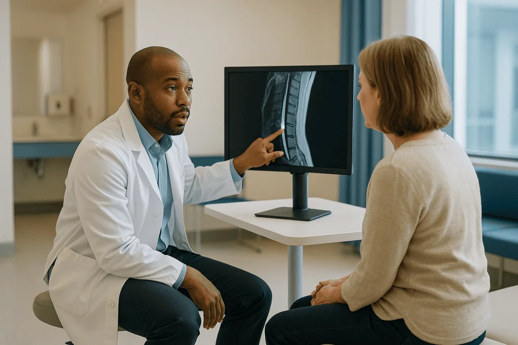
Transverse myelitis is inflammation across a segment of the cord. The National Institute of Neurological Disorders and Stroke (NINDS) describes symptoms that develop over hours to days, often with a band of altered sensation at the level of the inflamed segment, weakness below it and bladder or bowel changes. On MRI the affected segment may be swollen and bright, and a contrast injection may show enhancement, a term explained in the next section.
Demyelination means damage to myelin, the fatty insulation around nerve fibers. Multiple sclerosis (MS) typically produces short lesions, often less than one or two vertebral segments long, sitting toward the edge of the cord. Neuromyelitis optica spectrum disorder and a related antibody-driven condition tend to produce longer lesions extending over several segments and swelling the cord more centrally. Radiologists describe lesion length and position for exactly this reason, and those descriptions steer the choice of blood tests.
Infections, some vitamin deficiencies, reduced blood flow to the cord and certain autoimmune diseases can also create bright signal without compression. A brain MRI is frequently added, because finding lesions in the brain as well as the cord changes the list of likely explanations. The scan tells the team that the cord is inflamed; the tests that follow are what tell them why, and treatment decisions rest on that combination rather than on the image alone.
MRI with contrast for spine: when gadolinium is added and why
Contrast means an injection given through a small cannula in a vein during the scan. The agents used for MRI are based on gadolinium, a metal that alters the magnetic signal of nearby tissue. Where the normal barrier between blood and nerve tissue is intact, gadolinium stays in the bloodstream. Where inflammation, infection or a tumor has made that barrier leaky, gadolinium seeps into the tissue and lights it up on the T1 image. Radiologists call this enhancement.
Enhancement helps in several ways. It suggests a lesion is active rather than old scarring, which matters for inflammatory conditions. It outlines tumors and helps separate them from surrounding swelling. It can show an abscess wall, and it makes tiny deposits of infection or cancer along the cord’s surface more visible. Not every spinal MRI needs contrast, and the radiologist often decides after reviewing the first set of images whether adding it will answer a question the plain images cannot.
Safety questions are common and reasonable. The NHS notes that side effects from gadolinium are uncommon and usually mild, such as a brief feeling of warmth or a metallic taste. Serious allergic reactions are rare. Kidney function is often checked first, because the agent leaves the body through the kidneys and people with severely reduced kidney function are at risk of a rare complication affecting skin and connective tissue. Mayo Clinic points out that trace amounts of gadolinium can remain in the body after scans, and that no harm from this retention has been established to date; regulators advise using contrast when it adds diagnostic value rather than routinely.
Pregnancy, breastfeeding, prior contrast reactions and kidney disease are all things to tell the team before the appointment so the choice can be made with full information.
Who is usually offered MRI for spinal cord disorders, and who is asked to wait
Imaging follows the clinical picture, not the other way round. Certain findings push a spinal MRI up the queue: new weakness in the arms or legs, a change in bladder or bowel control, numbness with a clear level on the trunk, unsteady walking with brisk reflexes, back pain accompanied by fever or a history of cancer, and any spinal symptom after a significant injury. In these situations the scan is often arranged urgently, sometimes the same day.
People with ordinary low back pain and no neurological signs are usually not sent for MRI, at least not early. The NHS advises that scans are not routinely needed for back pain, because they rarely change management and frequently reveal age-related changes that can be misread as the cause. Waiting a few weeks while staying active is the guideline-level position for uncomplicated back pain, and it is a sign of good care rather than neglect.
Some people are asked to wait or to consider an alternative test for safety reasons. The magnet interacts with metal. Older pacemakers and some implanted defibrillators, cochlear implants, certain aneurysm clips, metal fragments in the eye and some pumps or stimulators can be hazardous or malfunction inside the scanner. Many modern devices are labelled as conditionally safe, meaning the scan can go ahead under specific settings, but that has to be confirmed in advance with the device documentation.
Pregnancy is not an absolute barrier; the NHS notes MRI is generally considered safe in pregnancy, though it is often postponed in the first trimester unless the need is pressing. Severe claustrophobia is real and manageable: wider-bore scanners, lying feet first, music and, in some cases, a sedative arranged by the team can make the scan possible. None of these decisions belong to the scanner booking desk; they sit with the referring clinician and the radiology team together.
Spinal cord MRI vs CT scan and other imaging: a plain comparison
Patients often ask why a CT was done in the emergency department and an MRI was still needed afterward, or why an X-ray was skipped. Each test answers a different question.
| Test | What it shows best | Radiation | Typical role in cord problems |
|---|---|---|---|
| MRI | The cord, discs, ligaments, fluid, inflammation, tumors | None | First-line test for suspected cord disorder |
| CT scan | Bone detail, fractures, bony narrowing, calcified discs | Yes | Trauma, surgical planning, when MRI is unsafe |
| CT myelogram | Outline of cord and roots using dye in the spinal fluid | Yes | Alternative when MRI cannot be done or is unclear |
| X-ray | Alignment, gross bone changes, instability on movement | Yes (low) | Screening after injury, checking alignment |
| Brain MRI | Lesions in the brain and optic nerves | None | Added when inflammation is suspected in the cord |
Computed tomography, or CT, stacks many X-ray beams into cross-sectional images. MedlinePlus describes it as fast and excellent for bone, which is why it dominates in trauma; it can show a fracture pressing into the canal within minutes. What it cannot do well is show the cord’s own tissue. Soft structures appear as similar shades of gray, so inflammation or a small tumor inside the cord may be invisible.
A CT myelogram adds contrast dye injected into the spinal fluid through a lumbar puncture before the CT, so the cord and nerve roots appear as a silhouette against bright fluid. It is more invasive than MRI and is reserved for people who cannot enter a magnet or whose MRI is degraded by metal hardware from previous surgery.
Plain X-rays show alignment and instability but not the cord, and ultrasound has almost no role in the adult spine because bone blocks the sound waves. MRI and CT are complementary rather than competing; the team chooses based on what needs to be seen and how quickly.
Lumbar puncture after MRI: what spinal fluid adds to the picture
When MRI shows inflammation without a compressive cause, the next question is what the spinal fluid contains. A lumbar puncture, sometimes called a spinal tap, is a procedure in which a thin needle is passed between two lower lumbar vertebrae into the fluid-filled sac below the end of the spinal cord, and a small sample of cerebrospinal fluid is withdrawn.
The order matters. Mayo Clinic notes that imaging is often performed first to check for a mass or raised pressure in the head or spine, because removing fluid below a blockage can be dangerous. That is one reason the scan generally precedes the tap rather than following it.
The fluid is analyzed for several things. A raised white cell count points toward inflammation or infection. Protein levels rise when the barrier around the nervous system is disrupted. Oligoclonal bands are proteins made by immune cells within the nervous system; their presence in the fluid but not in the blood supports a diagnosis such as MS. Tests for specific viruses, bacteria and fungi may be run, and in some cases the fluid is checked for cancer cells. The opening pressure can also be measured.
The procedure itself takes roughly 30 to 45 minutes including preparation, according to Mayo Clinic, with the needle in place for only a few minutes. Local anesthetic numbs the skin. Some people feel pressure or a brief electric sensation down a leg if a nerve root is brushed. Afterward, a headache that is worse when upright is the most common side effect; lying flat and drinking fluids are commonly advised, and the team will explain what to expect and when to report a headache that does not settle.
Results for cell counts come back quickly, while antibody and culture results may take days to weeks, which shapes the follow-up timeline discussed later.
Blood tests that commonly follow an abnormal cord MRI
Blood tests rarely diagnose a spinal cord disorder on their own, but they narrow the field quickly and cheaply relative to further imaging. Which ones are ordered depends on the MRI pattern and the history.
Vitamin and metabolic checks come first for many patients. Vitamin B12 deficiency can damage the cord, particularly the columns at the back that carry position sense, and produces a characteristic pattern on MRI. The NIH Office of Dietary Supplements describes neurological symptoms of deficiency including numbness and balance problems. Copper deficiency can mimic it. Blood glucose, thyroid function and a full blood count are often included.
Antibody tests follow when the scan suggests inflammation. Aquaporin-4 antibodies are linked to neuromyelitis optica spectrum disorder; myelin oligodendrocyte glycoprotein (MOG) antibodies mark a related condition. Both are associated with long cord lesions, and identifying them changes long-term management, so the treating team may request them even before the lumbar puncture results return. Broader autoimmune screens, including antinuclear antibodies and markers of inflammation such as erythrocyte sedimentation rate and C-reactive protein, look for systemic conditions that can involve the cord.
Infection screens are standard. HIV and syphilis can both affect the spinal cord, sometimes years after initial infection, and are treatable, so they are checked routinely rather than only when suspected. Depending on travel and exposure history, tests for tuberculosis, Lyme disease or certain viruses may be added.
Kidney function is checked before contrast, and clotting tests may be run before a lumbar puncture. Results that come back normal are informative too: a clean metabolic and infection screen alongside a clear inflammatory pattern on MRI tilts the team toward an immune-mediated cause and toward the nerve studies and follow-up scans that confirm it.
Nerve and muscle tests: EMG, nerve conduction and evoked potentials
MRI shows structure; these tests measure function, and they help answer a question the scan cannot: is the problem in the spinal cord, in the nerve roots leaving it, or in the peripheral nerves and muscles further out?
Electromyography, or EMG, records the electrical activity of muscles through a fine needle electrode. A nerve conduction study, usually done in the same session, delivers small electrical pulses to a nerve and measures how fast and how strongly the signal travels. Cleveland Clinic describes the combined test as taking about 30 to 90 minutes. The pulses feel like brief static shocks and the needle causes a short pinch; most people find it uncomfortable rather than painful.
The pattern of results is what matters. Damage to a single nerve root produces changes in the specific muscles that root supplies. A widespread peripheral neuropathy slows conduction along the arms and legs symmetrically. A pure spinal cord problem often leaves these peripheral tests normal, which is itself useful information, because it directs attention back to the cord and away from a coexisting nerve condition that might otherwise muddy the picture.
Evoked potentials measure how quickly a signal reaches the brain. Somatosensory evoked potentials stimulate a nerve in the wrist or ankle and record the response from the scalp; a delay suggests the signal is slowed on its way through the cord. Visual evoked potentials flash a checkerboard pattern and record the response from the visual part of the brain, detecting past optic nerve inflammation that a person may not have noticed. Because MS and related conditions often involve the optic nerves, a delayed visual response can support a diagnosis when the cord scan alone is ambiguous.
None of these tests carry radiation, and none require contrast. They are scheduled when the results would change the interpretation, not as a routine step for everyone.
What the days and weeks after a spinal MRI usually look like
The scan ends and the waiting begins, which for many people is the hardest part. Knowing the usual rhythm helps.
Reporting comes first. A radiologist reviews the images and writes a report for the clinician who requested the scan. The NHS notes that results may take a week or two to reach you through that clinician, though urgent findings are flagged the same day and acted on immediately. If you have not heard anything and the referrer said it was urgent, it is reasonable to phone rather than assume.
The follow-up conversation shapes the next steps. If the scan shows a compressive cause, the discussion turns to management options, and additional tests are limited to those needed for planning. If it shows inflammation, a cluster of tests is usually arranged over the following one to three weeks: brain MRI if not already done, blood tests, and a lumbar puncture. Basic fluid results return within days; antibody tests and cultures can take longer. Nerve studies are often slotted in during this window.
Repeat imaging is common and is not a sign that the first scan failed. Inflammatory lesions evolve, enhancement fades, and a second scan weeks or months later helps distinguish a single event from a relapsing condition. The interval is set by the team based on the working diagnosis.
Recovery timelines vary enormously by cause. For transverse myelitis, NINDS notes that most people begin to recover within weeks of symptom onset, that the majority of improvement occurs within about three months, and that gains can continue for up to two years. Those are typical ranges from a named source, not a forecast for any individual; the team caring for you will set expectations against your own findings and trajectory.
What people often get wrong about spinal cord MRI
Myths gather around any test that produces frightening-looking pictures. A few deserve direct correction.
The first is that MRI shows pain. It does not. It shows structure, and structure and pain are loosely coupled. Mayo Clinic notes that disc herniations are frequently found in people with no symptoms. A report listing degenerative changes is describing the spine’s age, not necessarily the source of your symptoms, and a clinician has to match the picture to the examination before either means anything.
The second is the mirror image: that a normal scan means nothing is wrong. Early inflammation, small lesions and functional disorders of nerve conduction can all coexist with a scan that looks clean. A normal MRI narrows the list of causes; it does not close the case, and the follow-on tests exist precisely for this situation.
Third, people worry about radiation. MRI uses none. The magnet and radio waves have no known cumulative effect, and repeat scans do not add up the way repeated CT scans do.
Fourth, contrast is often assumed to be dangerous. Serious reactions are rare according to the NHS, and the agent is used only when it adds information. The kidney check beforehand is a precaution, not a sign of expected trouble.
Fifth, a single scan is treated as the final word. Inflammatory cord conditions are diagnosed over time, with repeat imaging, fluid results and antibody tests assembled into a pattern. Uncertainty after the first MRI is the normal state, not a failure of the test or the team.
Finally, some assume that finding a lesion means a specific diagnosis has been made. A bright spot in the cord is a description, not a name. Until the supporting tests are in, the honest position is that the cord is affected and the cause is being established.
Questions to ask your care team about MRI for spinal cord disorders
A short list carried into the appointment tends to produce a more useful conversation than trying to remember everything in the room. These are the questions that most often clarify the path ahead.
- Which part of my spine will be scanned, and will my brain be scanned at the same time or later?
- Will contrast be used, and is there anything about my kidneys, allergies or pregnancy status you need to know first?
- Do I have any implants, devices or metal that need to be checked for scanner safety, and who confirms that?
- How urgent is this scan, and what should I do if my symptoms change before the appointment?
- Who will explain the results to me, and roughly when should I expect to hear?
- If the scan shows inflammation, which tests are likely to follow, and in what order?
- Would a lumbar puncture be needed, and what would it tell you that the scan cannot?
- Is a nerve conduction study or EMG planned, and what question is it meant to answer?
- Will I need a repeat scan, and what interval are you considering?
- Are there activities I should avoid while we are waiting for a diagnosis?
- What symptoms should prompt me to call you or go to an emergency department right away?
Ask, too, for a copy of the report and for the images to be shared with any other specialist involved, so that no test has to be repeated for want of a file. If the report contains terms you do not recognize, ask for them to be translated into plain language during the conversation rather than afterward; radiology vocabulary is dense, and the clinician who ordered the scan is the right person to interpret it against your examination.
Bring someone with you if you can. Results conversations move quickly, and a second set of ears catches what anxiety filters out.
When to call your doctor
Spinal cord symptoms can change quickly, and some changes cannot wait for a scheduled scan or a results appointment. The NHS guidance on cauda equina syndrome and the NINDS description of transverse myelitis both emphasize speed when certain signs appear.
Seek emergency care immediately if you notice new or rapidly worsening weakness in your legs or arms; difficulty passing urine, inability to feel the bladder filling, or new loss of bladder or bowel control; numbness around the genitals, buttocks or inner thighs, sometimes described as saddle numbness; sudden severe back or neck pain with fever, especially if you have had a recent infection, injection or procedure near the spine; or numbness or weakness after a fall or accident. These patterns can indicate compression of the cord or the nerve roots below it, and the window for preserving function may be short.
Call your doctor the same day if numbness or tingling is climbing steadily up your legs or trunk over hours, if walking has become noticeably unsteady, or if a band of altered sensation has appeared around your chest or abdomen.
After a contrast injection, contact the team or emergency services for swelling of the face or throat, difficulty breathing, widespread rash or hives, or a racing heartbeat. After a lumbar puncture, report a headache that does not settle with lying flat and fluids within a day or two, a fever, worsening back pain, or any drainage from the puncture site.
If you are simply unsure, calling is the right choice. The people arranging your tests would rather hear from you unnecessarily than learn afterward that a warning sign was waited out. Every decision about what happens next, from repeating a scan to starting a treatment, rests with the team who can examine you and see your results together.
Frequently asked questions
What does a spinal MRI show that a CT scan does not?
A spinal MRI shows the soft tissue of the cord itself, including swelling, inflammation, tumors, fluid cavities and thinning, along with the discs and ligaments around it. CT excels at bone and is faster, which suits trauma, but soft tissues appear as similar shades of gray on CT, so problems inside the cord can be invisible. The two tests are complementary and are often both used.
Why would I need MRI with contrast for spine imaging?
Contrast is added when the radiologist needs to know whether a lesion is active, to outline a tumor, or to find infection or tiny deposits along the cord surface. Gadolinium leaks into tissue where the normal barrier is damaged and lights it up. Kidney function, allergies and pregnancy status are checked beforehand, and the team decides whether contrast will answer a question the plain images cannot.
How long does an MRI of the spinal cord take?
According to the NHS, an MRI scan usually lasts between 15 and 90 minutes depending on how much of the body is imaged. The neck, upper back and lower back are often scanned as separate regions, so a full spine study with contrast sits toward the longer end. Lying still throughout is the most important contribution you can make to image quality.
Is spinal cord MRI vs CT scan a matter of safety?
Partly. MRI uses a magnet and radio waves with no radiation, while CT uses X-rays. MRI is unsafe for some implanted devices and metal fragments, so CT or CT myelography is chosen in those cases. Otherwise the choice rests on what needs to be seen: the cord and soft tissue favor MRI, bone detail and speed after injury favor CT.
Why is a lumbar puncture after MRI sometimes needed?
When MRI shows inflammation without anything pressing on the cord, spinal fluid analysis helps identify the cause. It measures white cells, protein, oligoclonal bands linked to multiple sclerosis, and signs of infection or cancer cells. Mayo Clinic notes imaging is often done first to exclude a mass or raised pressure, which is why the tap follows the scan rather than preceding it.
Can an MRI miss a spinal cord problem?
Yes. Very small lesions, inflammation in its first day or two, and conditions affecting nerve function rather than structure can all coexist with a normal scan. A normal MRI narrows the list of possible causes but does not close the case. That is why repeat imaging, blood tests, lumbar puncture and nerve studies are arranged when symptoms persist despite a reassuring first scan.
What is the difference between a cord lesion in MS and in neuromyelitis optica?
Radiologists describe lesion length and position because the two conditions tend to differ. Multiple sclerosis often produces short lesions, typically under one or two vertebral segments, sitting toward the edge of the cord. Neuromyelitis optica spectrum disorder more often produces longer lesions spanning several segments with central swelling. Blood tests for aquaporin-4 and MOG antibodies help confirm the distinction.
What blood tests usually follow an abnormal spinal MRI?
Commonly requested tests include vitamin B12 and copper, a full blood count, inflammatory markers, screens for HIV and syphilis, and autoimmune antibodies such as aquaporin-4, MOG and antinuclear antibodies. Kidney function is checked before contrast and clotting before a lumbar puncture. The selection depends on the pattern seen on the scan and on your medical and travel history.
Does a normal EMG mean my spinal cord is fine?
Not necessarily. EMG and nerve conduction studies mainly assess nerve roots, peripheral nerves and muscles. A pure spinal cord problem can leave these tests normal, which is itself useful because it points attention back to the cord. Evoked potentials, which time how quickly signals travel through the cord to the brain, are the functional tests that more directly reflect cord conduction.
How soon after a spinal MRI will I get results?
The NHS notes that results typically take a week or two to reach you through the clinician who requested the scan, while urgent findings are flagged and acted on the same day. If you were told the scan was urgent and have not heard, it is reasonable to call. Follow-on tests such as blood work and lumbar puncture are usually arranged over the following one to three weeks.
References
- NHS: MRI scan
- NIH NINDS: Transverse Myelitis
- Cleveland Clinic: EMG (Electromyography)
- MedlinePlus: CT scan
This article is for general information only and is not a substitute for professional medical advice. Please consult a qualified doctor about your individual situation.

More from the Blog

Is Everyday Life With HSP Manageable? Stairs, Driving, Work and Pacing Strategies
For most people, daily life with HSP (hereditary spastic paraplegia) is manageable, though it usually needs adjusting over time. HSP causes gradually increasing stiffness…

Trigger Assessment Before Migraine Treatment: What Your Neurologist Wants Tracked
A migraine trigger assessment is a structured period, usually several weeks to a few months, of recording attacks alongside sleep, meals, stress, menstrual cycle,…

What to Ask Your Neurologist When an Autonomic Neuropathy Plan Involves Several Specialists
When several specialists share an autonomic neuropathy plan, ask your neurologist who coordinates the whole plan, which symptom each clinician owns, how test results…

Spinal Cord Disorder Follow-Up: Which Bladder, Bowel or Weakness Changes Need an Urgent Call
After a spinal cord disorder, call your care team the same day for any new loss of bladder or bowel control, difficulty passing urine,…

Fetal Surgery or Repair After Birth for Spina Bifida: What Does Each Pathway Involve?
Fetal surgery for spina bifida closes the spinal opening before birth, usually between about 19 and 26 weeks of pregnancy, through an incision in…

Daily Life With ALS: Assistive Devices, Nutrition and Energy Conservation Explained
Daily life with ALS changes gradually as motor neurons weaken muscles, but much of it can be planned for and managed. Assistive devices such…