

How Deep Brain Stimulation Surgery Is Performed: Electrodes, Testing and the Pulse Generator
Key Takeaways
- DBS surgery is typically staged: electrodes are placed in the brain first, then a pulse generator is implanted under the collarbone and connected by a wire tunneled beneath the scalp and neck.
- The brain has no pain receptors, so awake patients usually feel nothing during electrode insertion; discomfort comes mainly from frame pins, the drilling vibration and later soreness along the neck and chest.
- Real-time testing in the operating room checks both benefit and the threshold at which side effects such as tingling, speech change or double vision appear, helping confirm the lead is centered on target.
- The device is usually switched on a few weeks after surgery, once swelling settles, and is fine-tuned over several programming visits rather than at a single appointment.
- A good response to levodopa is one of the strongest predictors of a good response to stimulation in Parkinson's disease, which is why an on-and-off medicine test is part of evaluation.
- Non-rechargeable generators typically need replacing after several years in a minor chest procedure, while rechargeable units last longer but require regular charging through the skin.
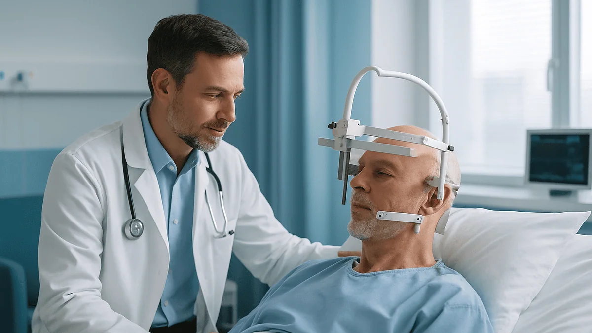
Deep brain stimulation surgery is usually performed in stages. Surgeons use MRI and CT images to plan a target deep in the brain, open a small hole in the skull, guide a thin electrode into place while recording brain signals and testing stimulation, then implant a pulse generator under the skin of the chest and connect it by a wire tunneled beneath the scalp and neck. The device is switched on and adjusted at later visits.
The question usually arrives at the kitchen table, not in a clinic. Someone who has lived with Parkinson’s disease or essential tremor for years watches their hand shake through a cup of coffee, then asks a spouse: what would it actually be like to have wires put in my brain? The idea sounds like science fiction. The reality is a careful, well-rehearsed operation that neurosurgeons have refined over decades.
Understanding how DBS surgery is performed takes much of the fear out of it. There is no cutting of brain tissue in the way most people imagine. A needle-thin electrode is guided along a planned path to a spot roughly the size of a pea, and the team checks the position in real time. Later, a small battery-powered generator, similar to a cardiac pacemaker, is placed under the collarbone.
This explainer walks through each stage, from the head frame to the first programming visit, and is honest about discomfort, risks and the things that can go wrong.
What deep brain stimulation actually does inside the brain
Deep brain stimulation, or DBS, delivers gentle electrical pulses to a specific cluster of nerve cells deep in the brain. Those pulses do not destroy anything. They change the abnormal firing pattern of circuits that control movement, a bit like adding a steady metronome to an orchestra whose rhythm section has drifted off tempo.
In Parkinson’s disease, the loss of dopamine-producing cells leaves the basal ganglia, a set of movement-regulating structures near the center of the brain, sending out noisy, poorly timed signals. That noise shows up as tremor, stiffness and slowness. Stimulation applied to the subthalamic nucleus (a small structure that acts as a brake on movement) or the globus pallidus interna (an output station of the basal ganglia) appears to smooth those signals. For essential tremor, the usual target is the ventral intermediate nucleus of the thalamus, a relay station for movement information, according to Mayo Clinic.
The exact mechanism is still debated. Researchers once assumed high-frequency stimulation simply switched off overactive cells; the current view is more nuanced, involving changes to the timing of signals across whole networks. What matters for a patient is that the effect is adjustable and reversible. Turn the device down and the effect fades. Turn it off and the brain returns to its prior state. That reversibility is the main reason DBS replaced older operations that permanently burned or froze small areas of brain tissue.
DBS does not treat every symptom. It tends to help symptoms that also respond to levodopa, the standard medicine class for Parkinson’s, while doing little for problems such as balance loss, memory change or speech softening. Your neurologist will explain which of your symptoms fall on which side of that line.
Who is usually offered DBS, and who is asked to wait
DBS is not a first treatment. Mainstream guidance, including the NHS, describes it as an option when medicines no longer control symptoms smoothly. For Parkinson’s disease that usually means motor fluctuations: good hours when medicine works, then wearing-off periods, sometimes with involuntary writhing movements (dyskinesia) at peak medicine levels. People with severe essential tremor or certain forms of dystonia (sustained, twisting muscle contractions) may also be considered. Some centers also use DBS for selected cases of epilepsy or obsessive-compulsive disorder that have not responded to standard care.

A good response to levodopa is one of the strongest predictors of a good response to stimulation in Parkinson’s disease. That is why evaluation includes a formal test of movement with and without medicine. If medicine barely helps, stimulation is unlikely to help either.
Several groups are usually asked to wait, or are advised that DBS is not suitable:
- People whose diagnosis is still uncertain, because conditions that mimic Parkinson’s respond poorly to stimulation.
- People with significant memory or thinking problems, since surgery and stimulation can worsen these.
- People with untreated depression or other unstable mental health conditions.
- People with medical conditions that make anesthesia or brain surgery unusually risky, such as uncontrolled bleeding disorders.
- People whose main complaints are balance, falls or speech, which DBS generally does not improve.
Age alone is not a firm barrier, though older adults are assessed carefully for frailty and cognition. The decision is made by a multidisciplinary team, typically a movement disorder neurologist, a neurosurgeon, a neuropsychologist and often a psychiatrist, and it rests with that team in discussion with you.
Before the operation: evaluation, imaging and planning
Weeks before anyone touches a scalpel, the groundwork is laid. The neuropsychological assessment often surprises people: a few hours of memory, attention and language tests, designed to confirm that thinking is robust enough to tolerate surgery and to establish a baseline for later comparison. A psychiatric review checks mood and coping.
The medicine on-and-off test follows. You arrive having held your usual Parkinson’s medicine overnight, so the team can score your movement in the unmedicated state. Then you take your normal dose and are re-examined once it takes effect. The gap between the two scores gives a realistic preview of what stimulation might achieve, notes Cleveland Clinic.
Imaging is the surgeon’s map. A high-resolution MRI, often obtained days or weeks in advance, shows the deep nuclei and the blood vessels that must be avoided. On the day of surgery, a CT scan taken with the head frame or skull markers in place is merged with that MRI on planning software. The surgeon then draws a trajectory: an entry point near the top of the head, a path that skirts the fluid-filled ventricles and surface veins, and a target coordinate accurate to about a millimeter.
Practical preparation matters too. You will be asked about blood thinners, supplements and any devices already implanted. Hair is usually trimmed or shaved only along the incision lines. Most people are asked not to eat or drink for a set period beforehand, and your team will tell you exactly which regular medicines to take that morning and which to hold. Do not change anything on your own.
Step one on surgery day: fixing the head and confirming the target
Precision is everything in this operation, and precision begins with keeping the head perfectly still. The traditional method is a stereotactic frame, a lightweight metal ring fixed to the skull at four points with pins. Local anesthetic numbs each pin site, so the sensation is pressure rather than pain, though many people describe the frame as the strangest part of the day. Newer frameless systems use small anchors screwed into the skull days earlier, or a custom-printed platform, to achieve the same fixed reference.
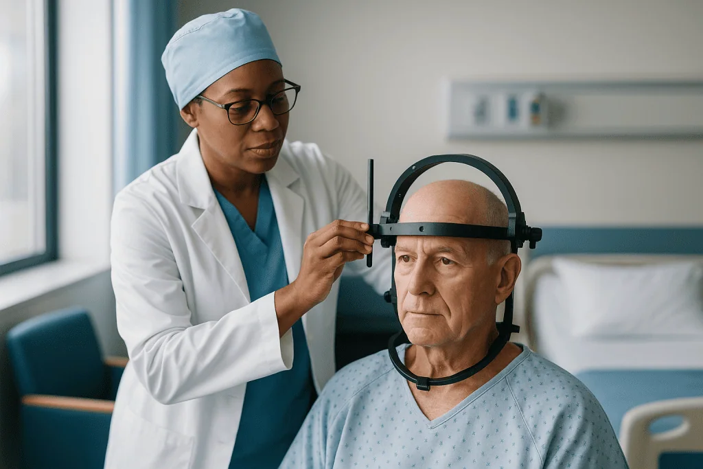
With the frame on, you are taken for a CT scan. The software fuses this scan with your earlier MRI, so that every point in the brain now has a coordinate the frame can find. The surgeon reviews the planned path one last time.
Back in the operating room, you lie on a table, usually semi-reclined. Warm blankets, a quiet room and a nurse at your side are standard. If the surgery is being done awake, sedation is used while the scalp is opened and lightened when the team needs your cooperation. If it is being done asleep, general anesthesia is started now.
The scalp incision is small, typically a curved cut a few centimeters long on one side of the head, sometimes both. The skull beneath is opened with a burr hole, a round opening about the width of a coin, described by MedlinePlus as a small hole through which the electrode will pass. The brain itself has no pain receptors, so this stage causes no pain even when awake. A plastic anchoring ring is fitted to the hole, ready to hold the lead in place later.
Awake or asleep: how DBS surgery is performed with different anesthesia
People are often astonished to learn that much of this surgery can happen while they are awake and talking. The reason is functional: an awake patient can move an arm on request, report tingling or say a sentence so the team can hear whether speech changes. That live feedback helps confirm the electrode sits in the intended spot rather than a few millimeters off, where side effects live.
Awake surgery does not mean feeling everything. Sedation keeps you drowsy during the uncomfortable parts. Local anesthetic handles the scalp. Many people remember the day as long and odd rather than painful, with periods of dozing punctuated by a voice asking them to tap their fingers.
Asleep surgery under general anesthesia has grown steadily as imaging has improved. Some centers place electrodes with intraoperative MRI or CT, checking position by pictures rather than by patient response. Johns Hopkins Medicine notes that the approach chosen depends on the target, the condition being treated and patient factors such as anxiety, severe tremor that makes lying still difficult, or breathing problems.
Neither method has been shown in mainstream reviews to be clearly superior for all patients. Awake surgery offers real-time symptom testing; asleep surgery offers comfort and can suit people who could not tolerate hours of stillness. If you have a strong preference, say so early. The surgical team will explain which option their program uses, why, and whether your situation allows a choice. Whatever the method, Parkinson’s medicine is usually held that morning so the team can see the untreated symptoms clearly.
Placing the electrodes: recording brain signals along the path
The electrode, more properly called a lead, is a flexible insulated wire thinner than a strand of spaghetti, with a small number of metal contacts at its tip. Before that lead goes in, many teams first pass a microelectrode, a recording probe finer than a human hair, along the planned trajectory.
Nerve cells make characteristic sounds when their electrical activity is played through a speaker. The subthalamic nucleus crackles like radio static; the border zones sound different again. Neurophysiologists listen and watch the traces as the probe advances a fraction of a millimeter at a time, mapping where the target begins and ends. If the recording suggests the path has clipped the edge rather than the center, the surgeon can shift by a millimeter or two and try again.
Once the team is satisfied, the permanent lead is lowered along the same path and its depth is fixed. The anchoring ring at the burr hole holds it firmly against the skull, and the excess wire is coiled beneath the scalp.
Several practical points reassure patients at this stage:
- The brain does not feel the probe. Awake patients typically notice nothing during insertion.
- Many people need leads on both sides of the brain, because symptoms on the left of the body are controlled by the right hemisphere and vice versa. Some centers do both sides in one session; others stage them.
- Bleeding is the main risk during insertion. Careful planning around blood vessels and a slow, steady advance are the chief safeguards.
Imaging, whether a repeat CT in the operating room or an MRI afterward, confirms the final position and rules out bleeding before the next stage begins.
Testing in the operating room: what the team is looking for
Here is where the awake approach earns its keep. With the lead in place, a temporary external stimulator is connected and switched on at low settings. The team watches and asks.
Two things are being judged at once. The first is benefit: does the tremor settle when the current passes, does the wrist loosen when the examiner rocks it back and forth, do finger taps speed up? Even a few minutes of stimulation can produce visible change in Parkinson’s tremor and rigidity, and that change, while temporary, tells the team the contacts sit inside a responsive zone.
The second is the threshold for side effects. Because the target nuclei are surrounded by other structures, stimulation that spreads too far produces telltale signs, described by Mayo Clinic as possible stimulation-related effects: tingling in the face or hand, a pulling sensation in the mouth or eye, double vision, slurred speech, or a wave of unexpected emotion. If those appear at settings only slightly above the helpful level, the lead is probably a little off center. A wide gap between the setting that helps and the setting that causes side effects is what the team wants to see.
In asleep surgery, this behavioral testing is replaced by imaging confirmation and, in some programs, recordings of brain activity that do not require patient cooperation.
Once testing is complete, the external stimulator is disconnected. The lead is capped, the scalp closed over it and dressed. In a single-stage operation the team then moves straight to the chest; in a staged operation you may go to recovery with the lead in place and return days later for the generator.
Implanting the pulse generator and connecting the system
The pulse generator is the battery and computer of the system, a flat, palm-sized case about the thickness of a stack of a few coins. This stage is always done under general anesthesia, because tunneling wires beneath the skin is uncomfortable.
A pocket is made under the skin just below the collarbone, occasionally in the abdomen. An extension wire is then threaded under the scalp, behind the ear and down the side of the neck to the chest, using a long hollow tunneling tool. The lead from the brain is connected to one end of the extension near the top of the head; the other end plugs into the generator, which is placed in the pocket and secured. The system is checked for electrical continuity, and the incisions are closed.
| Component | Where it sits | What it does |
|---|---|---|
| Lead (electrode) | Deep in the brain, anchored at the burr hole | Delivers pulses through small contacts at its tip |
| Extension wire | Under the scalp and neck skin | Carries current from generator to lead |
| Pulse generator | Under the skin below the collarbone | Houses battery and circuitry; stores the programmed settings |
| Patient controller | Carried by the patient | Checks battery, switches on or off, adjusts within limits set by the clinician |
Generators come in two broad types. Non-rechargeable units run for several years before the whole generator is swapped in a minor operation; MedlinePlus gives a typical range of roughly 3–5 years depending on settings. Rechargeable units last considerably longer but require regular charging through the skin with a wireless charger. Which type suits you is a discussion for your team, weighing convenience against the ability to manage a charging routine.
How painful is DBS surgery, and how long does it take?
Pain is the fear behind most of the questions people type into a search engine. The honest answer is that DBS is less painful than its description suggests, but it is a long day and the neck and chest incisions can be sore afterward.
During the brain stage, the only sensations most awake patients report are the pressure of the frame pins going in under local anesthetic, an odd vibration when the burr hole is drilled (bone conducts sound, so it is loud rather than painful) and stiffness from lying still for hours. The brain itself registers nothing. Headache is common for a day or two afterward and is usually managed with the ordinary pain relief your team prescribes.
The generator stage produces more conventional post-surgical soreness. The path where the extension wire was tunneled along the neck can feel tight or bruised for a week or two, and turning the head may pull at first. Many people describe the chest pocket as the most tender spot, particularly when lying on that side.
Mayo Clinic describes the electrode placement as taking several hours, with the generator implantation a shorter separate procedure. Time in the department is longer still, because frame placement, imaging and recovery add to the total. If both sides are done in one sitting, expect most of a day.
Hospital stay is short by neurosurgical standards. MedlinePlus describes a stay of about a day after lead placement, and many programs plan one to two nights, longer if anything needs watching. You will be asked to arrange for someone to drive you home and stay with you for the first days.
DBS surgery recovery time: the first days and weeks
The first week is about healing skin and letting the brain settle. Stitches or staples along the scalp and chest come out or dissolve within about two weeks, per the wound-care advice on MedlinePlus. Keep the incisions dry as instructed, sleep with the head slightly raised if that eases headache, and avoid bending, straining or heavy lifting until cleared.
A curious phenomenon often appears in these early days: symptoms may improve before the device is even switched on. This is called the microlesion or stun effect, a temporary result of tiny swelling around the new lead. It fades over days to weeks. Do not read it as a preview of the final result, and do not reduce medicine because of it; your neurologist manages that.
Fatigue is near universal. So is a feeling of mental fogginess for a short spell, and some people notice low mood or irritability. These usually pass, but they are worth mentioning at follow-up, because the team wants to distinguish ordinary post-surgical tiredness from anything that needs attention.
A typical recovery pattern, with the caveat that yours may differ:
- Days 1–7: rest at home, gentle walking, wound checks, no driving.
- Weeks 2–4: wounds healed, light activities resume, first programming visit scheduled.
- Weeks 4–8: gradual return to normal routines; Mayo Clinic describes a return to usual activities over a few weeks.
Return to work depends on the job. Desk-based roles often resume sooner than physical ones. Ask your surgeon about driving, which is usually restricted until healing is confirmed and stimulation is stable, and follow local rules for medical conditions and driving.
Switching on and programming: why the real work starts after surgery
Most people are surprised that they leave hospital with the device off. Swelling around the lead needs to subside so that settings chosen now will still be right later. Mayo Clinic describes the system being activated a few weeks after implantation.
At the first programming session, the neurologist or a specialist nurse holds a wand or tablet near the generator and, one contact at a time, tests how your symptoms respond and where side effects begin. It is a methodical process. Each contact is tried at gradually rising intensity while the clinician checks rigidity, tremor, finger taps and speech. Results are recorded, and a starting configuration is chosen: which contact or combination is active, how strong each pulse is, how wide, and how many pulses per second.
Expect this to take an hour or more, and expect it to feel like a fitting rather than a finished product. Settings are fine-tuned over several visits across the following months, as the brain adapts and as medicines are adjusted alongside. Reducing Parkinson’s medicine is often possible after successful stimulation, but the timing and amount are decided by the prescribing clinician; making changes yourself risks both under-treatment and side effects.
Newer generators allow the clinician to steer current in particular directions or to program different settings for day and night. Your patient controller will let you check the battery, switch the device on or off in an emergency and, if enabled, nudge intensity within a range set by the clinician.
Patience is the skill required here. People who expect a single switch to transform them are often disappointed at visit one, then pleasantly surprised at visit three.
Is DBS a high-risk surgery? The downsides in plain terms
Any operation inside the skull carries real risk, and no responsible clinician will describe DBS as minor. It is, though, a procedure with a long track record and safeguards built into every stage. Mayo Clinic lists the main surgical risks as bleeding in the brain, stroke, infection, breathing problems, nausea, heart problems and seizure. Bleeding is the most serious and is uncommon; ask your team for their own outcome data rather than relying on general figures.
Risks divide into three groups. Surgical risks are those above, plus wound problems and, occasionally, a lead that ends up slightly off target and needs repositioning. Hardware risks come later: infection around the generator or wire, a wire fracturing or eroding through thin skin, or a generator that fails or needs replacing. Stimulation-related effects are the third group and are usually reversible by reprogramming: tingling, muscle tightness, speech changes, balance disturbance, double vision, and mood or behavior changes such as impulsivity, apathy or low mood.
The downsides people underestimate are the commitments. You are tied to a specialist center for programming and battery care for life. Some medical procedures, including certain MRI scans and diathermy (deep heat treatment used in some physical therapies), require precautions or are off limits. And there is the psychological adjustment of carrying a device; some people find the chest lump or the visible scalp scars harder than expected.
Set against this is the reversibility. Unlike older lesioning operations, the entire system can be turned off or removed. That does not undo a bleed or an infection, but it does mean stimulation side effects are not permanent.
Life after DBS surgery: batteries, MRI scans, airports and daily routines
Once healed and programmed, most people forget the hardware for long stretches. A few realities of living with a stimulator are worth knowing before you say yes.
Battery care is the most regular task. Non-rechargeable generators are checked at visits and replaced when they approach the end of their life, a short operation on the chest only; MedlinePlus gives the typical lifespan as several years. Rechargeable units need charging on a schedule your team will set, often while reading or watching television with a charger held over the chest.
Magnetic and electrical environments deserve respect but not fear. Airport security arches and shop anti-theft gates are generally passable; walk through at a steady pace and carry your device identification card in case the alarm sounds. Household appliances, mobile phones and induction stoves are usually fine at normal distances. Strong industrial magnets, arc welding and some medical equipment are different. Always tell any clinician, dentist or physical therapist about the device, and never have an MRI without the DBS team confirming that your specific system and settings allow it. Cleveland Clinic stresses this precaution.
Exercise, swimming, travel and intimacy all resume once wounds have healed and your team agrees. Contact sports that could hit the chest or head merit a conversation.
Symptoms of the underlying condition continue to evolve. DBS treats symptoms; it does not halt disease progression. Over years, settings and medicines are adjusted, and some symptoms that stimulation does not touch, such as balance or speech changes, may become more prominent. Ongoing follow-up with the movement disorder team, and often with physical, speech and occupational therapists, is part of the long game.
What people often get wrong about deep brain stimulation
Myths gather around any surgery involving the brain. Here are the ones movement disorder teams hear most, and what the evidence actually says.
It is a last resort for the very late stages. The reverse is closer to the truth. Candidates are typically people who still respond well to medicine but suffer fluctuations, and who are cognitively well. Very advanced disease with dementia is usually a reason not to operate.
It stops the disease. DBS relieves symptoms; the underlying condition continues at its own pace. No mainstream guideline claims it slows or halts Parkinson’s disease or essential tremor.
You will come off all medicine. Some people reduce their Parkinson’s medicine after surgery, and that reduction can ease dyskinesia. Many still need medicine. Changes are made gradually by the prescribing clinician, never abruptly.
You feel the pulses. At therapeutic settings you should feel nothing. Tingling or pulling means the settings need adjusting.
The surgery changes who you are. Personality change is not an expected outcome. Mood, motivation and impulse control can shift with stimulation or with medicine changes, which is why teams screen and monitor for them; these effects are usually adjustable.
It only works for Parkinson’s. Essential tremor and dystonia are established indications, and other conditions are treated in specialized programs, as Johns Hopkins Medicine outlines.
It is experimental. DBS has been in mainstream use for movement disorders for decades and is recommended in national guidelines when medicine is no longer adequate. The experimental label applies only to newer targets and conditions under study.
Questions to ask your care team before agreeing to DBS
A consultation goes better when you arrive with questions written down. These are the ones experienced patients wish they had asked.
- Which of my symptoms do you expect stimulation to help, and which do you expect it to leave unchanged?
- Which target are you recommending for me, and why that one rather than the alternative?
- Will my surgery be awake or asleep, in one stage or two, and one side or both?
- How many of these procedures does your team perform, and what are your own rates of bleeding, infection and lead revision?
- What happens if the lead is not in the ideal position? How would you know, and what would you do?
- Rechargeable or non-rechargeable generator: which do you suggest for me and what are the trade-offs?
- When will the device be switched on, how many programming visits should I plan for, and who will do them?
- How will my medicines change after surgery, and who makes those decisions?
- Which imaging, dental and therapy procedures will need precautions once I have the device?
- What should I do if the device switches off unexpectedly or I lose the controller?
- Who do I call, day or night, if I develop a fever or a wound problem?
- What are the alternatives if I decide not to proceed, including medicine adjustments and focused ultrasound where available?
Bring a family member or friend. Two sets of ears catch more, and the person who will drive you home and sit with you in the first week deserves to hear the plan directly. Ask for written information and, if the center offers it, to speak with someone who has been through the procedure. Do not feel pressured to decide on the day. This is an elective operation, and a good team will expect you to take time.
When to call your doctor after DBS surgery
Most recoveries are uneventful, but a few signs need a same-day call to the surgical team, and some need emergency services without delay.
Call emergency services immediately for any of the following, which can indicate bleeding, stroke or seizure:
- Sudden severe headache unlike your usual post-operative ache, especially with vomiting or drowsiness.
- New weakness or numbness of the face, arm or leg, particularly on one side.
- Sudden difficulty speaking, understanding speech or seeing.
- A seizure, or a period of confusion or unresponsiveness.
- Chest pain, breathlessness or a rapid, irregular heartbeat.
Call your surgical team the same day for signs of infection or hardware trouble:
- Fever, or an incision that becomes increasingly red, warm, swollen or begins to leak fluid or pus.
- Skin over the generator or along the wire that looks thin, discolored or is breaking down.
- A sudden return of symptoms that suggests the device has switched off or a wire has failed.
- New or worsening tingling, muscle pulling, double vision, slurred speech or balance problems that do not settle when the device is turned off with your controller.
- A marked change in mood, new thoughts of self-harm, unusual impulsive behavior or persistent confusion.
Keep the team’s contact numbers on your phone and on the fridge, and carry your device identification card at all times. MedlinePlus and your discharge paperwork will list the local specifics. When in doubt, call; teams would far rather hear about a false alarm than a missed infection or bleed.
Frequently asked questions
How painful is DBS surgery?
Less painful than most people expect, though it is a long day. The brain itself feels nothing, and local anesthetic numbs the scalp and the frame pin sites. Afterward, headache for a day or two is common, and the neck and chest can feel bruised for a week or two where the wire was tunneled and the generator placed. Your team will prescribe pain relief and explain what to expect.
Is DBS a high-risk surgery?
It is brain surgery, so it carries real risks, including bleeding, stroke, infection and seizure, but serious complications are uncommon in experienced programs. Most problems are either treatable or reversible by adjusting or removing the hardware. Ask your surgical team for their own complication rates rather than relying on general figures, and weigh those against how much your symptoms currently limit your life.
What is life like after DBS surgery?
For many people, daily life becomes more predictable once programming is optimized, with fewer swings between good and bad hours. You remain under the care of a specialist team for programming and battery checks, carry a device card, and take precautions around MRI and strong magnets. The underlying condition continues to progress, so medicines and settings are adjusted over the years.
What is the downside of DBS surgery?
The main downsides are surgical risk, the lifelong commitment to a specialist center for programming and battery care, and possible stimulation side effects such as speech change, tingling or mood shifts. Hardware can become infected or fail and need revision. DBS also does not help every symptom, particularly balance, memory and speech, and it does not slow the disease itself.
What is DBS surgery recovery time?
Most people stay in hospital one to two nights after electrode placement, have wounds healed within about two weeks, and return to usual activities over a few weeks, according to Mayo Clinic and MedlinePlus. The device is usually switched on a few weeks after surgery, and the full benefit unfolds over months of programming visits.
Are you awake during deep brain stimulation surgery?
Often, but not always. Many programs place the electrodes with the patient sedated but rousable, so the team can test movement and speech in real time. Others perform the operation under general anesthesia using intraoperative imaging to confirm position. The generator stage is always done asleep. Which approach you have depends on the target, your condition and your center’s practice.
How long does DBS surgery take?
Electrode placement typically takes several hours, with the generator implantation a shorter separate procedure, per Mayo Clinic. Frame placement, imaging and recovery add to the overall time in the department, so plan on most of a day, especially if both sides of the brain are treated in one session.
Can you feel the stimulation once the device is on?
At correctly adjusted settings you should not feel anything. If you notice tingling, a pulling sensation in the face or limbs, or changes to your speech or vision, the current is probably spreading beyond the intended area, and the settings need adjusting. Report such sensations to your programming team rather than putting up with them.
Does DBS mean I can stop my Parkinson's medicine?
Not necessarily. Some people are able to reduce their medicine after stimulation is optimized, which can ease involuntary movements, but many continue to take some. Any change is made gradually by the prescribing clinician, based on how you respond to programming. Stopping or cutting medicine on your own can cause a sharp worsening of symptoms and other complications.
Can I have an MRI scan after DBS?
Sometimes, under strict conditions. Many modern systems allow certain MRI scans, but only with specific scanner settings and with the device placed in a safe mode beforehand. Always tell any clinician about your stimulator and ask your DBS team to confirm what is permitted for your exact system before any scan is booked.
References
- MedlinePlus Medical Encyclopedia: Deep brain stimulation
- Cleveland Clinic: Deep Brain Stimulation (DBS)
- NHS: Parkinson's disease, Treatment
This article is for general information only and is not a substitute for professional medical advice. Please consult a qualified doctor about your individual situation.

More from the Blog

Why Gamma Knife Results Take Months to Show and How Follow-Up MRI Tracks the Change
Gamma Knife radiosurgery rarely produces visible results right away. The radiation damages the DNA of target cells so they lose the ability to divide,…

Why Prompt Reporting of New Symptoms Matters in Demyelinating Disease Follow-Up
Reporting new or changed symptoms promptly in a demyelinating disease such as multiple sclerosis lets the care team decide quickly whether it is a…

Who Is a Candidate for Deep Brain Stimulation in Parkinson Disease? What the Evaluation Tests
A person is usually a candidate for deep brain stimulation (DBS) in Parkinson disease when they have confirmed idiopathic Parkinson disease, still respond clearly…

What Happens During a TMS Session: Coil Placement, Pulses and How Long You Are in the Chair
During a TMS session you sit awake in a reclining chair while a clinician positions an electromagnetic coil against your scalp, most often over…

Daily Life With a Movement Disorder: Rehabilitation, Fall Safety and Caregiver Routines
Living with a movement disorder usually means combining three things: regular rehabilitation with physical, occupational and speech therapists; a home and daily routine arranged…

Broca’s, Wernicke’s and Global Aphasia: How the Type Changes the Therapy Approach
Therapy for aphasia is matched to the pattern of language loss. In Broca's aphasia, understanding is relatively preserved, so therapy drills spoken output, often…