

What Is an EEG? How Doctors Read the Brain’s Electrical Weather
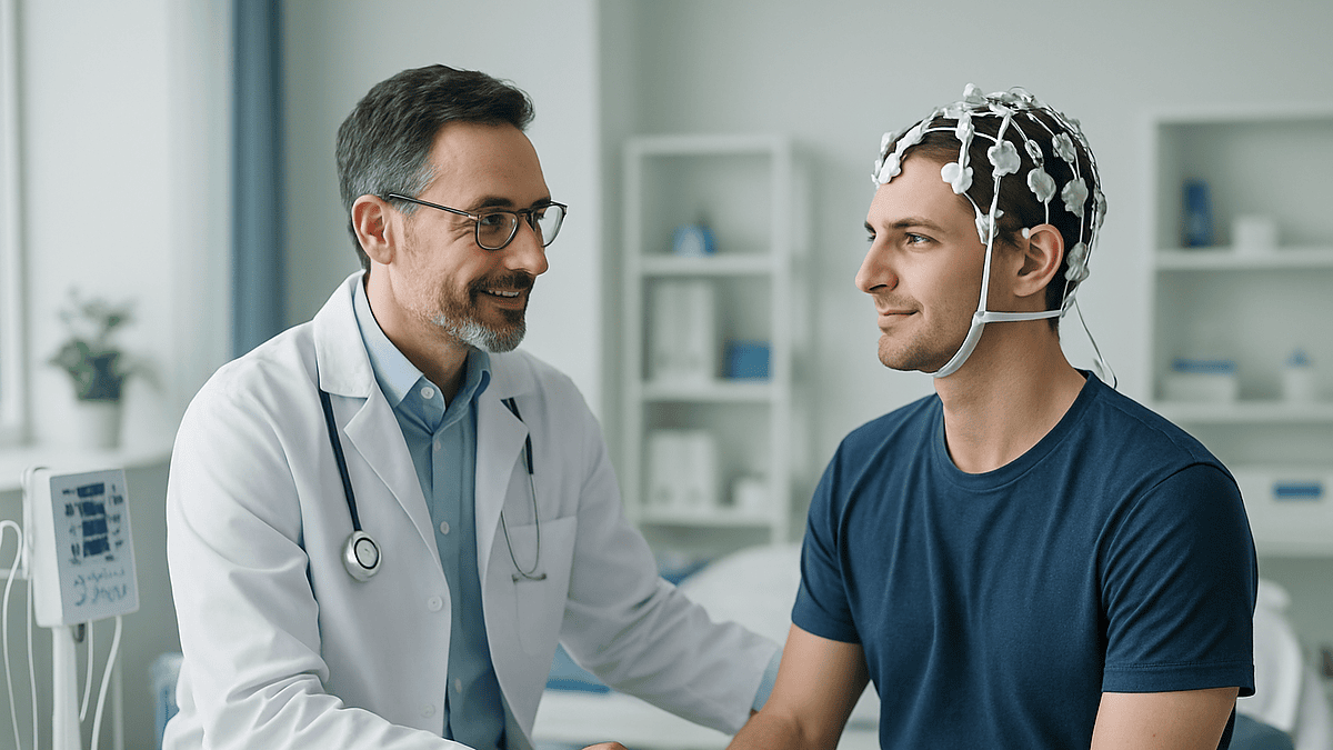
Key Takeaways
- An EEG records brain signals measured in millionths of a volt, amplified thousands of times into readable wave patterns, nothing electrical ever flows into your head.
- A routine EEG samples only 20–40 minutes of brain activity, so a single normal result detects epilepsy-related discharges in only about half of people who have the condition.
- EEG and MRI answer different questions: the EEG tracks brain function in milliseconds, while MRI photographs anatomy in millimeters, which is why doctors often order both.
- Deep breathing and flashing lights during the test are deliberate provocation maneuvers designed to draw out abnormal activity that stays hidden at rest.
- Clean, product-free hair and normal meals (no caffeine, no fasting) are the two preparation steps that most affect recording quality.
- Any seizure lasting longer than five minutes, or a second seizure before the person regains awareness, is a call-911 emergency, not a wait-and-see situation.
Quick Answer
An EEG (electroencephalogram) is a painless, safe test that records the brain’s electrical activity through small sensors placed on the scalp. It captures brain waves in real time, which helps doctors evaluate seizures, epilepsy, sleep disorders, and unexplained changes in consciousness. A routine recording lasts about 20 to 40 minutes, though the full appointment usually takes closer to an hour, and longer versions exist.
The first thing most people notice is the tape measure. Before a single wire goes on, a technologist measures your head the way a tailor measures a sleeve, front to back, ear to ear, marking small spots with a soft wax pencil. Then comes a dab of gritty paste at each mark, a sensor pressed into it, and finally the quiet instruction: close your eyes and relax.
On the screen behind you, something remarkable starts scrolling. Twenty-odd wavy lines, each one tracing the electrical chatter of billions of neurons, updating many times per second. Meteorologists read pressure systems; neurologists read this: the brain’s electrical weather, complete with calm fronts, sudden storms, and patterns that repeat night after night.
If your doctor has ordered one, or you’re simply curious how anyone eavesdrops on a living brain without opening it, here is what the recording actually captures, what it can and cannot tell you, and how to get the cleanest possible result.
What is an EEG, exactly?
Your brain runs on electricity, not much of it, but constantly. Roughly 86 billion neurons communicate by firing tiny electrical impulses, and when large groups of them fire in rhythm, the combined signal is just strong enough to reach the surface of the scalp. An electroencephalogram, or EEG, detects that signal using small metal discs attached to the skin with conductive paste.
The voltages involved are minuscule, measured in millionths of a volt, thousands of times weaker than what powers a watch battery. The EEG machine amplifies them enormously and draws them as continuous wavy lines, one for each recording site. Faster, tighter waves generally mean an alert, engaged brain region; slower, taller waves appear during drowsiness and deep sleep. Abnormal shapes, sharp spikes, sudden bursts, slowing where there shouldn’t be any, are what the neurologist is hunting for.
The idea is nearly a century old. German psychiatrist Hans Berger recorded the first human EEG in the 1920s, and the core principle hasn’t changed since: listen to the brain’s electricity from outside, without breaking the skin. What has changed is everything around it, digital amplifiers, video synchronization, wearable recorders you can take home for days.
One crucial point sets the eeg test apart from nearly every other brain study: it measures function, not structure. A scan photographs the brain; an EEG listens to it. That distinction explains most of what follows.
Why would an EEG be ordered?
The single most common reason is a suspected seizure. When someone loses consciousness, convulses, or has an unexplained staring spell, doctors need to know whether abnormal electrical discharges in the brain were responsible, and an EEG is the only routine test that can show those discharges directly.
Seizures are far from the whole story, though. According to MedlinePlus and Mayo Clinic, doctors also order an eeg brain scan to help evaluate:
- Epilepsyconfirming the diagnosis, identifying the seizure type, and locating where in the brain seizures begin, which shapes treatment decisions.
- Fainting and blackoutsdistinguishing a seizure from a cardiac or blood-pressure cause, since the two can look alike to bystanders.
- Encephalopathythe diffuse brain dysfunction that can accompany infections, liver or kidney problems, or certain medications; the EEG often shows characteristic generalized slowing.
- Sleep disordersEEG is a core component of overnight sleep studies, because sleep stages are defined by their brain-wave signatures.
- Monitoring in critical caretracking brain activity in patients who are sedated, in a coma, or at risk of silent seizures after a brain injury or stroke.
- Confirming the absence of brain activityin specific, carefully regulated circumstances, an EEG contributes to the evaluation of brain death.
Notice the pattern: every item on that list involves a question about how the brain is working right now, not what it looks like. When the question is anatomical, a tumor, a bleed, a structural abnormality, imaging takes the lead instead.
What does an EEG show, and what does it miss?
An EEG shows electrical patterns: their speed, their size, their symmetry between the left and right hemispheres, and how they change when you open your eyes, breathe deeply, or drift toward sleep. Against that backdrop of normal rhythms, several findings stand out to a trained reader.
Spikes and sharp waves, brief, jagged deflections, suggest a brain region prone to seizures, even between episodes. Rhythmic, evolving discharges captured during an actual event are close to definitive. Slowing in one area can point to localized dysfunction; slowing everywhere suggests a generalized process affecting the whole brain, from deep sedation to metabolic illness.
Now the honest limits, because they matter just as much. An EEG cannot read thoughts, measure intelligence, or diagnose mental health conditions. It records mainly from the brain’s outer surface, so activity in deep structures can be muffled or invisible. It photographs nothing: a small tumor, an aneurysm, or early structural disease can sit beneath a perfectly normal recording.
And it is a snapshot in time. If the abnormal activity your doctor suspects didn’t happen during the 20 to 40 minutes of recording, the tracing may look unremarkable, which is why, as the NHS notes, a normal result does not by itself rule out epilepsy. Understanding what does an eeg show really means understanding this trade-off: extraordinary detail about brain timing, purchased at the cost of seeing only the window you record.
What can an EEG see that an MRI cannot?
Time. That’s the short answer, and it’s worth unpacking, because “which test is better?” is one of the most common questions patients ask, and it’s the wrong question.
An MRI produces exquisitely detailed still images of brain anatomy, resolving structures down to about a millimeter. But it says little about what those structures are doing from one moment to the next. An EEG is the mirror image: it can’t show you a millimeter of tissue, but it tracks electrical activity on the scale of milliseconds, fast enough to catch a seizure discharge as it ignites and spreads.
Picture a house with faulty wiring. The MRI is the architectural blueprint: every wall, every room, beautifully drawn. The EEG is the live readout on the electrical panel, flickering the instant a circuit misbehaves. A blueprint can look flawless while the lights strobe; a panel can read normal in a house with a cracked foundation. You often need both documents to understand the house.
In practice, that’s exactly how neurologists use them. A person with new seizures typically gets an EEG to characterize the electrical event and an MRI to search for a structural cause: a scar, a malformation, an old injury. Each test answers a question the other cannot. Cleveland Clinic and Johns Hopkins both describe them as complementary rather than competing, and any evaluation of unexplained seizures that skips one of them is usually incomplete.
The EEG procedure, step by step
Knowing the choreography in advance removes most of the mystery. A routine eeg procedure at an outpatient lab unfolds in a predictable sequence.
Setup (20–30 minutes). The technologist measures your head using a standardized grid: the international “10–20 system”, so that each sensor lands in the same relative position on every patient, every time. Around 20 small discs are attached with conductive paste, sometimes after a gentle scrub of each spot to improve contact. Some labs use a snug cap with the sensors pre-mounted instead.
Recording (20–40 minutes). You recline in a quiet, often dimly lit room. Mostly you rest with your eyes closed; periodically you’ll be asked to open and close them, since a healthy brain shifts rhythms noticeably when it does. Two “activation” maneuvers are standard: several minutes of deep, rapid breathing, and a strobe light flashing at varying speeds. Both can coax out abnormal activity that stays hidden at rest: that’s the point, and staff are trained and equipped in case a seizure occurs.
Wrap-up (10–15 minutes). The sensors come off, the paste gets wiped away (a shower at home finishes the job), and you leave with no restrictions unless you were deliberately sleep-deprived, in which case someone else should drive.
Throughout, the machine records only. Nothing flows into your head: a fact worth repeating, because the word “electrodes” makes plenty of reasonable people picture the opposite.
How many hours does an EEG take?
It depends entirely on which kind you’re having, and the range is wider than most people expect, from under an hour to several days. Doctors match the recording length to the question: a brief study may suffice for a clear-cut case, while rare or hard-to-capture events demand a longer net.
| Type of EEG | Recording time | Where it happens | Best suited for |
|---|---|---|---|
| Routine EEG | 20–40 minutes (about 1 hour total appointment) | Outpatient lab | First evaluation of suspected seizures or unexplained spells |
| Sleep-deprived EEG | 1–2 hours, often including a nap | Outpatient lab | Cases where a routine EEG was normal; sleep loss makes abnormalities more likely to appear |
| Ambulatory EEG | 24–72 hours, continuous | Home, wearing a portable recorder | Infrequent events that a short recording is unlikely to catch |
| Video EEG monitoring | Several days | Hospital epilepsy monitoring unit | Capturing actual events on synchronized video and EEG; surgical planning |
The logic behind the ladder is simple probability. If your episodes happen once a month, a 30-minute window will almost certainly miss one; a three-day home recording, or a monitored hospital stay where doctors can safely observe an event, stacks the odds in your favor. Most people start at the top of the table and only move down if the answer stays elusive: the NHS and Cleveland Clinic both describe this stepwise approach as standard practice.
What should you not do before an EEG test?
Preparation is refreshingly low-tech, and most of it concerns your hair. Sensors need clean contact with the scalp, so anything that coats it, conditioner, oil, gel, hairspray, leave-in product, degrades the signal. Wash your hair the night before or morning of, then apply nothing afterward. Skip hairpieces, extensions, and elaborate styles that block access to the scalp. You will not need to shave anything.
Beyond hair, a short list of don’ts, drawn from Mayo Clinic and Johns Hopkins guidance:
- Don’t have caffeine on the day of the test. Coffee, tea, cola, energy drinks, and chocolate can all shift brain-wave patterns enough to muddy the recording.
- Don’t fast. This one surprises people. Low blood sugar changes the EEG, so eat normally before you arrive.
- Don’t stop any medication on your own. Some medicines influence brain waves, and your care team needs to know what you take, but only they should decide whether anything is adjusted beforehand. Never change a regimen without explicit instructions.
- Don’t sleep normally if you’ve been told not to. For a sleep-deprived EEG, you may be asked to stay up much of the night. Follow the instructions exactly, and arrange a ride, since driving drowsy is genuinely dangerous.
One more practical note: wear a shirt that buttons or zips rather than one that pulls over your head, so you don’t disturb the sensors: a small courtesy your technologist will appreciate more than you’d guess.
Alpha, beta, theta, delta: reading the brain’s weather patterns
Every EEG report speaks the language of frequency, how many times per second a wave cycles, measured in hertz. Four bands do most of the descriptive work, and knowing them turns a report from jargon into something closer to a forecast.
Beta waves (roughly 13–30 Hz) are the fast, low chop of an alert, thinking brain, dominant when your eyes are open and you’re engaged with the world. Alpha waves (8–13 Hz) roll in when you close your eyes and relax; they’re strongest over the back of the head and famously vanish the moment you open your eyes again, a reflex so reliable that technologists use it to check the recording. Theta waves (4–8 Hz) belong to drowsiness and light sleep. Delta waves (under 4 Hz), slow, tall swells, dominate deep sleep in adults and are normal in young children even while awake.
Context decides whether a pattern is weather or storm damage. Delta waves at 3 a.m. in a sleeping adult are exactly what should be there; the same waves parked over one region of an awake adult’s brain suggest that area isn’t functioning properly. Symmetry matters too: the two hemispheres should broadly mirror each other, and a persistent mismatch draws the reader’s eye immediately.
This is why EEG interpretation resists automation more than you might think. The reader isn’t matching shapes to a chart; they’re asking whether this pattern makes sense for this person, at this age, in this state of wakefulness. Age, sleep stage, and even eye position rewrite what “normal” means.
Is an EEG safe? Does it hurt?
An EEG is among the safest tests in medicine, and it does not hurt. The sensors record electricity; they never deliver it. No needles, no radiation, no contrast dye, no magnets. Mayo Clinic and MedlinePlus both classify the standard test as essentially risk-free, and it’s used routinely in newborns: a fair indicator of how gentle it is.
What you might actually feel is mundane: cool paste on your scalp, mild pressure from the discs or cap, and perhaps some skin redness where sensors sat, which fades within hours. The most persistent aftereffect is paste in your hair, defeated by an ordinary shower.
The one caveat deserves plain language rather than fine print. The deep-breathing and flashing-light portions of the test are designed to provoke abnormal activity, and in a small number of people, mainly those with photosensitive epilepsy, they can trigger an actual seizure during the recording. That sounds alarming; in context, it isn’t. It happens in a controlled setting with trained staff present, and a captured event is often diagnostically valuable, sometimes answering in minutes a question that months of normal recordings could not. Labs screen for known photosensitivity in advance and can modify or skip the flash sequence.
For longer studies, the risks shift from medical to practical: skin irritation from days of adhesive, and the awkwardness of sleeping and showering around a head full of wires. Annoying, certainly. Dangerous, no.
What conditions are diagnosed with an EEG?
Epilepsy sits at the center of EEG’s diagnostic territory, and honesty requires a nuance up front: the test rarely delivers a diagnosis alone. It confirms, refines, and localizes, one strong voice in a conversation that also includes your history, an eyewitness account of your episodes, imaging, and blood work.
Within that frame, the eeg test contributes most decisively to:
- Epilepsy and seizure disorders. Characteristic spike patterns support the diagnosis, distinguish generalized epilepsy (involving the whole brain at once) from focal epilepsy (starting in one region), and, for people considering surgery, help map exactly where seizures originate.
- Encephalitis and encephalopathy. Brain inflammation and diffuse dysfunction produce recognizable slowing and, in some conditions, distinctive periodic patterns that can shift the diagnostic odds meaningfully.
- Sleep disorders. Overnight polysomnography leans on EEG to stage sleep, which underpins the evaluation of conditions like narcolepsy and abnormal behaviors during sleep.
- Nonepileptic events. Sometimes the most useful finding is a normal EEG recorded during an episode, which tells doctors the event, however dramatic, was not an epileptic seizure and redirects the workup toward cardiac, circulatory, or psychological causes.
- Critical illness. Continuous monitoring detects subtle or nonconvulsive seizures in intensive care, where a patient may seize with no outward sign at all.
What the EEG generally does not diagnose: headaches, dementia subtypes, mental health conditions, or learning differences. It may occasionally contribute a supporting detail in those settings, but no responsible clinician hangs such diagnoses on brain waves alone.
What does an abnormal, or normal, EEG result actually mean?
Here’s where a little statistical honesty saves a lot of anxiety, in both directions.
An abnormal result does not automatically mean epilepsy or brain disease. A small percentage of perfectly healthy people show minor irregularities on EEG, patterns a cautious reader will flag as nonspecific rather than diagnostic. Medications, sleep deprivation, and even normal variants that run in families can all nudge a tracing outside textbook limits. Interpretation always circles back to the clinical question: does this finding explain your symptoms?
The reverse cuts just as deep. A normal routine EEG does not rule out epilepsy, not even close. Because a standard recording samples only half an hour of brain activity, a single routine EEG captures epilepsy-related discharges in only about half of people who genuinely have the condition. Between seizures, many brains with epilepsy hum along looking entirely ordinary. That’s precisely why the diagnostic ladder exists: repeat studies, sleep-deprived recordings, and multi-day monitoring each raise the odds of catching what a snapshot missed.
When your report arrives, expect measured language. Neurologists write phrases like “epileptiform discharges over the left temporal region” or “mild diffuse slowing, nonspecific”, descriptions, not verdicts. The verdict comes when your doctor weighs the tracing against everything else they know about you. If a result confuses or worries you, ask two questions: Does this finding explain my symptoms? and What would change if we repeated the test? Good answers to both usually dissolve most of the worry.
Living through a longer EEG: ambulatory and video monitoring
When short recordings keep coming back normal but the spells keep happening, doctors extend the net, and daily life gets temporarily strange.
An ambulatory EEG sends you home wearing the study. Sensors are glued on more durably, wires gather into a small recorder worn on a belt or in a pouch, and a snug head wrap keeps everything in place for one to three days. You’ll keep a diary of symptoms, sleep, and activities, and press an event button whenever something happens so the reader can zero in on that exact moment. The rules are mostly common sense: no showers or swimming, no chewing gum for hours on end (jaw muscles create electrical noise), and stay clear of things that generate strong interference. Otherwise, you live your life, which is exactly the point, since your episodes happen in your life, not in a lab.
A video EEG, usually in a hospital epilepsy monitoring unit, pairs continuous brain-wave recording with synchronized video over several days. Watching what your body does at the precise instant the tracing changes is the gold standard for classifying difficult events, and it’s essential groundwork for anyone being evaluated for epilepsy surgery. Under close supervision, doctors may deliberately increase the chance of capturing an event: a controlled version of the very thing you’ve been trying to avoid, turned into diagnostic gold.
Neither experience is glamorous. Both exist because a captured event is worth more than a hundred normal recordings, and patients who understand that trade-off tend to tolerate the wires with far better humor.
When should you see a doctor, and when is it an emergency?
An EEG is a tool for answering questions, but recognizing that a question exists comes first. Make an appointment promptly if you or someone close to you experiences any of the following:
- A first-ever seizure or convulsion, even a brief one that resolved completely
- Unexplained blackouts, falls, or gaps in memory
- Staring spells, in children especially, episodes of abrupt unresponsiveness lasting seconds, sometimes mistaken for daydreaming
- Repeated jerking movements on waking, or nighttime events like tongue-biting or waking with unexplained soreness
- Fainting during exercise, or fainting preceded by palpitations, which may point toward the heart rather than the brain, another reason evaluation matters
Call emergency services immediately if a seizure lasts longer than five minutes, if a second seizure follows without the person regaining awareness, if breathing doesn’t resume normally afterward, if the seizure happens in water, if the person is pregnant or has diabetes, or if they’re injured during the event. Those thresholds come from standard first-aid guidance, and the five-minute rule in particular is worth memorizing: a prolonged seizure is a medical emergency, not something to wait out.
Bring a witness or a phone video to the appointment if you possibly can. A thirty-second clip of an actual episode is often worth more to a neurologist than any single test, and it frequently determines whether an EEG is ordered at all, and which kind.
Getting your results: who reads an EEG, and how long it takes
The technologist who ran your study won’t tell you the results, and not out of secrecy, interpretation legally and practically belongs to a physician, typically a neurologist with specific training in reading these tracings. They review the recording wave by wave, correlate it with the technologist’s notes (“patient coughed,” “eyes opened,” “drowsy here”), and dictate a formal report.
Expect results within a few days to a couple of weeks for a routine study, per Cleveland Clinic’s typical timelines; multi-day recordings take longer simply because there are more hours of data to review: a 72-hour ambulatory study contains over a hundred times the data of a routine one. If your situation is urgent, hospital-based EEGs can be read within hours.
The report usually travels to the doctor who ordered the test, who then discusses it with you in context. That extra step frustrates people who want the answer now, but it exists for a good reason: an EEG finding means little in isolation. “Mild slowing” reads very differently in a sleep-deprived college student than in someone with new confusion, and only a clinician who knows your whole story can weigh it properly.
When you do sit down for that conversation, three questions extract the most value: What did the recording show, in plain words? Does it explain my symptoms, or do we need more information? And what happens next, another test, a longer recording, or reassurance? A good clinician welcomes all three, and the answers will tell you far more than the report’s technical vocabulary ever could.
Frequently asked questions
What can an EEG see that an MRI cannot?
An EEG captures the brain’s electrical activity in real time, down to the millisecond, including seizure discharges as they happen, which no MRI can show. MRI photographs anatomy in fine detail but reveals almost nothing about moment-to-moment function. A brain can look structurally perfect on MRI while its electrical activity is clearly abnormal on EEG, and vice versa. Neurologists treat the two tests as complementary and often order both.
How many hours does an EEG procedure take?
A routine EEG appointment takes about an hour: 20–30 minutes of setup and 20–40 minutes of recording. A sleep-deprived version runs one to two hours, often including a nap. Ambulatory EEGs record continuously for 24 to 72 hours while you go about life at home, and video EEG monitoring in a hospital can last several days. Your doctor chooses the length based on how often your symptoms occur.
What should you not do before an EEG test?
Avoid caffeine on the day of the test, don’t skip meals (low blood sugar alters brain waves), and don’t apply any hair products after washing your hair, conditioner, gel, oil, and spray all interfere with sensor contact. Never stop or change medications on your own; only your care team should adjust anything beforehand. If you’ve been asked to sleep-deprive for the test, follow those instructions exactly and arrange a ride.
Why would an EEG be ordered?
The most common reason is to evaluate a suspected seizure or diagnose epilepsy. Doctors also order EEGs to investigate unexplained fainting, confusion, or blackouts; to assess encephalopathy or brain inflammation; to stage sleep during sleep studies; and to monitor brain activity in intensive care, where silent seizures can occur without visible signs. In every case, the underlying question is about how the brain is functioning rather than what it looks like.
Does an abnormal EEG always mean epilepsy?
No. A small percentage of healthy people show minor EEG irregularities, and sleep deprivation, medications, and harmless inherited variants can all produce findings outside textbook norms. Neurologists distinguish nonspecific changes from truly epileptiform patterns like spikes and sharp waves, and even those are weighed against your symptoms and history. An abnormal tracing is a clue that requires context, not a diagnosis by itself.
Can a normal EEG rule out seizures or epilepsy?
No. A routine EEG records only a short window, and between seizures many people with epilepsy show completely normal brain activity: a first routine study catches epilepsy-related discharges only about half the time. That’s why doctors escalate to sleep-deprived, ambulatory, or multi-day video EEG when suspicion remains despite a normal result. A normal tracing lowers the odds but never closes the question on its own.
Is an EEG painful or dangerous?
No: the test is painless and considered essentially risk-free, safe enough for newborns. Sensors only record electricity; they never deliver any. The one caveat: the flashing-light and deep-breathing portions can trigger a seizure in a small number of susceptible people, but this occurs in a controlled setting with trained staff present, and a captured event is often diagnostically valuable. Afterward, expect nothing worse than paste in your hair and possibly mild skin redness.
Can you sleep during an EEG?
Yes, and it’s often encouraged. Some abnormal patterns appear only during drowsiness or sleep, so falling asleep during the recording can make the test more revealing, not less. For sleep-deprived EEGs, staying up the night before is specifically intended to help you doze off in the lab. Overnight sleep studies and multi-day monitoring deliberately record entire nights, since sleep stages themselves are defined by their brain-wave signatures.
Do you have to shave your head for an EEG?
No. Sensors attach to the scalp through your hair using conductive paste or a fitted cap, and technologists work around hair of any length or texture. What does matter is that hair is clean and completely free of products, conditioner, oils, gels, and sprays all weaken the signal. Braids, extensions, and weaves that block access to the scalp may need to be removed before the appointment, so ask the lab in advance.
How long does it take to get EEG results?
Typically a few days to about two weeks for a routine study. A neurologist trained in EEG interpretation reviews the entire recording, writes a formal report, and sends it to the doctor who ordered the test, who then discusses it with you in the context of your symptoms. Multi-day recordings take longer to read because they contain vastly more data, while urgent hospital-based EEGs can be interpreted within hours.
References
- Electroencephalogram (EEG): What It Is, Procedure & Results: Cleveland Clinic
- Electroencephalogram (EEG): NHS
This article is for general information only and is not a substitute for professional medical advice. Please consult a qualified doctor about your individual situation.

More from the Blog

AI in Healthcare: What It Already Does in Hospitals, and Where It Falls Short
AI in healthcare currently helps hospitals read medical images, flag patients at risk of deterioration such as sepsis, draft clinical notes from recorded visits,…

Home Blood Pressure Monitors: Wrist vs Arm and How to Take a Reading That Counts
For most adults, the best home blood pressure monitor is an automatic upper-arm cuff that has passed independent validation testing and fits your arm…
Telehealth: What Can Be Diagnosed on a Video Call and What Cannot
Telehealth can reliably assess conditions a clinician diagnoses mostly by history and by looking: many rashes, mental health concerns, medication follow-ups, chronic disease check-ins…

Recovering from Radiotherapy: How Long Until You Feel Better
Most people start to feel noticeably better within a few weeks of their last radiotherapy session, although side effects such as fatigue and skin…

Patient Portals: How to Read Your Own Lab Results Without Panicking
A patient portal is the secure website or app your health system uses to show you appointments, messages, and test results, and since 2021…

Over-the-Counter CGMs: What Glucose Data Means for People Without Diabetes
Over-the-counter continuous glucose monitors such as Stelo and Lingo show adults without diabetes how blood sugar rises and falls through the day. For most…