

When Does a Lung Abscess Need Drainage or Surgery? What Your Team Watches For
Key Takeaways
- Published reviews report that roughly 80 to 90 percent of lung abscesses resolve with antibiotics and positioning alone, with surgery needed in about 10 to 15 percent.
- A cavity of about six centimeters or more, persistent fever despite weeks of appropriate antibiotics, heavy bleeding, rupture into the chest space or suspected tumor are the usual triggers for considering drainage or surgery.
- Fever often improves within the first several days of effective antibiotics, but the cavity on imaging can take weeks to months to shrink, so a persistent shadow in someone who feels better is expected.
- Percutaneous drainage suits abscesses lying against the chest wall, while central cavities are more often approached through a bronchoscope or, if needed, surgery.
- Empyema is pus around the lung rather than inside it, and it is treated with a chest tube; a simple lung abscess usually is not.
- Recurrence most often follows an unaddressed cause such as ongoing aspiration, dental disease or heavy alcohol use, or an antibiotic course stopped early.
Most lung abscesses are treated with several weeks of antibiotics and do not need a procedure. A care team usually considers drainage or surgery when fever and infection markers fail to settle despite appropriate antibiotics, when the cavity is very large, when there is heavy bleeding, a rupture into the chest space, a blocked airway or suspected tumor, or when the person is too unwell to wait. The treating team makes that judgment case by case.
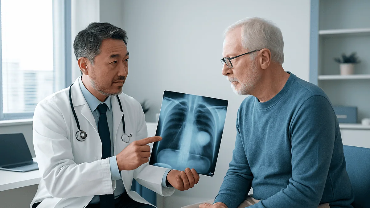
The chest scan is up on the screen, and the pulmonologist is pointing at a dark hollow in the right lung with a flat line across it, like a half-filled bottle tipped on its side. The patient has been coughing up foul-tasting sputum for three weeks and running fevers every evening. The question forming in the room is a practical one: is this something antibiotics can handle, or is someone going to have to go in there?
It is one of the most common questions families ask once the words “lung abscess” have been said out loud. The honest answer is that most people never need a needle, a tube or an operating room, and that the cases that do are recognizable by a fairly consistent set of signals.
This article walks through when does a lung abscess need surgery, what drainage actually involves, why time is often the first treatment, and what your team is quietly watching on the ward and on repeat scans while deciding.
What is a lung abscess, in plain terms?
A lung abscess is a pocket of pus inside lung tissue, walled off by inflamed lung that has broken down around it. The pus is dead tissue, white blood cells and bacteria. Because the cavity often connects to an airway, air gets in and sits above the fluid, which is why radiologists talk about an “air-fluid level”: the flat line where gas meets liquid on a scan.
Doctors sort abscesses in two main ways. A primary abscess forms in an otherwise reasonably healthy lung, most often after aspiration, meaning stomach contents or mouth secretions slipping down into the lungs during a period of reduced consciousness, such as heavy alcohol use, a seizure, anesthesia or a swallowing problem. A secondary abscess develops on top of something else: a blocked airway from a tumor or inhaled object, a lung already damaged by bronchiectasis, a weakened immune system, or infection carried through the bloodstream from elsewhere, such as an infected heart valve.
The distinction matters because it shapes the plan. A primary aspiration abscess is typically caused by mixed mouth bacteria, many of them anaerobes (bacteria that thrive without oxygen), and a review in Annals of Translational Medicine notes that these respond to antibiotics alone in the large majority of cases. A secondary abscess sitting behind a blockage may not drain no matter which antibiotic is chosen, because the road out is closed.
The time course also matters. An acute abscess has been present under about six weeks; a chronic one has persisted longer, and chronic cavities develop thicker, more fibrous walls that are slower to collapse and heal. That thickened wall is one reason older cavities are more likely to end up in the surgical conversation.
How serious is an abscess in the lung?
Serious enough that it is always treated in hospital, at least initially, and rarely so dangerous that decisions must be made within hours. That middle ground is worth understanding, because it explains why teams often choose to wait and watch rather than act fast.

The immediate dangers are the ones you might expect from any deep infection: it can spread into the bloodstream and cause sepsis, the body’s overwhelming and life-threatening response to infection. Pus can also break through the lung surface into the pleural space, the thin gap between lung and chest wall, producing an empyema, which is pus trapped around the lung rather than inside it. Rarely, an abscess erodes into a blood vessel and causes heavy coughing of blood.
Against that, most people who develop a primary lung abscess improve on antibiotics without any procedure. The Annals of Translational Medicine review referenced throughout this article reports that medical treatment succeeds in roughly 80 to 90 percent of cases, with surgery reserved for the remaining 10 to 15 percent. Those figures come from published series rather than from a promise about any individual, but they set the tone: this is usually a slow, treatable infection, not an emergency.
Where the outlook is genuinely more guarded, the same review and the NIH-hosted StatPearls summary point to consistent factors: a very large cavity, an abscess caused by a tumor blocking an airway, a weakened immune system, advanced age, and infection with certain organisms such as Pseudomonas, Staphylococcus aureus or Klebsiella. None of those means surgery is inevitable. Each simply raises the team’s alertness and lowers their threshold for stepping in.
When does a lung abscess need surgery? The short version
Surgery is the exception, not the rule, and it almost always follows a failed trial of antibiotics rather than replacing it. Understanding that sequence takes much of the fear out of the question.
Guideline-level practice, summarized in both the StatPearls review and the Annals of Translational Medicine paper, describes a fairly consistent list of triggers that move a team from “continue treatment” toward “consider a procedure”:
- Fever, rising inflammatory markers or a growing cavity despite several weeks of appropriate antibiotics.
- A large abscess, with a diameter of about six centimeters or more often quoted as the size at which antibiotics alone are less likely to succeed.
- Heavy or repeated coughing of blood that does not settle.
- Rupture into the pleural space with empyema or a bronchopleural fistula, an abnormal channel between airway and chest cavity.
- Suspicion that the cavity is actually a tumor that has broken down, or an abscess sitting behind a tumor that blocks drainage.
- Sepsis that is not responding to medical treatment, where the pocket of pus is judged to be driving the illness.
Even when one of these is present, “surgery” is not automatically the next step. Many teams first try a less invasive route: a drain placed through the skin under imaging guidance, or drainage through a bronchoscope, a thin flexible camera passed down the airway. Formal surgery to remove part of the lung is typically kept for cases where those options are unsuitable or have not worked.
So the honest answer to when does a lung abscess need surgery is this: when antibiotics have been given a fair chance and the infection is still winning, when a complication makes waiting unsafe, or when something other than a simple abscess is suspected.
How lung abscess treatment usually begins: antibiotics and patience
The first treatment for nearly every lung abscess is not a scalpel but an intravenous line. Antibiotics chosen to cover anaerobic mouth bacteria and the common aerobic organisms are started, often before culture results are back, and adjusted once the lab identifies what is growing.

The mechanism is straightforward: drugs reach the inflamed rim of the cavity through its rich blood supply, kill or suppress the bacteria, and allow the body to reabsorb the pus and collapse the space. What surprises many patients is how slowly that plays out. The StatPearls review notes that fever commonly improves within the first several days of effective treatment, while the cavity itself may take weeks to shrink and chest imaging can lag well behind how the person feels. Antibiotic courses are correspondingly long; the reviews cited here describe typical durations in the range of four to six weeks, sometimes longer, with the switch from intravenous to oral treatment made once the person is stable. How long, and which agents, is entirely the prescribing team’s decision.
Alongside antibiotics, teams encourage what is sometimes called postural drainage: positioning the body so gravity helps the abscess empty into the airways to be coughed out. It sounds low-tech because it is, and it works because most abscesses already have an airway connection.
Doctors judge the response with a few simple measures: temperature trend, white cell count and inflammatory markers, appetite and energy, the volume and smell of sputum, and a repeat scan after a couple of weeks. A person who is eating, afebrile and producing less sputum is on the right path even if the picture on the X-ray still looks alarming. That mismatch between feeling and film is one of the most common reasons families ask about surgery too early.
What does it mean if a lung abscess appears on a CT scan?
It usually means the radiologist has seen a rounded cavity with a thick, irregular wall, often containing that telltale air-fluid level, surrounded by inflamed lung. A CT scan (computed tomography, a rotating X-ray that builds cross-sectional images) is the standard way to confirm the diagnosis because a plain chest X-ray can miss smaller cavities or confuse them with other shadows.
The scan does more than confirm. It answers several questions that steer treatment:
- Size: measured in centimeters, because large cavities drain less well on their own.
- Wall thickness and shape: a very thick, lumpy or nodular wall raises concern that the cavity could be a tumor that has broken down rather than a simple infection.
- Location: whether the abscess is near the lung surface, which makes a needle drain feasible, or deep and central, where a needle would have to cross healthy lung.
- Neighbors: whether there is fluid or pus in the pleural space, a blocked airway, an inhaled foreign object, or multiple abscesses suggesting spread through the bloodstream.
- Distinguishing from empyema: the MedlinePlus empyema entry describes pus around the lung, which on CT tends to form a smooth, lens-shaped collection that compresses lung rather than sitting inside it. This matters because empyema is treated primarily with tube drainage, while a simple abscess is not.
A lung abscess on CT scan is therefore a starting point, not a verdict. The same image that alarms a patient often reassures a specialist: a modest cavity, thin wall, clear air-fluid level and an obvious aspiration story is exactly the picture most likely to resolve with antibiotics. The features that prompt closer attention are size, an oddly thick wall, and anything suggesting the cavity cannot empty through the airways.
What happens during lung abscess drainage?
Drainage sits between antibiotics and surgery, and in practice it is where many stubborn abscesses are dealt with. Three routes are used, and the choice depends on where the cavity sits and what the team suspects is keeping it from emptying.
Percutaneous drainage means through the skin. With local anesthetic and CT or ultrasound guidance, a radiologist passes a needle, then a thin tube, into the cavity and leaves it in place to drain over days. It suits abscesses lying against the chest wall, where the needle does not have to cross healthy lung. The reviews cited here describe it as the usual first procedural step when antibiotics alone are not enough, particularly for people too unwell for an operation.
Bronchoscopic drainage uses a flexible camera passed through the mouth or nose into the airways under sedation. The team can suction pus, wash out the cavity, take samples for culture, and, importantly, look for a blockage such as a tumor or an inhaled object that explains why the abscess is not emptying. Some centers also place a small catheter through the scope for ongoing drainage of central cavities that a skin needle cannot safely reach.
Tube thoracostomy, a chest tube placed between the ribs into the pleural space, is not really abscess drainage at all; it treats empyema, the complication in which pus has escaped around the lung. The NHS empyema page describes this as the mainstay of empyema management alongside antibiotics.
None of these is trivial. A skin drain carries a risk of bleeding, collapsing the lung, or spreading infection into the pleural space. That is precisely why teams weigh location and wall thickness so carefully before choosing, and why a drain is a decision, not a reflex.
When does a lung abscess need surgery rather than a drain?
Drains buy time and remove pus; they do not remove destroyed lung. Surgery becomes the conversation when the problem is structural rather than simply a full cavity.
The reviews cited in this article describe surgical resection as appropriate in a recognizable set of circumstances:
- Antibiotics, with or without drainage, have been tried for an adequate period and the cavity persists or enlarges. Chronic abscesses with thick fibrotic walls are especially unlikely to collapse on their own.
- Bleeding is significant and recurrent, because pus and dead tissue eroding into a vessel cannot be reliably controlled from outside.
- A bronchopleural fistula or empyema has developed and simpler drainage has not resolved it.
- Imaging or bronchoscopy raises real suspicion of cancer, either as the cause of the cavity or hidden behind it. Removing the segment both treats the infection and provides a definitive tissue diagnosis.
- The abscess sits centrally, so a needle drain would have to cross a large amount of healthy lung, and bronchoscopic drainage is not feasible.
- The abscess is very large, the person is deteriorating, and the team judges that the infected tissue itself is driving sepsis.
Notice what is missing from that list: size alone. A large cavity in someone who is improving is usually left to keep improving. Duration alone is also absent; the team looks for a persistent cavity plus ongoing symptoms, not merely a slow X-ray.
Thoracic surgeons also weigh the person’s fitness, because lung resection under general anesthesia asks a lot of the heart and remaining lung. Someone with severe emphysema or heart failure may be steered toward a prolonged drain and antibiotics precisely because their body may not tolerate an operation. In this way, the decision about when does a lung abscess need surgery is as much about the whole person as about the shadow on the scan.
Who is usually asked to wait, and who is usually moved along faster?
Waiting is an active decision. The patient who is asked to continue antibiotics and return for a scan in two weeks is not being neglected; the team has judged that the odds favor resolution and that a procedure would add risk without clear benefit.
People who are typically asked to wait share a profile drawn from the cited reviews: a clear aspiration story, a single cavity of modest size with a relatively thin wall, fever already trending down after the first days of treatment, stable oxygen levels, no blood in the sputum, no fluid around the lung, and no reason to suspect a blocked airway. Their labs are improving even if the X-ray has not changed much. For this group, the reviews describe the great majority resolving on antibiotics alone.
People who tend to be moved toward drainage or surgery sooner look different. Their fever persists past the first week or two of appropriate treatment. Their cavity is large or enlarging. They cough blood. Their scan shows pus around the lung, a very thick or irregular wall, several abscesses at once, or a mass behind the cavity. They may have a weakened immune system that limits how well their body walls off infection, or they may be sick enough that another few weeks of waiting is itself the risk.
A third group sits in between and generates most of the honest uncertainty: the person who is somewhat better but not clearly well, with a cavity that is neither shrinking nor growing. Here teams often extend antibiotics, repeat imaging, and may perform bronchoscopy to make sure nothing is blocking drainage before committing to anything larger. Asking your team which of these three groups they think you fall into is one of the most useful questions you can pose, because the answer tells you what they are watching for next.
Antibiotics, drainage or surgery: how the options compare
The table below summarizes what each approach is trying to achieve, where it fits, and the trade-offs your team weighs. It is a map of typical practice described in the cited reviews, not a prescription for any individual.
| Approach | What it does | Usually considered when | Main trade-offs |
|---|---|---|---|
| Antibiotics with postural drainage | Kills bacteria at the cavity rim; body reabsorbs pus and collapses the space | First step for nearly all abscesses, especially primary aspiration abscesses with an airway connection | Slow; courses run weeks; requires monitoring for lack of response |
| Percutaneous (through-the-skin) drain | Removes pus directly through a catheter placed under imaging guidance | Antibiotics failing; abscess lies against the chest wall; person too unwell for surgery | Risk of lung collapse, bleeding or spreading infection into the pleural space; not suitable for central cavities |
| Bronchoscopic drainage and inspection | Suctions pus via the airway; identifies blockage such as tumor or foreign body | Central abscess; suspicion of airway obstruction; need for cultures | May not fully empty a large cavity; sedation required |
| Chest tube for empyema | Drains pus from around the lung, not from inside it | Abscess has ruptured into the pleural space | Treats the complication, not the original cavity |
| Surgical resection (segment or lobe) | Removes the destroyed, infected lung tissue outright | Persistent or enlarging cavity despite the above; severe bleeding; fistula; suspected cancer | General anesthesia; loss of lung volume; longer recovery; requires adequate fitness |
Two patterns are worth drawing out. First, the options form a ladder more than a menu; teams usually climb it step by step unless a complication forces a jump. Second, the deciding factors are rarely about the abscess alone. Location dictates whether a needle is safe. Overall fitness dictates whether an operation is. The cause dictates whether removing pus will be enough or whether a tumor or blockage means the lung tissue itself has to go.
What lung abscess surgery involves, and what the risks are
When surgery is chosen, the goal is to remove the diseased portion of lung while sparing as much healthy tissue as possible. The two common operations are segmentectomy, removal of the anatomical segment containing the abscess, and lobectomy, removal of the whole lobe. The right lung has three lobes and the left has two; each is divided into segments with its own airway and blood supply, which is what allows a surgeon to remove one piece cleanly.
The approach may be through a traditional incision between the ribs or through several small cuts using a camera, known as video-assisted thoracoscopic surgery. Dense scarring around a chronic abscess sometimes makes the keyhole approach impractical, and surgeons may convert to an open operation if needed. Either way, general anesthesia is used, and one or more chest tubes are left in afterward to drain air and fluid while the remaining lung re-expands.
Risks are those of any major chest operation, and your surgical team will lay them out in detail: bleeding, wound or chest-space infection, a persistent air leak from the cut surface of the lung, blood clots, pneumonia in the remaining lung, and the general risks of anesthesia in someone already weakened by weeks of infection. Removing lung tissue permanently reduces reserve, which is why breathing tests are done beforehand to estimate how well someone will cope.
Alternatives are always discussed at the same time. Prolonged antibiotics with a long-term skin drain is the usual alternative for people too frail for resection. In selected cases surgeons have described creating a controlled opening through the chest wall into the cavity to let it drain externally over weeks, a historical technique that still has a place when resection is unsafe. Which path fits is a judgment your treating team makes with you, weighing infection control against the demands of the operation.
How long is a hospital stay for a lung abscess?
There is no single number, and anyone who quotes one confidently is guessing. The length of a lung abscess hospital stay depends on how quickly fever settles, whether intravenous antibiotics can be switched to oral treatment, whether a drain or operation is needed, and how well the person was before the infection began.
What can be said with reference to the cited reviews is the shape of the timeline. Antibiotic treatment as a whole typically runs for weeks, in the range of four to six or longer, but not all of it needs to happen in hospital. The usual pattern is an initial inpatient period for intravenous antibiotics and close observation, followed by discharge on oral treatment once the person is eating, afebrile and clinically stable, with outpatient follow-up and repeat imaging. People who need a percutaneous drain often stay while the drain is in and output is monitored. Those who have surgery stay while chest tubes are draining and until pain control, breathing and mobility are acceptable for going home.
Certain things reliably lengthen a stay: sepsis at admission, a weakened immune system, a large or multiple abscesses, complications such as empyema, and the need to change antibiotics after culture results. Certain things shorten it: a clear cause that is addressed, a small cavity, and a rapid fever response.
A more useful question to ask your team than “how many days” is “what has to happen before I can go home.” The answer, whether it is a certain number of fever-free days, a stable white cell count, a drain removed or a scan repeated, gives you concrete milestones to watch rather than an arbitrary date that may shift and cause needless worry.
What the following weeks usually look like after treatment
Recovery from a lung abscess is measured in weeks and often months, and the sequence is fairly predictable even if the pace varies.
In the first days of effective antibiotics, fever and sweats usually ease and appetite starts to return. The StatPearls review describes this early clinical response as the first sign the chosen antibiotic is working. Sputum may temporarily increase as the cavity drains through the airways; that is often a good sign rather than a setback, although any blood should be reported.
Over the following two to three weeks, energy improves and cough lessens. Imaging at this stage often still shows the cavity, sometimes barely changed. The reviews are clear that radiographic healing lags weeks to months behind clinical improvement, and a persistent shadow in someone who feels well is expected, not alarming. Some cavities leave a thin-walled scar or a small residual space that never fully disappears and is harmless.
If a percutaneous drain was placed, it stays until output drops and imaging shows the cavity has collapsed, then is removed at the bedside. After surgery, the chest tube typically comes out within the first week or so once air leak and fluid have settled, and the incision heals over several weeks. Breathing exercises, walking early and gradually increasing activity all help the remaining lung expand and reduce clot risk.
Throughout, oral antibiotics continue at home for the duration the prescribing team sets, with clinic visits and a follow-up scan to confirm the cavity is shrinking. The most important task in this phase is also the least glamorous: finishing the course. Stopping early because you feel better is one of the commonest reasons an abscess recurs and a procedure that was avoided the first time becomes necessary.
What people often get wrong about lung abscess surgery
“An abscess has to be drained or it will never heal.” True for many abscesses elsewhere in the body, not for most in the lung. A lung abscess usually already drains itself through the airway it formed next to, which is why coughing up pus is part of the illness. The reviews cited here describe most primary abscesses resolving with antibiotics and positioning alone.
“The X-ray still shows it, so the antibiotics aren’t working.” Imaging lags behind clinical improvement by weeks to months. Teams judge response mainly by fever, blood tests and how the person feels, and only worry about a persistent cavity when symptoms persist alongside it.
“Bigger cavity means immediate surgery.” Size lowers the odds of antibiotics alone succeeding and raises the team’s alertness, but a large abscess in someone who is clearly improving is usually watched, not operated on.
“Surgery removes the infection so antibiotics can stop.” Resection removes destroyed tissue; antibiotics continue afterward for the period the team sets, because infected lung and bloodstream do not clear the moment the specimen leaves the room.
“If it needs a procedure, that means the doctors got it wrong at the start.” A trial of antibiotics first is the recommended sequence in the cited reviews. Needing a drain later is not a failure of care; it is the ladder working as designed.
“A lung abscess means I have cancer.” Most do not. Cancer is one cause a team must rule out, especially in a smoker or when the cavity wall is thick and irregular, which is why bronchoscopy is sometimes done. Ruling it out is not the same as suspecting it.
“Once it’s gone, it’s gone.” Recurrence happens most often when the underlying cause is not addressed: ongoing aspiration from a swallowing problem, untreated dental disease, or continued heavy alcohol use. Treating the cause is as much a part of care as treating the cavity.
Questions to ask your care team
Bring these to the ward round or clinic. They are designed to surface the reasoning behind the plan rather than to challenge it, and most teams welcome them.
- Do you think this is a primary abscess from aspiration, or is something else causing it? Has a blocked airway or tumor been considered and how was it ruled out?
- How large is the cavity, and where does it sit? Is it near the chest wall where a drain would be feasible, or central?
- What signs are you watching to decide whether the antibiotics are working, and roughly when would you expect to see them?
- When is the next scan planned, and what would you need to see on it to keep going as we are, versus to consider a drain?
- If a drain becomes necessary, which route would you likely use and who would perform it?
- What specifically would make you recommend surgery, and how would you assess whether I am fit enough for it?
- Have my lungs and heart been tested with surgery in mind, and what did those tests show?
- What has to happen before I can go home, and how will the antibiotics continue afterward?
- Do I need bronchoscopy, and if so what are you hoping to learn from it?
- What can I do myself: positioning, breathing exercises, dental care, changes to swallowing or alcohol use?
- Who do I call if I get worse at home, and what counts as worse?
Write down the answers. Lung abscess care unfolds over weeks, often across several clinicians, and having the reasoning recorded lets you notice when the plan shifts and ask why. It also helps family members who join later understand that waiting was a choice, made for reasons, and not an absence of action.
When to call your doctor
Most of the recovery from a lung abscess happens quietly at home on oral antibiotics, and most days will feel like slow improvement. Certain changes, however, should prompt a same-day call to your care team, and a few should send you straight to emergency care.
Seek emergency care immediately if you have:
- Coughing up more than a streak or two of blood, or any large amount of blood.
- Sudden severe chest pain or sudden worsening breathlessness, which can signal the abscess has ruptured into the chest space or the lung has collapsed.
- Signs of sepsis described by the NHS: confusion or slurred speech, mottled or bluish skin, a rash that does not fade when pressed, very fast breathing, or feeling that you are dying or extremely unwell.
- Fainting, or lips and fingertips turning blue.
Call your team the same day if you notice:
- Fever returning after it had settled, or persisting beyond the point your team said to expect improvement.
- Sputum increasing, becoming markedly fouler, or turning rusty or bloody.
- Worsening breathlessness, new chest pain on breathing, or pain at a drain or incision site with spreading redness or discharge.
- Persistent vomiting or inability to keep antibiotics down, which can undermine the entire course.
- A drain that stops draining suddenly, leaks around the skin, or comes loose.
- New swelling or pain in a calf, which can indicate a clot in someone who has been immobile.
Do not adjust or stop antibiotics on your own, even if a side effect is troubling you; call and ask. The decision about every change, and about whether any of these signs means a procedure is now needed, rests with the treating team, who know your scans, your cultures and your history. Your job is to notice and report, and to do it early rather than waiting for the next scheduled appointment.
Frequently asked questions
How long is a hospital stay for a lung abscess?
It varies widely and no fixed number applies to everyone. People usually stay for an initial period of intravenous antibiotics and observation, then go home on oral treatment once fever has settled and they are eating and stable. Antibiotic courses overall run for weeks, but most of that time is at home. A drain or operation extends the stay. Asking what milestones must be met before discharge is more informative than asking for a number of days.
How serious is an abscess in the lung?
It is a serious infection that is always managed by a hospital team, but most cases are treatable with antibiotics rather than surgery. The main dangers are sepsis, rupture into the chest space causing empyema, and rarely heavy bleeding. Published reviews describe a more guarded outlook with very large cavities, tumor-related abscesses, weakened immunity, advanced age and certain organisms, but these raise vigilance rather than making a procedure inevitable.
What does it mean if a lung abscess appears on a CT scan?
It means imaging has shown a cavity in lung tissue, typically with a thick wall and an air-fluid level. The scan also tells the team its size, whether it sits near the chest wall where a drain is feasible, whether pus has escaped around the lung, and whether the wall looks suspicious for a tumor. A modest cavity with a clear aspiration story is the picture most likely to resolve with antibiotics alone.
What is the average life expectancy for someone with a lung abscess?
There is no meaningful average, because outcomes depend almost entirely on the cause and the person’s underlying health. Most people with a primary aspiration abscess recover fully with antibiotics and return to normal life expectancy. The factors that published reviews associate with worse outcomes are an abscess caused by a tumor, a weakened immune system, very large or multiple cavities, and certain aggressive bacteria. Your team can speak to your individual situation.
What is lung abscess drainage and does it hurt?
Lung abscess drainage means removing pus from the cavity, either through a thin tube placed through the skin under imaging guidance or through a bronchoscope passed down the airway under sedation. Local anesthetic numbs the skin for a percutaneous drain, so the placement is uncomfortable rather than painful for most people, and soreness at the site is managed afterward. Bronchoscopic drainage is done under sedation and is usually well tolerated.
Can a lung abscess heal on its own without antibiotics?
Occasionally a small abscess drains through the airway and resolves, but relying on that is unsafe and not recommended. Untreated abscesses can enlarge, rupture into the chest space, spread infection to the bloodstream or cause bleeding. Standard care described in the cited reviews is a prolonged course of antibiotics chosen for mouth and airway bacteria, with positioning to help the cavity drain, and monitoring to catch the minority that need a procedure.
Why do lung abscess antibiotics take so many weeks?
The cavity has a poor blood supply at its center and is filled with dead tissue where bacteria hide from both the immune system and drugs. Antibiotics act at the inflamed rim, and the body then has to reabsorb the contents and collapse the space, which is slow. Reviews describe typical courses of about four to six weeks or longer. Stopping early because you feel better is a leading reason abscesses recur.
What is the difference between a lung abscess and empyema?
A lung abscess is pus inside lung tissue; empyema is pus in the pleural space around the lung, between the lung surface and the chest wall. The distinction matters for treatment. Empyema is usually managed with a chest tube plus antibiotics, whereas a simple abscess is treated with antibiotics first and drained only if that fails. An abscess can rupture and cause empyema, which is one of the complications teams watch for on repeat scans.
Will part of my lung have to be removed?
For most people, no. Surgical removal of a segment or lobe is reserved for the minority whose abscess persists despite antibiotics and drainage, who have severe bleeding or a fistula, or in whom a tumor is suspected. When it is needed, surgeons remove as little lung as possible, and fitness is assessed beforehand with breathing tests. People too frail for surgery are usually managed with a longer-term drain and antibiotics instead.
How can I prevent a lung abscess from coming back?
Address the cause that let it form. That often means good dental care, since mouth bacteria seed most aspiration abscesses, treating swallowing difficulties, reducing or stopping heavy alcohol use, managing reflux, and controlling conditions such as diabetes that weaken defenses. Finish the full antibiotic course your team prescribes and attend the follow-up scan. If an airway blockage or tumor caused the abscess, treating that underlying problem is the main prevention.
References
- Lung abscess: etiology, diagnostic and treatment options (Annals of Translational Medicine, via PubMed Central)
- Lung Abscess (StatPearls, NIH National Library of Medicine Bookshelf)
- Empyema (MedlinePlus Medical Encyclopedia)
- Aspiration pneumonia (MedlinePlus Medical Encyclopedia)
This article is for general information only and is not a substitute for professional medical advice. Please consult a qualified doctor about your individual situation.

More from the Blog

Recovering From a Mediastinoscopy: Neck Soreness, Wound Care and When to Resume Lifting
Most people go home the same day or the morning after a mediastinoscopy and feel close to normal within a few days. Neck soreness…

How Are COPD Stages Determined? Spirometry Results and What They Mean for Treatment
COPD stages are determined mainly by spirometry, a breathing test that measures how much air you can force out of your lungs and how…

Living With Asbestosis: Pulmonary Rehabilitation, Vaccinations and Protecting Your Lungs
Living with asbestosis means managing permanent lung scarring rather than reversing it. The evidence supports four things most: joining a supervised pulmonary rehabilitation program,…

Is a Bronchoscopy Done Under General Anesthesia? Sedation Options Explained
Most bronchoscopies are not done under general anesthesia. A flexible bronchoscopy is usually performed with local anesthetic sprayed into the nose and throat plus…

How Family Can Support Someone Through a Smoking Cessation Program Without Nagging
The most effective way to support someone quitting smoking is to ask how they want help, then follow their lead: listen without lecturing, keep…

What Does Asbestosis Follow-Up Involve? Lung Tests, Imaging and Complication Checks
Asbestosis follow-up monitoring usually combines a clinical review, breathing tests such as spirometry and gas transfer, a check of resting and exertional oxygen levels,…