

When Pneumonia Does Not Improve With Treatment: What Doctors Check Next

Key Takeaways
- Clinicians generally judge whether an antibiotic is working at around 72 hours, looking at fever, breathing rate, and oxygen level rather than at the cough or the X-ray.
- Antibiotics have no effect on viruses, which cause a substantial share of pneumonia, so a stalled response often reflects the wrong target rather than a weak drug.
- Infected fluid trapped between the lung and chest wall (empyema) or a lung abscess is poorly reached by antibiotics in the blood and usually needs drainage to resolve.
- The NHS timeline expects fever to settle within a week, chest pain and mucus to ease by 4 weeks, and cough and breathlessness to be much reduced by 6 weeks.
- A pneumonia that clears and then recurs in the same part of the lung, especially in a smoker, is a reason to look for an obstruction rather than repeat the same course.
- Fatigue lasting a month or more after pneumonia is documented as typical and is not by itself a sign of treatment failure.
When pneumonia does not improve after a few days of antibiotics, doctors usually re-examine the patient, repeat blood tests and imaging, and look for an explanation: a virus or resistant bacterium the drug cannot touch, a pocket of infected fluid around the lung, a blocked airway, a weakened immune system, or a condition that only resembles pneumonia. The treatment plan is then adjusted by the care team.
Day four of the antibiotics, and the thermometer still reads 101. The cough has not loosened, the stairs still steal your breath, and the neighbor who had “the same thing” was back at work in a week. You start to wonder whether the pills are doing anything at all.
Pneumonia not improving with antibiotics is one of the most common reasons people return to a clinic or emergency department after a chest infection is diagnosed. It is also one of the most misunderstood. Sometimes the drug is simply not the right match. Sometimes the drug is fine but something is blocking it: fluid trapped between the lung and the ribcage, a plugged airway, a body too depleted to help. And sometimes the problem was never pneumonia at all.
Clinicians follow a fairly disciplined sequence when a chest infection stalls. This article walks through that sequence, what each test is looking for, and how to tell a slow recovery from a genuinely worsening one.
Pneumonia not improving with antibiotics: what "not improving" actually means
Doctors do not judge an antibiotic on the first morning. The medicine needs time to reach the lung tissue, and the immune system needs time to clear the debris of dead bacteria. What clinicians look for is a turn: fever coming down, breathing rate easing, oxygen levels steadying, appetite creeping back. The NHS advises that most people should start to feel better within a few days of starting treatment, even though the cough can linger for weeks.
Guideline groups in the United States and Europe use roughly 72 hours as the checkpoint. If, by that point, the fever has not broken, oxygen levels are still low, or the patient looks sicker rather than better, the situation is labeled treatment failure or non-responding pneumonia. That label is not a verdict. It is a trigger for a structured second look.
Two patterns fall under this umbrella, and they call for different thinking. Progressive pneumonia is the alarming version: the patient deteriorates within the first two or three days, often with falling oxygen or blood pressure, and it demands urgent reassessment. Non-resolving or slow-resolving pneumonia is the quieter version: the person is not worse, but the fever, cough, or X-ray shadow refuses to clear on the expected schedule.
A third pattern is easy to confuse with the other two. Some people feel dramatically better in the first week, then hit a plateau of tiredness and breathlessness that lasts a month or more. Mayo Clinic notes that fatigue commonly persists well after the infection itself has been controlled. That plateau is frustrating, but it is usually recovery, not failure. Knowing which of the three patterns you are in shapes everything that follows.
How antibiotics work against pneumonia, and why timing matters
Pneumonia is inflammation of the tiny air sacs of the lung, the alveoli, which fill with fluid and pus so that oxygen cannot cross into the blood as easily. Bacteria are the most frequent cause in adults, but viruses and, less often, fungi can produce the same picture, according to MedlinePlus.

Antibiotics act only on bacteria. Broadly, they either break down the bacterial cell wall so the organism bursts, or they jam the machinery bacteria use to build proteins and copy their genetic material. Neither mechanism touches a virus, which has no cell wall and borrows the human cell’s machinery instead. That single fact explains a large share of “the antibiotics didn’t work” stories: they were never aimed at the culprit.
When the drug does match the bug, killing happens quickly, but symptoms lag. Fever is driven by chemical signals the immune system releases, and those signals fade over a day or two once the bacterial load drops. Cough persists because the airways are still clearing the sticky mixture of mucus, dead cells, and immune debris. The chest X-ray lags furthest behind; shadows can take weeks to fade even in a textbook recovery, which is why the NHS advises follow-up imaging around 6 weeks for some groups rather than immediately.
The first antibiotic is almost always chosen empirically, meaning before any test has identified the organism. Clinicians pick a class likely to cover the common local bacteria, weighing age, other illnesses, recent antibiotic use, and whether the infection began at home or in a healthcare setting. That is an educated bet, and most of the time it pays off. When it does not, the reassessment begins with the question: was the bet wrong, or is something blocking a correct bet from working?
Why would antibiotics not work on pneumonia? The main reasons
Clinicians tend to sort the explanations into four groups, and they run through all four rather than stopping at the first plausible one.
The wrong target. The infection may be viral, including influenza, respiratory syncytial virus, or the virus that causes COVID-19. It may be an “atypical” bacterium, such as Mycoplasma or Legionella, that some common antibiotic classes do not cover well. Or it may be a bacterium that has developed resistance, meaning it has acquired the ability to disable or pump out the drug. The CDC tracks resistance in Streptococcus pneumoniae, the leading bacterial cause, and notes it complicates treatment for a meaningful share of infections.
The right drug, blocked. Antibiotics travel through the blood. If infection has collected in a walled-off pocket, such as a lung abscess or a pus-filled collection between the lung and chest wall called an empyema, blood flow into that pocket is poor and the drug cannot reach it in useful amounts. A tumor, inhaled object, or thick mucus plug blocking an airway has the same effect, leaving a segment of lung that cannot drain.
The host cannot help. Antibiotics reduce the bacterial load, but the immune system finishes the job. People with poorly controlled diabetes, advanced kidney or liver disease, cancer treatment, long-term steroid use, HIV, or simply very advanced age may respond more slowly, as Mayo Clinic lists among its risk factors.
It is not pneumonia. Heart failure, a blood clot in the lung, an inflammatory lung disease, or certain cancers can shadow an X-ray and cause fever and breathlessness. No antibiotic will fix those.
A fifth, humbler reason sits alongside these: the medicine is not getting in. Vomiting, missed doses, or an inability to absorb tablets can all mimic drug failure. Doctors ask about this directly, without judgment, because the answer changes the plan.
Signs of worsening pneumonia versus a slow recovery
The distinction matters more than any single test, because it decides whether reassessment happens today or at a routine follow-up.
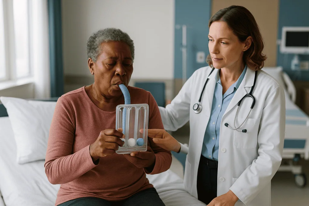
A slow recovery has a recognizable texture. The fever trends downward even if it has not vanished. Breathing at rest feels easier than it did two days ago. You can hold a conversation without stopping. You are eating a little more. The cough is annoying, perhaps productive, but not accompanied by new chest pain. The NHS timeline reflects this shape: fever should have settled within about a week, chest pain and mucus production should be much reduced by 4 weeks, and cough and breathlessness should have eased noticeably by 6 weeks.
Worsening pneumonia moves in the other direction. Fever climbs again after a brief improvement, or never falls. Breathing becomes faster at rest, and speaking a full sentence requires a pause. Confusion or unusual drowsiness appears, particularly in older adults, where it can be the only obvious sign. Lips or fingertips take on a bluish or grayish tinge. Sharp chest pain on breathing in suggests inflammation of the lung lining, and a sudden increase in foul-smelling or blood-streaked sputum can point to an abscess or a complicated collection.
Doctors also watch numbers that patients may not notice. A resting heart rate that stays high, blood pressure that drifts down, oxygen saturation that dips below the patient’s earlier readings, or urine output that falls all point toward the body losing ground. Cleveland Clinic lists rapid breathing, low oxygen, and confusion among the features that can signal severe disease.
None of these signs alone proves failure, but any of them appearing after treatment has started is a reason to be re-examined promptly rather than to wait out the course.
What doctors check first: the bedside reassessment
Before ordering anything, a good clinician goes back to the beginning. The reassessment is partly detective work and partly a check on the original diagnosis.
The history is retaken with new questions. Has the fever pattern changed? Is the sputum different in color, volume, or smell? Any recent travel, contact with birds, exposure to hot tubs or air-conditioning systems (a classic route for Legionella), or time in a healthcare facility? Has the person recently choked on food or drink, which raises the possibility of aspiration pneumonia, where stomach or mouth contents have been inhaled? Have all doses actually been taken, and has anything been vomited? Is there a history of smoking, previous cancer, or a weakened immune system?
The examination focuses on the chest and the whole body. Listening with a stethoscope can reveal an area where breath sounds have gone quiet, which may indicate fluid collecting outside the lung. Tapping on the chest wall produces a dull note over fluid. The clinician checks for swollen calves, since a clot in the leg can travel to the lung and mimic infection. They look at the heart rate, breathing rate, blood pressure, oxygen saturation, and temperature together rather than in isolation.
Severity is scored. Several validated tools combine age, confusion, breathing rate, blood pressure, and blood test results to estimate how ill someone is and whether hospital care is safer. The exact tool varies by country and hospital; the purpose is the same.
Finally, the original chest X-ray is reviewed with fresh eyes. It is not unusual for a subtle fluid collection, a small mass, or a shadow in an unexpected position to look different once the antibiotic has not worked. That review often decides which test comes next.
Imaging: what a repeat X-ray, CT scan, or ultrasound can reveal
The chest X-ray is the workhorse of pneumonia care, but it has limits, and those limits become important when treatment stalls.
A repeat X-ray is usually the first step. Doctors compare it to the original, looking not for complete clearance, which would be unrealistic within days, but for direction of travel. Is the shadow spreading? Has a new area appeared on the other side? Is there a fluid line at the base of the lung suggesting a pleural effusion, the medical term for fluid between the lung and the chest wall? Has a cavity formed inside the shadow, which can indicate an abscess or a less common infection?
A CT scan of the chest gives far more detail. It can distinguish a simple effusion from a thick, partitioned empyema. It can show a mass or an inhaled object obstructing an airway. It can reveal a pattern of inflammation that looks more like an autoimmune or drug-related lung condition than an infection. When combined with contrast dye, a CT can detect blood clots in the lung arteries, an important mimic. Mayo Clinic lists CT among the tests used when pneumonia is not clearing as expected.
Bedside ultrasound has become a valuable third option. It is quick, involves no radiation, and is particularly good at locating pockets of fluid so that a needle or drain can be placed safely. Many clinicians now use it routinely to guide sampling of an effusion.
Imaging rarely delivers a single answer on its own. What it does is narrow the field: a fluid collection sends the team toward drainage and sampling; a blocked airway sends them toward bronchoscopy; a picture that does not look infectious sends them toward the mimics. Each path has its own next step.
Cultures, viral tests, and blood markers: finding the actual organism
Once imaging has framed the problem, the search for the organism intensifies. This matters because the first antibiotic was a guess, and a second guess is weaker than a targeted choice.
Sputum culture asks the patient to cough deeply into a sterile container so the sample can be grown in the laboratory and tested against a panel of antibiotics. Results take days, and the sample is often contaminated by mouth bacteria, so a negative culture does not rule out bacterial infection. Blood cultures look for bacteria that have spread into the bloodstream; they are more reliable when positive but are often negative even in genuine pneumonia.
Urine antigen tests detect fragments of two important organisms, Streptococcus pneumoniae and Legionella, and can return results within hours. Nasal or throat swabs run through molecular panels can identify influenza, respiratory syncytial virus, the COVID-19 virus, and atypical bacteria such as Mycoplasma. A positive viral result reframes the whole situation.
Blood tests track the body’s response. The white cell count and inflammatory markers such as C-reactive protein tend to fall as infection is controlled; a level that stays high or climbs supports the idea that something is still active. Kidney and liver tests check whether the illness or the medicines are causing collateral damage. In people with a weakened immune system, additional tests for fungi and less common organisms may be added.
If a pleural effusion is present, fluid drawn from it is one of the most informative samples of all. Its acidity, sugar level, protein content, and cell count help distinguish a simple reactive effusion from an empyema that needs drainage. The Johns Hopkins overview of pneumonia describes these sampling approaches as standard tools when the diagnosis or response is uncertain.
Complications that stop pneumonia from clearing: effusion, empyema, and abscess
Three complications account for a large proportion of true antibiotic failure, and they share a theme: infection has found a place the drug cannot reach.
A parapneumonic effusion is fluid that collects in the pleural space next to an infected lung. In its early, thin form it often resolves as the pneumonia does. If bacteria invade the fluid, it thickens, becomes acidic, and eventually forms an empyema: a collection of pus that walls itself off with fibrous partitions. Antibiotics circulating in the blood penetrate this pocket poorly. The standard approach is to drain it, typically with a chest tube placed under imaging guidance, sometimes with medicines instilled through the tube to break up the partitions, and occasionally with surgery if the collection is too organized to drain. Antibiotics continue alongside, but drainage is what changes the trajectory.
A lung abscess is a cavity of pus inside the lung tissue itself, often following aspiration or infection with certain organisms. Abscesses tend to produce foul-smelling sputum and a prolonged fever. Many respond to a longer course of appropriate antibiotics because they can drain through the airways; some need a drainage procedure.
Less commonly, infection spreads beyond the chest. Bacteria in the bloodstream can seed the heart valves or joints. Sepsis, the body’s overwhelming response to infection, can cause blood pressure to fall and organs to fail, and it requires urgent hospital care. Mayo Clinic lists effusion, abscess, bacteremia, and respiratory failure among the complications that make pneumonia more dangerous.
The practical lesson is that persistent fever after several days on a sensible antibiotic should prompt imaging aimed at these collections. Changing the drug without looking for a pocket often leads to a second disappointment.
When it is not pneumonia at all: the mimics doctors have to rule out
An X-ray shadow with fever and breathlessness is strongly suggestive of pneumonia, but several other conditions produce the same trio and do not respond to any antibiotic.
Heart failure causes fluid to back up into the lungs, producing shadows, breathlessness, and sometimes a low-grade fever. It is more likely in older adults and in people with known heart disease. A blood test for a heart-strain hormone and an echocardiogram usually clarify the picture.
Pulmonary embolism, a blood clot lodged in a lung artery, can cause sharp chest pain, breathlessness, cough, and even a shadow where lung tissue has been starved of blood. It is a serious mimic, and doctors think about it especially in people who have been immobile, had recent surgery, or have swelling in one leg.
Lung cancer can present as a pneumonia that clears partially and then returns in the same spot, because a tumor is blocking the airway that segment drains through. Repeated infection in one location, particularly in a current or former smoker, is a reason to look more closely with CT and sometimes bronchoscopy.
Inflammatory and drug-related lung disease includes a range of conditions where the immune system inflames the lung without any infection. Some are triggered by medicines, some by inhaled substances, some by autoimmune conditions. They often improve with anti-inflammatory treatment rather than antibiotics, which is why identifying them matters.
Tuberculosis and fungal infections can look like an ordinary pneumonia that simply refuses to clear. They are considered more often in people who have lived in or traveled to areas where these infections are common, or who have weakened immunity.
Bronchoscopy, in which a thin flexible camera is passed into the airways to look and to collect samples, is the test that most often settles these questions when imaging and blood tests have not.
Who is usually escalated quickly, and who is usually asked to wait
Not every stalled pneumonia gets the full battery of tests on day three. Doctors weigh how unwell the person is against how likely a serious explanation is, and that judgment determines the pace.
Rapid escalation is the norm for anyone whose vital signs are deteriorating: falling oxygen saturation, rising breathing rate, low blood pressure, new confusion, or a fever that climbs after briefly settling. It is also the norm for people whose immune systems are compromised by chemotherapy, transplant medicines, uncontrolled HIV, or long-term steroid use, because unusual organisms are more likely and the margin for delay is smaller. Older adults, people with chronic lung disease such as COPD, and those who developed pneumonia in a hospital or care facility also tend to be reassessed early, since the CDC identifies these groups as being at higher risk of severe outcomes.
Watchful waiting, with a clear safety net, is more common in a younger, otherwise healthy adult whose fever is trending down, whose oxygen is normal, and whose main complaint is a stubborn cough and tiredness. In that situation, changing the antibiotic on day three would more likely add side effects than benefit. The plan is usually to complete the course, arrange a follow-up review, and return sooner if specific warning signs appear.
Between these two groups sit many people whose picture is mixed, and this is where clinical judgment, repeat examination over a day or two, and a single well-chosen test often do more than a broad sweep of investigations. A pregnant patient, someone with kidney disease, or a person taking medicines that interact with common antibiotic classes may also have their reassessment shaped by those specific constraints.
Whichever path applies, the decision belongs to the treating team, who can see the whole picture. Patients help most by reporting changes honestly and promptly.
How long is too long to have pneumonia? A typical recovery timeline
People often measure their illness against a friend’s and conclude something has gone wrong. A published timeline is a fairer yardstick. The NHS sets out the following expectations for an uncomplicated community pneumonia in an otherwise healthy adult.
| Time since starting treatment | What is typically expected (NHS) | What would prompt a check |
|---|---|---|
| First few days | Starting to feel better; fever beginning to fall | No change or worsening by about 72 hours |
| 1 week | Fever should have resolved | Persistent or returning fever |
| 4 weeks | Chest pain and mucus production much reduced | Ongoing pleuritic pain or increasing sputum |
| 6 weeks | Cough and breathlessness substantially reduced; follow-up X-ray for some groups | Breathlessness not improving; abnormal follow-up imaging |
| 3 months | Most symptoms resolved; tiredness may persist | New or recurring chest symptoms |
| 6 months | Most people feel back to normal | Any symptom that has plateaued or worsened |
Two points about this table deserve emphasis. First, the fatigue column is not a footnote. Mayo Clinic notes that tiredness can last a month or more, and many people find it the most disruptive part of recovery. Second, the X-ray lags behind the patient. A shadow that is still present at 4 weeks in someone who feels well is usually a slow-clearing picture, not a failed one, which is why follow-up imaging is timed around 6 weeks rather than earlier.
So how long is too long? Fever beyond the first week, breathlessness that is not improving by 6 weeks, or any symptom that reverses direction is the practical threshold for asking to be reassessed rather than waiting.
What the following days and weeks usually look like after a change of plan
Once the team has found a reason, the path forward depends on what that reason is, but a few patterns are common enough to describe.
If the organism was simply not covered, the antibiotic class is usually switched or broadened while culture results are awaited, then narrowed once the laboratory reports what grew and what it is sensitive to. Improvement, when the new choice is right, tends to follow the same rhythm as a first-time response: the fever turns first, breathing follows, and the cough trails behind.
If a viral infection was identified, antibiotics may be stopped, and care shifts to oxygen support, fluids, rest, and, for some viruses, specific antiviral treatment decided by the clinician. Recovery from viral pneumonia can be just as slow as from bacterial, and the plateau of fatigue is common.
If an effusion or empyema was drained, many people notice easier breathing within a day or two as the lung re-expands. The chest tube stays until drainage slows and imaging confirms the space has cleared, which can take several days. Antibiotics continue for longer than in an uncomplicated case, with the duration set by the team based on response.
If a mimic was found, the pneumonia label is set aside and treatment for the actual condition begins. That can be a relief and a shock at the same time, and it is reasonable to ask for a clear explanation of what changed and why.
Hospitalized patients are typically discharged once their fever has settled, oxygen is stable on room air or a plan for home oxygen exists, and they can eat, drink, and move safely. Follow-up within a few weeks, sometimes with repeat imaging around 6 weeks as the NHS suggests, closes the loop. Gentle, graded activity, adequate fluids, and not smoking support recovery; there is no evidence that pushing through exhaustion speeds it up.
What people often get wrong about pneumonia that will not clear
“The antibiotic must be too weak; I need a stronger one.” Antibiotics are not arranged on a ladder of strength. They cover different organisms. A broader drug is not automatically better, carries more side effects, and does nothing for a virus, an abscess, or a blood clot. Finding the reason for failure beats reaching for a bigger hammer.
“I still have a cough, so the infection is still there.” The NHS timeline expects cough to persist for up to 6 weeks in a normal recovery. A cough that is gradually improving in someone without fever is the airways clearing, not the infection persisting.
“My X-ray hasn’t cleared, so treatment failed.” Imaging lags weeks behind symptoms. Clinicians judge failure by the patient first and the picture second.
“I felt better, so I stopped the tablets.” Stopping early can allow surviving bacteria to rebound, and the recurrence can be harder to treat. Any decision about shortening or extending a course belongs to the prescriber; the MedlinePlus guidance is to complete the course as directed.
“Walking pneumonia is not real pneumonia.” The informal term describes a milder illness, often caused by Mycoplasma, in which people stay on their feet. It is genuine pneumonia, and it is one of the cases where a common antibiotic class may not be effective, which is why it sometimes appears in “antibiotics didn’t work” stories.
“Pneumonia can’t come back once treated.” It can, especially if the underlying reason, such as aspiration, a blocked airway, or weakened immunity, has not been addressed. Two episodes in the same part of the lung within a short period is a reason for further investigation rather than a second identical course.
“Tiredness this long means something is wrong.” Fatigue lasting a month or more is documented as typical by Mayo Clinic. It warrants a review if it plateaus or worsens, not alarm on its own.
Questions to ask your care team when pneumonia is not improving
A stalled recovery is disorienting, and appointments can feel rushed. Having a short list of questions helps you leave with a plan you understand rather than a vague instruction to “keep going.”
- Which pattern am I in: getting worse, not changing, or slowly improving? What specifically tells you that?
- Do you know what organism is causing this? If not, which tests will help, and when will the results be back?
- Could this be viral, and if so, does that change what I should be taking?
- Has my chest X-ray been compared with the first one? Is there any sign of fluid around the lung or a blocked airway?
- Do I need a CT scan or an ultrasound, and what would you be looking for?
- Is there anything about my other conditions or medicines that could be slowing recovery?
- If the plan is to wait, what exactly should make me call before my next appointment, and whom do I call after hours?
- When should my follow-up review be, and will I need a repeat X-ray?
- How long should I expect the cough and tiredness to last in my situation?
- Are there activities I should avoid, and when is it reasonable to return to work or exercise?
- Is there anything in my history, such as smoking or previous chest infections, that means I should be checked for something other than infection?
Write down the answers, or ask a companion to. If the explanation does not make sense, say so; a good clinician would rather repeat it than have you leave uncertain. Bring your thermometer readings and, if you have a home oxygen monitor, a log of the numbers, because trends over several days are more informative than a single reading in the clinic.
When to call your doctor: red-flag signs during pneumonia treatment
Most people with pneumonia recover at home with follow-up. Some develop signs that mean the plan needs to change today, not at the next scheduled visit. Contact your care team or seek urgent care the same day if you notice any of the following after starting treatment:
- Fever that returns after settling, or a fever that has not started to fall within about 72 hours of the first dose, particularly if you feel worse rather than the same.
- Breathing that is faster or harder at rest than it was two days ago, or needing to pause for breath mid-sentence.
- Oxygen saturation readings on a home monitor that are falling from your earlier readings, or that your clinician has told you are too low.
- New confusion, unusual drowsiness, or difficulty waking, especially in an older adult.
- Sharp chest pain that worsens on breathing in, or pain on one side that is new.
- Coughing up blood, or sputum that suddenly becomes much more abundant or foul-smelling.
- Being unable to keep fluids or medicines down because of vomiting.
- Passing very little urine, feeling faint on standing, or a racing heartbeat at rest.
Call emergency services immediately, rather than waiting for a callback, if lips or fingertips turn blue or gray, if breathing becomes severely labored, if someone collapses or cannot be roused, or if chest pain is severe and sudden. Cleveland Clinic and Mayo Clinic both list difficulty breathing, bluish skin, chest pain, and confusion as emergency features.
Two quieter situations also deserve a call, though not an ambulance: a cough or breathlessness that has stopped improving by around 6 weeks, and any symptom that had gone away and has come back. Both are reasons for a review and possibly repeat imaging.
Every decision about tests, changing or stopping a medicine, drainage, or admission rests with the clinicians who can examine you. Your role is to notice the direction of travel and report it early.
Frequently asked questions
How long is too long to have pneumonia?
Fever lasting beyond the first week of treatment, or breathlessness that is not improving by around 6 weeks, is longer than the NHS recovery timeline expects and warrants a review. Cough and tiredness commonly persist for 6 weeks to 3 months in an otherwise normal recovery. The key signal is direction: symptoms that plateau or reverse should prompt a check regardless of the calendar.
Why would antibiotics not work on pneumonia?
The most common reasons are that the infection is viral or caused by a bacterium the chosen drug does not cover, that infection has collected in a pocket such as an empyema or abscess where blood-borne drugs cannot reach, that the immune system is too weakened to finish the job, or that the illness is not pneumonia at all. Missed or vomited doses can also mimic failure.
What are the signs of worsening pneumonia?
Fever that returns or climbs, faster or more labored breathing at rest, falling oxygen readings, new confusion or drowsiness, sharp chest pain on breathing in, coughing up blood, or a bluish tinge to lips or fingertips all suggest worsening rather than slow recovery. Any of these after treatment has started is a reason to be re-examined the same day.
How long after antibiotics do you get better from pneumonia?
Most people start to feel better within a few days of an appropriate antibiotic, according to the NHS, with fever usually settling within the first week. Improvement in cough and breathlessness is slower, typically over 4 to 6 weeks, and tiredness can last a month or more. The chest X-ray lags furthest behind and may take weeks to clear.
Can pneumonia come back after antibiotics?
Yes. Recurrence can happen if the course was stopped early, if the organism was not fully covered, or if an underlying problem such as aspiration, a blocked airway, or weakened immunity has not been addressed. Two episodes in the same area of the lung within a short time usually prompts further investigation, often with CT and sometimes bronchoscopy.
What tests are done when pneumonia does not clear?
Doctors typically repeat the chest X-ray and compare it with the original, then consider a CT scan or ultrasound to look for fluid collections, abscesses, or blocked airways. Sputum and blood cultures, urine antigen tests, and viral swabs try to identify the organism. Blood markers track inflammation. If fluid is present around the lung, sampling it is often the most informative step.
What is the pneumonia recovery timeline after antibiotics for older adults?
Older adults often recover more slowly than the standard NHS timeline suggests, and confusion or reduced appetite may be more prominent than fever. Fatigue and breathlessness can take longer to settle, and follow-up imaging is more commonly arranged. Because older adults are at higher risk of complications, clinicians tend to reassess them earlier if progress stalls.
Does a chest X-ray that has not cleared mean treatment failed?
Not on its own. X-ray shadows typically lag weeks behind symptom improvement, which is why the NHS times follow-up imaging around 6 weeks for those who need it. Clinicians judge response primarily by fever, breathing, and oxygen levels. A shadow that is spreading or a new fluid line, however, would be taken seriously even if the person feels similar.
What is the difference between walking pneumonia and pneumonia that will not clear?
Walking pneumonia is an informal term for a milder pneumonia, often caused by Mycoplasma, in which people stay active. It is genuine pneumonia and may not respond to some common antibiotic classes, so it sometimes appears in stalled-recovery stories. Pneumonia that will not clear is any pneumonia, mild or severe, that fails to improve on the expected timeline.
Should I ask for a different antibiotic if I am not improving?
Ask to be reassessed rather than for a specific drug. Changing antibiotics without finding the reason for failure often leads to a second disappointment, because a fluid collection, a virus, or a mimicking condition will not respond to any antibiotic. Your clinician will decide whether to switch, broaden, stop, or continue based on examination, imaging, and test results.
References
This article is for general information only and is not a substitute for professional medical advice. Please consult a qualified doctor about your individual situation.

More from the Blog

Do Antibiotics Help Acute Bronchitis? Why Most Cases Are Viral and What Treatment Looks Like
Antibiotics usually do not help acute bronchitis, because the large majority of cases are caused by viruses that antibiotics cannot touch. Pooled trials suggest…

Which Chest Conditions Suit Robotic Thoracic Surgery? Lung Nodules, Mediastinal Tumors and More
Robotic thoracic surgery is most often considered for small lung nodules and early-stage lung cancer needing a wedge resection, segmentectomy or lobectomy, for tumors…
Does Acute Bronchitis Need a Chest X-Ray? How Doctors Rule Out Pneumonia
Most people with acute bronchitis do not need a chest X-ray. Doctors usually diagnose it from the story of the illness and a physical…

Questions to Ask Your Cystic Fibrosis Team: Lung Function, Infections and Nutrition Goals
Useful questions to ask a cystic fibrosis team fall into three groups: lung function (what my FEV1 is, how it has changed, what a…

How Long Does Lung Abscess Treatment Take? Why Antibiotic Courses Run for Weeks
Lung abscess treatment usually takes several weeks, not days. Published reviews describe antibiotic courses of roughly three to six weeks, sometimes extended to eight…

What Pulmonary Rehabilitation Offers People With Bronchiectasis: Realistic Expectations
Pulmonary rehabilitation for bronchiectasis is a supervised program of exercise training, airway clearance coaching, and education that usually runs for several weeks. Evidence from…