

Which Ear Reconstruction Framework Is Right: Rib Cartilage or a Porous Implant?
Key Takeaways
- Rib cartilage is the patient's own living tissue and heals after minor injury, while porous polyethylene cannot heal and must be watched for exposure over a lifetime.
- Johns Hopkins places typical rib cartilage reconstruction at roughly ages 6 to 10 because the ribs must grow enough cartilage, whereas a porous implant may be considered from around age 3.
- Rib cartilage reconstruction usually needs several staged operations months apart; porous implant reconstruction is often completed in one or two.
- Neither framework changes hearing, and the CDC and Johns Hopkins describe hearing assessment as the first priority in microtia care.
- The unique risk of rib cartilage is the chest donor site, including scar, contour change and rarely an air leak around the lung; the unique risk of an implant is skin breakdown that exposes the plastic.
- Comparative evidence is almost entirely single-center case series, so any complication percentage quoted in a consultation reflects that team's experience rather than a universal figure.
Neither framework wins outright in ear reconstruction rib cartilage vs implant decisions. Rib cartilage is the patient's own living tissue, tolerates injury well and is the longer-established option, but requires a chest incision and usually more stages. A porous polyethylene implant avoids the chest, allows earlier surgery and fewer stages, yet carries a lifelong small risk of exposure. The right choice depends on age, anatomy, priorities and the surgical team's experience.
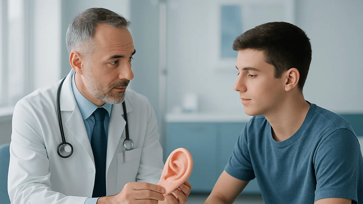
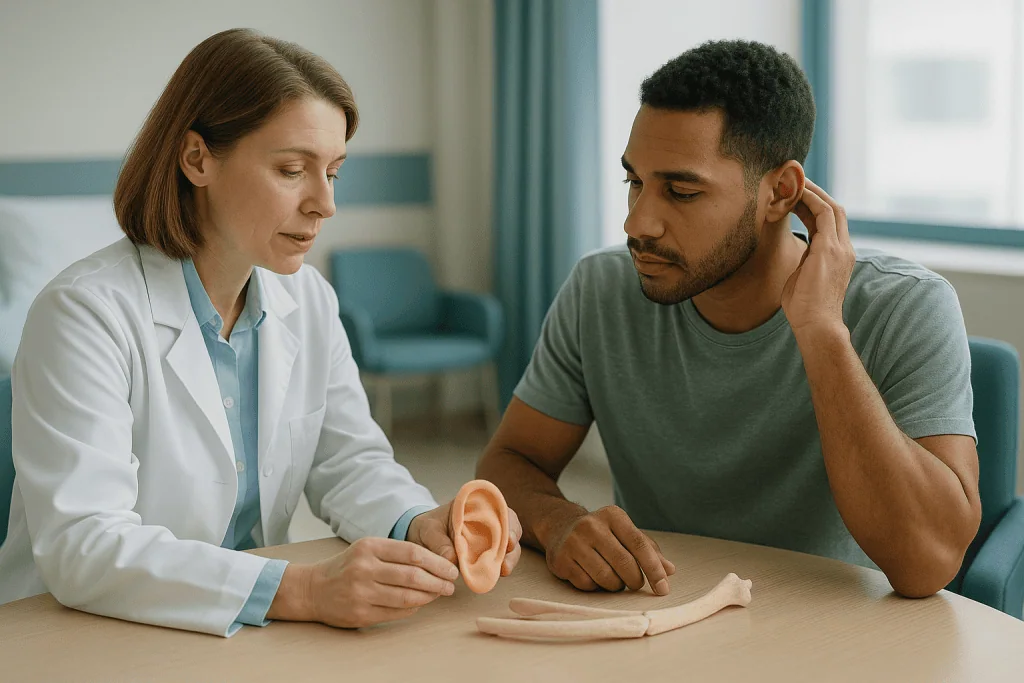
The two objects on the desk look nothing alike. One is a pale, slightly translucent curve of cartilage, carved by hand and photographed beside a ruler. The other is a smooth white scaffold with a matte, sponge-like surface, machined to the shape of an ear. The parents across the table have a seven-year-old in the waiting room who has started keeping his hair long on one side, and they have come with a single question: which of these should become his ear?
That question, ear reconstruction rib cartilage vs implant, sits at the center of most microtia consultations and quite a few conversations after trauma or skin cancer surgery. It is rarely answered in one visit, and it should not be. Both approaches can produce a convincing ear. Both carry trade-offs that only make sense once you understand what the surgeon is actually building underneath the skin.
This explainer walks through what each framework is, how the operations differ, what the recovery feels like, and what the evidence does and does not tell us. The decision itself belongs with the family and the treating team.
Ear reconstruction rib cartilage vs implant: what the two frameworks actually are
Every reconstructed ear has two parts: a framework that provides the shape, and a covering of living skin that hides it. The framework is the part under debate. The skin, whether stretched local skin, a thin flap of tissue from the scalp, or a skin graft, is broadly similar whichever framework sits beneath it.
Rib cartilage reconstruction uses autologous tissue, meaning tissue taken from the patient’s own body. A surgeon removes segments of cartilage from where several lower ribs meet the breastbone, then carves and stitches them into a three-dimensional ear shape. Because it is living tissue with a blood supply that re-establishes over time, it heals like the rest of the body and is recognized by the immune system as self.
Porous polyethylene is an alloplastic material, meaning a manufactured substance implanted into the body. It is a medical-grade plastic with tiny interconnected pores. Those pores matter: blood vessels and fibrous tissue grow into them, which anchors the implant and helps it resist infection compared with a solid, non-porous material. The implant arrives pre-shaped in a range of sizes and is usually covered with a thin layer of tissue lifted from beneath the scalp before skin is placed over it.
The Centers for Disease Control and Prevention estimates that microtia, an ear that is small and underdeveloped from birth, affects roughly 1 in every 2,000 to 10,000 babies in the United States. Most of the comparative experience with these two frameworks comes from that population, usually children. Johns Hopkins Medicine describes both approaches as accepted options, and that neutrality reflects the evidence: published comparisons are mostly single-center case series rather than randomized trials, so no guideline declares one superior.
How rib cartilage ear reconstruction works, step by step
The operation begins at the chest, not the head. Through an incision a few centimeters long over the lower ribs, usually on the side opposite the affected ear so the natural curve of the cartilage matches, the surgeon harvests pieces of costal cartilage. Costal cartilage is the flexible tissue that joins the bony ribs to the breastbone. The bony ribs themselves are left in place.
Carving follows, and it is genuinely sculptural work. A larger block becomes the base plate. A thinner strip is bent to form the helix, the outer rim of the ear. Smaller fragments build the inner folds. Pieces are joined with fine wire or sutures, and the surgeon constantly compares the result against a template traced from the other ear. This stage can take several hours in a single operation.
The framework then goes into a pocket created under the skin at the ear site. Suction drains often sit under the skin for a few days so that the covering conforms tightly to every ridge and valley of the carving. Without that close contact, detail is lost.
Later stages add what a flat framework cannot: the ear is lifted away from the head with a wedge of cartilage or other tissue behind it, and the gap is covered with a skin graft. Some surgeons also reposition the earlobe and deepen the entrance to the ear canal for realism. Johns Hopkins notes that rib cartilage reconstruction commonly involves multiple operations, spaced months apart to let each stage heal.
Once healed, the cartilage is living tissue. Minor bumps and scrapes tend to heal the way skin over any cartilage does, which is one of the reasons the technique has remained the reference standard for decades.
How a porous polyethylene ear implant reconstruction is done
There is no chest incision. That single difference shapes almost everything else about this approach.
The implant arrives as two pieces, a base and a helical rim, or as a single unit depending on the design. The surgeon selects a size matched to the opposite ear, warms and adjusts the pieces slightly, and joins them. Because the shape is manufactured, the surgeon spends less time carving and more time on the soft tissue that will conceal the implant.
That soft tissue is the critical step. A thin, well-vascularized sheet of tissue called the temporoparietal fascia is lifted from beneath the scalp above the ear, still attached to its blood supply, and draped over the implant like a living blanket. Vascularized means the tissue keeps its own circulation. The fascia gives the plastic a layer of living cells to grow into and provides the blood flow that helps the skin above it survive. Skin, either local or grafted from elsewhere, then covers the fascia.
Because the implant is already three-dimensional and does not need to be lifted off the head in a separate operation, the projection of the ear is created at the first stage. Johns Hopkins describes porous implant reconstruction as typically requiring fewer operations than rib cartilage, often one or two.
Timing also differs. The implant does not depend on how much cartilage a child’s ribs have grown, so Johns Hopkins notes it may be considered from around age 3, earlier than rib cartilage is usually offered. That appeals to families who want reconstruction completed before a child starts school.
The trade-off is permanence of a different kind. A plastic framework cannot heal itself. If the skin over it breaks down, the implant beneath is exposed and needs surgical attention, sometimes years later.
Who each option is usually for, and who is usually asked to wait
Age and rib development shape the rib cartilage conversation. The ribs must have grown enough cartilage to carve a full-sized ear, which is why Johns Hopkins places typical rib cartilage reconstruction at roughly ages 6 to 10. Operating earlier risks a framework that is too small or too fragile, and a child whose ribs are not yet ready is usually asked to wait rather than compromise.

Chest history matters too. Previous chest surgery, a significant chest wall deformity, or a condition affecting cartilage quality can make harvesting less attractive. In those situations a porous implant, or a prosthetic ear, may be discussed instead.
Scalp and skin quality drive the implant conversation. A porous implant depends on a healthy temporoparietal fascia flap and thin, mobile skin to cover it. Extensive scarring from previous surgery or burns, prior radiation to the area, or very thin tissue can all raise the risk of exposure. Surgeons may steer these patients toward rib cartilage, which tolerates a less-than-perfect cover more forgivingly.
Adults facing reconstruction after trauma or cancer surgery are assessed differently. Their ribs are mature, so rib cartilage is technically available, but adult cartilage is stiffer and more likely to be calcified, and adults may weigh a chest scar and a longer recovery against their work and family commitments.
Children with syndromic microtia deserve a wider view. Craniofacial microsomia, a condition in which one side of the face develops smaller, often includes microtia alongside jaw and eye-socket differences, according to MedlinePlus Genetics. Reconstruction may need sequencing around jaw surgery, and the team will often ask families to wait until the overall plan is clear.
Anyone with an active scalp infection, poorly controlled diabetes, or who smokes is typically asked to address those first. Healing of a skin graft over a framework is unforgiving of poor blood flow.
Ear reconstruction rib cartilage vs implant: a side-by-side comparison
Laid out side by side, the trade-offs become clearer. The table below summarizes general features described by Johns Hopkins Medicine and the wider surgical literature; individual surgeons and patients will vary.
| Feature | Rib cartilage framework | Porous polyethylene implant |
|---|---|---|
| Material | Patient’s own living cartilage | Manufactured porous plastic |
| Typical starting age (microtia) | Roughly 6 to 10 years, once ribs have grown | May be considered from around 3 years |
| Donor site | Chest incision; possible chest wall contour change | None |
| Number of operations | Usually multiple stages, months apart | Often one or two |
| Response to injury | Heals like living tissue | Cannot heal; exposure needs surgery |
| Shape control | Depends on surgeon’s carving skill | Pre-formed; relies on soft-tissue cover |
| Long-term track record | Decades of published follow-up | Shorter follow-up history |
| Reversibility | Difficult; cartilage integrates | Implant can be removed if needed |
Two points deserve emphasis. First, the “number of operations” row is often the one families remember, but fewer operations does not automatically mean fewer visits or less overall risk. Second, the long-term track record row is not a verdict against implants. It simply reflects that rib cartilage has been used for far longer, so surgeons can describe how a cartilage ear looks fifty years on with more confidence than they can for any manufactured framework.
What the table cannot show is the surgeon’s experience with each method. Published outcomes for both frameworks come from surgeons who perform that particular technique often, and that consistency is itself part of the evidence.
What is an ear implant called? Sorting out the names you'll hear
The phrase “ear implant” causes real confusion, because it describes at least three unrelated things.
The framework discussed here is usually called a porous polyethylene ear implant, an alloplastic auricular framework, or simply an auricular implant. Auricular refers to the auricle, the visible outer ear. You may also hear a brand name in clinic; this article uses only the generic material name.
A cochlear implant is entirely different. It is an electronic hearing device with an internal receiver placed under the skin behind the ear and an electrode threaded into the cochlea, the snail-shaped organ of the inner ear. It treats certain kinds of severe hearing loss and has nothing to do with the shape of the outer ear.
A bone-anchored or bone-conduction hearing implant is different again. A small titanium fixture is placed in the skull bone behind the ear, and an external processor sends sound through the bone to the inner ear. This is commonly discussed for people with microtia, because many also have aural atresia, an ear canal that is narrow or absent, so sound cannot reach the eardrum in the usual way. Johns Hopkins notes that hearing assessment and management are part of microtia care alongside reconstruction of the outer ear.
Then there are osseointegrated prosthetic anchors. These are also small titanium fixtures in the skull bone, but they hold a removable silicone prosthetic ear in place with magnets or clips rather than transmitting sound.
Why does this matter? Because a family reading online about “ear implants” may find survival statistics for cochlear devices, cost debates about hearing aids, or discussion of dental implants that share the word but nothing else. When someone on the care team says implant, it is reasonable to ask which of these four they mean.
Why hearing is assessed before any ear reconstruction
Parents often arrive focused on appearance and leave thinking about sound. That reorientation is intentional.
The CDC describes microtia as a spectrum, from a slightly small but fully formed ear to anotia, where no external ear is present. In the more significant forms the ear canal is often narrow or absent, and the small bones of the middle ear may be malformed. The result is conductive hearing loss, meaning sound is blocked from reaching a working inner ear rather than the inner ear itself failing. In children with one affected ear the other typically hears normally, but even one-sided loss can affect where sounds come from and how well speech is heard in noisy classrooms.
Hearing tests therefore come first, often in infancy, long before any framework is chosen. Options may include a bone-conduction hearing device worn on a soft headband in early childhood, a surgically anchored version later, or in selected cases an operation to open the ear canal, called atresia repair.
The sequence matters for reconstruction. A bone-anchored fixture placed carelessly can sit exactly where a surgeon later needs healthy skin and fascia for the framework. Conversely, a rib cartilage or implant framework placed first can complicate canal surgery. Coordinated planning between the ear, nose and throat team and the plastic surgery team avoids one operation compromising the other. Johns Hopkins emphasizes this combined approach.
There is also a practical point about expectation. Neither rib cartilage nor a porous implant improves hearing. Both rebuild the visible ear only. Families who understand that distinction early tend to make calmer decisions about sequencing, and they are better placed to ask how each framework choice fits with the hearing plan rather than treating the two as separate problems.
Microtia surgery options beyond these two: prosthetics and choosing not to operate
The question “which framework” assumes a framework is wanted. Two other paths deserve equal airtime.
A prosthetic ear is a removable silicone ear, hand-tinted to match skin, held on either with medical adhesive or by clips and magnets attached to small titanium fixtures in the skull bone. The fixtures require a minor operation; the prosthesis itself involves no reconstruction of living tissue. Detail can be exquisite because an artist paints it rather than a surgeon carving it under time pressure. The trade-offs are daily handling, periodic replacement as silicone discolors and skin tone changes with age, care of the skin around the fixtures, and the psychological reality of an ear that comes off at night. For some adults after cancer surgery, especially where radiation has damaged local tissue, a prosthesis is the option with the least surgical risk.
Choosing not to reconstruct is a legitimate decision, not a failure to decide. Many people with microtia grow up, style their hair as they like, and never pursue surgery. Others wait until adulthood and choose for themselves. Surgeons who work in this field generally support letting an older child have a genuine voice in the decision, because the operations ask a lot of the patient and the result is theirs to live with.
Some families also consider staged thinking: hearing management now, a decision about the outer ear later. Nothing about a bone-conduction device forces the framework question, provided the teams plan skin and fixture placement together.
When someone asks what the alternatives to implants are, the honest answer is: living cartilage, a prosthesis, or no surgery at all. Each is used by real people who are content with their choice, and none of them is a compromise by definition.
Risks compared: the chest, the skin, infection and exposure
Every operation carries the general risks of anesthesia, bleeding and infection. The frameworks differ in their specific risks, and being candid about them is more useful than reassurance.
Rib cartilage carries the risks of a chest donor site. These include pain in the days after surgery, a visible scar, and sometimes a subtle indentation or contour change in the chest wall as the child grows. A rare but serious risk is pneumothorax, a puncture of the lung lining during harvest, which surgeons check for at the end of the operation and manage if it occurs. At the ear, the main early risk is skin breakdown over a carved edge, and the main long-term risk is resorption, where part of the cartilage is gradually absorbed and detail softens. Cartilage can also warp slightly as it heals, altering the shape.
Porous polyethylene avoids the chest entirely. Its defining risk is exposure: the skin and fascia over the implant break down, the plastic becomes visible, and bacteria gain access to a surface that has no immune cells of its own. Early exposure usually follows a problem with the fascia flap or graft. Late exposure, sometimes years afterwards, can follow trauma, pressure from headphones or helmets, or thinning skin. Small exposures may be covered with local tissue; larger ones may require removing the implant. Fracture of the implant rim after a direct blow is also reported.
Both frameworks share risks of infection, poor scar quality, asymmetry with the other ear, and disappointment with the result. For adults, MedlinePlus notes that smoking, diabetes and poor nutrition all slow wound healing, which is why teams address them before surgery.
Published complication rates for each method come from individual centers and vary widely with technique and follow-up length, so any single percentage quoted in a consultation should be understood as that team’s experience rather than a universal figure.
Rib graft ear reconstruction recovery: what the first days and weeks look like
Recovery from rib cartilage reconstruction happens in two places at once, and the chest usually complains louder than the ear.
In hospital, the first day or two revolve around chest discomfort with deep breaths and coughing, managed by the anesthesia and surgical team with a combination of local anesthetic techniques and pain relief chosen for the child’s age and weight. Breathing exercises and getting up to walk are encouraged early, both to keep the lungs clear and to reduce clot risk in older patients. Small suction drains under the ear skin typically stay for a few days so the skin settles onto the carving, and a protective bolster dressing shields the ear from pressure.
Once home, the ear is often bruised and swollen and looks nothing like the final result. Sleeping on the other side or on the back is standard advice. The NHS guidance on ear correction surgery describes wearing a soft protective headband at night for several weeks after ear surgery, and avoiding contact sports and swimming until the surgeon advises, generally a matter of weeks. School or work is usually possible once drains are out and pain is controlled with ordinary measures, typically within one to two weeks, though the chest may ache with vigorous movement for longer.
Porous implant recovery skips the chest entirely, which most families notice immediately. The scalp donor site for the fascia flap feels tight and numb for a time, and the skin graft site, often the groin or the other side of the scalp, needs its own dressing care. The ear itself has projection from the start, so the appearance at the first dressing change is closer to the final shape. Protection from pressure remains critical for months, because the covering tissue is thin while it matures.
For both, swelling settles over months, not weeks, and surgeons often decline to judge the result before six months to a year.
How does a reconstructed ear age over decades?
A child who has this operation at seven may live another eighty years with the result, so the question of aging is not academic.
Rib cartilage has the advantage of history. Surgeons have followed cartilage ears for decades and report that the framework generally keeps its shape, with some softening of fine detail from resorption and occasional slight warping. Living cartilage also grows a little in children, though not as much as the normal ear, so surgeons deliberately carve the framework slightly larger than the opposite ear to allow for growth. Skin over the framework ages like skin elsewhere, sagging and thinning with time, but because the cartilage beneath can tolerate minor injury and heal, the long-term maintenance burden is usually low.
Porous polyethylene does not grow and does not change, which is both its strength and its concern. The shape achieved at surgery is the shape at fifty. The uncertainty lies in the covering tissue. As skin thins with age, sun damage and weight change, the layer between the world and the plastic becomes narrower. Whether that translates into more late exposures over a lifetime is exactly the question longer-term follow-up is still answering; most published series follow patients for years rather than decades. Surgeons who favor implants argue that exposures are manageable and that a lifetime of avoided chest surgery is worth it; surgeons who favor cartilage point to the absence of that worry. Both positions are defensible on current evidence.
Practical habits matter more than framework choice for longevity. Sun protection, avoiding prolonged pressure from headphones, helmets and eyewear on the reconstructed side, and prompt attention to any skin change all help either type of ear last.
Families sometimes ask whether a cartilage ear could later be replaced with an implant, or vice versa. Revision is possible in both directions but is more complex than a first operation, and the team will explain what scarring from the first approach means for the second.
When the defect is not microtia: trauma, dog bites and skin cancer
Not everyone facing this decision was born with the need. Ears sit exposed on the side of the head and are among the most common facial sites for skin cancer, road and sports injuries, and animal bites.
Partial defects change the calculation. If the earlobe or a portion of the rim is missing, surgeons often use local flaps, or a small piece of cartilage taken from the other ear’s concha, the bowl-shaped hollow near the canal, rather than rib cartilage or a full implant. The framework debate applies mainly to total or near-total losses.
Timing after trauma matters. A fresh injury is contaminated and swollen; surgeons generally clean and close what can be saved, then plan reconstruction once tissues have settled over months. Scar tissue from the injury and any earlier repairs reduces the quality and mobility of the skin available for coverage, which tends to push the discussion toward rib cartilage or a prosthesis rather than a porous implant, since the implant relies on healthy fascia.
Skin cancer reconstruction carries an additional factor: radiation. If radiotherapy has been given or may be needed, the tissue’s blood supply is permanently reduced, and both skin grafts over cartilage and fascia flaps over implants heal less reliably. Many teams favor a prosthetic ear in this setting, or delay reconstruction until surveillance for recurrence is complete.
Adults also bring adult cartilage. Costal cartilage stiffens and may calcify with age, making it harder to carve and more prone to fracture during shaping. Surgeons usually assess this with imaging or at the time of harvest, and some adults are advised that an implant or prosthesis is the more predictable route.
The decision, once again, is individual. A twenty-year-old cyclist and a seventy-year-old after cancer surgery may reasonably receive different recommendations for the same-sized defect.
What people often get wrong about ear reconstruction
Myths cluster around this surgery, partly because so much online discussion of “implants” concerns teeth or contraception rather than ears. A few corrections.
“An implant is the modern option and cartilage is old-fashioned.” Both are in current use by experienced surgeons. Rib cartilage is older, and that history is a form of evidence, not a mark against it.
“Fewer operations means less risk.” Fewer stages is a real advantage for many families, but the implant’s risk profile is spread over a lifetime rather than concentrated in the surgical years. Total risk is measured differently, not eliminated.
“The reconstructed ear will hear.” Neither framework affects hearing. Hearing is addressed separately, and the CDC and Johns Hopkins both describe hearing assessment as an integral part of microtia care.
“The chest scar is a minor detail.” For most patients it fades to a thin line, but chest wall contour changes are reported and a small number of people find the scar bothers them more than the ear did. Ask to see the surgeon’s typical donor-site outcomes.
“A porous implant is like a silicone breast implant and will need swapping.” Porous polyethylene is a rigid plastic that tissue grows into; it is not a fluid-filled device and has no planned replacement schedule. It is removed only if exposed or damaged.
“The ear will look perfect immediately.” Swelling and bruising conceal the shape for weeks, and surgeons commonly wait months before judging the result. Photographs of early results mislead in both directions.
“If the child has microtia, surgery is urgent.” The urgent step is hearing. The outer ear can wait for rib growth, for the child’s own opinion, or indefinitely.
“Same operation, same result anywhere.” Outcomes for both frameworks are strongly linked to how often a surgeon performs that specific technique, which is why asking about experience is not rude, it is essential.
Questions to ask your care team before choosing a framework
A good consultation should leave you with fewer myths and more specific answers. These questions tend to surface what matters.
- Which framework do you perform more often, and roughly how many of each do you do in a year?
- May I see photographs of your own results, including donor sites and any complications, at least a year after surgery?
- For my child’s age and rib development, would you advise waiting, and what would we gain by doing so?
- How many operations do you anticipate, and what does each stage involve?
- How does your plan coordinate with the hearing team, and does the order of hearing and reconstructive surgery matter in our case?
- What is your experience managing exposure of a porous implant, and how often has that meant removal?
- What chest wall changes have you seen after cartilage harvest, and how do you minimize them?
- If the first framework fails, what are the realistic options for a second attempt?
- What activities, headwear and sports restrictions apply, and for how long?
- Who do we call, day or night, if something looks wrong in the first weeks?
- Is a prosthetic ear or no surgery a reasonable choice in our situation, and why or why not?
- Would you be willing for us to seek a second opinion, and is there anything specific we should ask?
Write the answers down or ask permission to record the conversation. Consultations about a child’s face are emotionally loaded, and details slip. Bring the child into the discussion in an age-appropriate way; older children who help choose tend to cope better with the demands of recovery.
Finally, notice how the surgeon talks about the framework they use less often. A balanced description of both options, including the weaknesses of the one they favor, is a good sign that the recommendation you receive is tailored rather than habitual.
When to call your doctor after ear reconstruction
Most recoveries are uneventful, but a reconstructed ear depends on thin tissue surviving over a hard framework, and problems caught early are far easier to manage than problems noticed late. Contact the surgical team promptly, at any hour, if you notice any of the following.
- Skin over the ear turning dusky, purple, gray or black, or a patch that looks pale and does not pink up when touched; this can signal compromised blood supply to the covering tissue.
- Any area where the framework, whether cartilage or implant, becomes visible through the skin, even a pinpoint.
- Increasing redness, warmth, swelling or pain at the ear, chest, scalp or skin graft site after the first few days, which MedlinePlus lists among the signs of a surgical wound infection.
- Pus, cloudy fluid or a foul smell from any wound or drain.
- Fever, chills or feeling generally unwell.
- Bleeding that soaks through the dressing or does not stop with gentle pressure.
- A sudden change in the shape or position of the ear, especially after a knock.
- After rib cartilage harvest: new or worsening shortness of breath, sharp chest pain on breathing, or a racing heartbeat, which need emergency assessment for a possible air leak around the lung.
- Calf pain, swelling or shortness of breath in older patients, which can indicate a blood clot.
- Numbness, tingling or severe pain under a dressing that feels too tight.
Call emergency services rather than the clinic for breathing difficulty, chest pain, heavy bleeding or a child who is drowsy, floppy or hard to rouse.
In the longer term, report any new sore, thinning or shiny patch over a porous implant, or any persistent redness on the ear, even years afterwards. Late exposures are often preceded by weeks of subtle warning that a quick clinic review can address. When in doubt, the team would always rather hear from you than not.
Frequently asked questions
What is an ear implant called?
The framework used to rebuild the outer ear is generally called a porous polyethylene ear implant or an alloplastic auricular framework; auricular simply means relating to the visible outer ear. The phrase “ear implant” also covers cochlear implants and bone-conduction hearing implants, which are electronic hearing devices and have nothing to do with the ear’s shape. Ask your team which type they mean.
What are the alternatives to implants for ear reconstruction?
The main alternatives are rib cartilage reconstruction using the patient’s own tissue, a removable silicone prosthetic ear held on with adhesive or small titanium anchors, and choosing not to operate at all. Each is used by many people who are content with the choice. For partial defects, surgeons often use local skin flaps or a small piece of cartilage from the other ear rather than any full framework.
What are the main microtia surgery options for a young child?
Hearing management comes first, often a bone-conduction device on a soft headband in infancy. For the outer ear, options are rib cartilage reconstruction once the ribs have grown, typically around ages 6 to 10 according to Johns Hopkins; porous polyethylene implant reconstruction, which may be considered from around age 3; a prosthetic ear; or waiting so the child can take part in the decision later.
Is rib graft ear reconstruction recovery worse than implant recovery?
It is different rather than simply worse. Rib cartilage recovery includes chest discomfort with deep breathing for the first days and a chest scar, alongside the ear itself, and is repeated over several stages. Implant recovery avoids the chest but includes a scalp donor site and a skin graft site, with strict protection of the thin covering tissue for months. Both need weeks of activity limits per NHS guidance on ear surgery.
Does a porous polyethylene ear implant need to be replaced?
No planned replacement is required. Porous polyethylene is a rigid plastic that fibrous tissue and blood vessels grow into, so it stays in place indefinitely unless a problem arises. It is removed or repaired only if the skin over it breaks down and exposes the material, or if a direct blow fractures the rim. Long-term follow-up beyond a couple of decades is still limited.
Which is better for rhinoplasty, silicone or cartilage, and does the same logic apply to ears?
For the nose, many surgeons prefer the patient’s own cartilage because it resists infection and extrusion better than solid silicone, though implants are used where donor cartilage is limited. The ear debate rhymes with this but is not identical: porous polyethylene is not solid silicone, and its pores allow tissue ingrowth. In both sites, thin skin and prior scarring make manufactured materials riskier.
Will ear reconstruction improve my child's hearing?
No. Both rib cartilage and porous implant frameworks rebuild only the visible outer ear. Hearing loss in microtia usually comes from a narrow or absent ear canal and middle ear differences, and is managed separately with bone-conduction hearing devices or, in selected cases, surgery to open the canal. Teams coordinate the two plans so that one operation does not compromise the other.
Can a reconstructed ear be damaged by sports or headphones?
Yes, particularly in the first year while the covering tissue matures, and for implants throughout life. A direct blow can crack or displace either framework, and prolonged pressure from headphones, helmets or eyewear can thin the skin over a porous implant and lead to exposure. Surgeons typically restrict contact sports for a set period and advise lifelong sensible protection of the reconstructed side.
How long before the reconstructed ear looks like the final result?
Longer than most families expect. Bruising and swelling hide the shape for weeks, and surgeons commonly wait six months to a year before assessing the outcome, because swelling settles and skin grafts mature slowly. Rib cartilage ears also gain projection only at a later stage, so early photographs can look flat. Early appearance is a poor guide to the eventual result in either direction.
Can you switch from rib cartilage to an implant, or the other way, if the first attempt fails?
Revision in either direction is possible but more complex than a first operation. Scar tissue from the earlier surgery reduces the quality and mobility of skin and fascia available for cover, which particularly affects the chances of a porous implant succeeding. Some patients after a failed reconstruction are advised that a prosthetic ear is the most predictable route. The treating team will assess the tissues individually.
References
- CDC: About Anotia/Microtia
- MedlinePlus Genetics: Craniofacial microsomia
- NHS: Ear correction surgery
- MedlinePlus: Surgical wound infection – treatment
This article is for general information only and is not a substitute for professional medical advice. Please consult a qualified doctor about your individual situation.

More from the Blog

Preparing for Face Aesthetics Surgery: Blood Tests, Anesthesia Review and Medicine Changes
Preparing for facial surgery usually means a pre-assessment visit where blood tests check clotting, blood count and kidney function, an anesthesia review covers your…

Supporting Someone After a Neck and Face Lift: Drains, Dressings and the First Days Home
In the first days after a neck and face lift, a helper's main tasks are keeping the head raised, emptying and recording any drains,…

Facial Augmentation With Fat Transfer or Implants: Which Option Suits Which Face?
Facial fat transfer uses a person's own fat, taken by gentle liposuction and injected into the face, to add soft, natural-looking volume; facial implants…

How Surgeons Sequence Combined Face Aesthetics Procedures: One Operation or Several?
Surgeons combine facial procedures in one operation when the areas heal on a similar timetable, the patient is medically well, and total anesthesia time…

How Surgeons Sequence Combined Face Aesthetics Procedures: One Operation or Several?
Surgeons may combine facial procedures such as a facelift, eyelid surgery and fat grafting in one operation when a person is healthy, does not…

Are Facial Implants Right for Your Face? Bone Structure, Skin Thickness and Age Considered
Facial implants are usually considered for adults whose bones have finished growing, who have a specific structural gap such as a recessed chin or…