

Which Size of Fibroid Is Dangerous? Size, Symptoms and When Treatment Is Advised
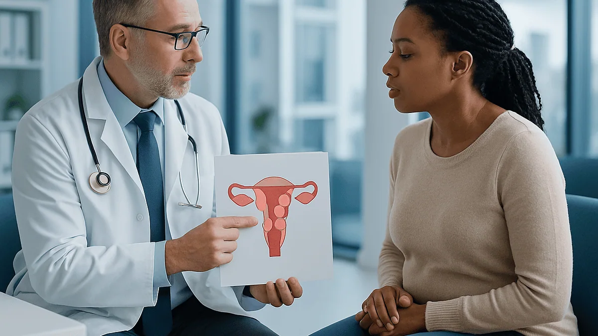
Key Takeaways
- Around 2 in 3 women develop a fibroid at some point, but only about 1 in 3 ever have symptoms, so most fibroids never need treatment.
- A fibroid of 1 to 3 cm inside the uterine cavity can cause more harm through heavy bleeding and anemia than a 6 cm fibroid in the outer wall.
- UK guidance uses roughly 3 cm as the point where specialist referral is considered for symptomatic fibroids, which is a referral trigger rather than a danger threshold.
- A 7 cm fibroid weighs about 180 grams, roughly a large peach, while a 10 cm fibroid approaches half a kilogram.
- Fibroids are benign and almost never become cancerous; the rare look-alike cancer is estimated at well under 1 in 1,000 growths and is flagged by behavior such as growth after menopause, not by size.
- Kidney compression from a large pelvic fibroid is rare but often silent, which is why a bulky uterus sometimes prompts a kidney ultrasound even without symptoms.
No single fibroid size is officially dangerous. Doctors judge risk by symptoms and location, not centimeters alone. Fibroids larger than about 5 cm are more likely to cause pressure, and those of 10 cm or more can press on the bladder, bowel or kidneys, but a small fibroid inside the uterine cavity can cause more harm through heavy bleeding. Sudden growth, severe pain or anemia matter more than size.
The ultrasound report lands in a patient portal at 9:40 on a Tuesday night. One line stands out: “intramural fibroid, 6.2 cm.” The rest of the report is gray text. That number, though, glows. Is six centimeters a lot? Is it dangerous? A search bar is open before the kettle boils.
That scene plays out thousands of times a week, because fibroid reports are written for clinicians and read by anxious people. The measurement feels like a verdict. It isn’t. A 6 cm fibroid sitting quietly in the muscular wall of the uterus can be less troublesome than a 2 cm one bulging into the lining, where it soaks a bathroom floor with blood every month.
This guide takes the size question seriously, because it is the question people actually ask, and then does what the top search results mostly skip: it explains what the evidence says about when a fibroid genuinely threatens health, and when the right response is watching, not cutting.
Is there a fibroid size that is officially "dangerous"?
Search the major clinical guidelines and you will not find a line that says “fibroids above X centimeters are dangerous.” That absence is not an oversight. Fibroids, also called leiomyomas or myomas, are non-cancerous growths of the muscle and connective tissue of the uterus, and the harm they do depends on what they press against, how they affect bleeding, and how fast they change. Size is one input among several.
Consider the numbers. The NHS estimates that around 2 in 3 women develop at least one fibroid at some point, yet only about 1 in 3 ever have symptoms. Most fibroids are discovered by accident during a scan for something else, and most of those never need anything more than a follow-up conversation.
What clinicians actually weigh is a short list: whether periods are heavy enough to drain iron stores, whether the fibroid is causing pressure symptoms such as urinary frequency or constipation, whether it is growing quickly, whether it is affecting fertility or a pregnancy, and whether it is in a location that could obstruct the ureters or the cervix. A fibroid that scores on none of those can be large and still be, in the clinical sense, harmless.
So the honest answer to “which size is dangerous” is that the word dangerous belongs to the symptom, not the centimeter. That said, size does shift the odds, and the next sections explain how.
How are fibroids measured, and what do small, medium and large mean?
Fibroids are measured on ultrasound, and sometimes MRI, as the longest diameter in centimeters, with two other dimensions often listed. Radiologists also describe the whole uterus, frequently borrowing pregnancy language: a uterus enlarged “to 12-week size” means it is roughly as big as it would be at 12 weeks of pregnancy, about the size of a grapefruit. The Mayo Clinic notes that fibroids range from seedlings invisible to the naked eye to bulky masses that distort and enlarge the uterus.
There is no universal grading system, but clinicians commonly group fibroids into rough bands, and the Cleveland Clinic describes the span as anything from a seed to a watermelon. The table below reflects how those bands are typically discussed and what they tend to mean in practice.
| Descriptor | Approximate size | Everyday comparison | Typical significance |
|---|---|---|---|
| Small | Under 5 cm | Pea to a plum | Often symptom-free unless inside the cavity |
| Medium | 5 to 10 cm | Lemon to an orange | Pressure or bulk symptoms become more common |
| Large | 10 cm and above | Grapefruit to a melon | Visible abdominal swelling, organ compression more likely |
Keep two caveats in mind. First, ultrasound measurements carry a margin of error of several millimeters, so a fibroid “growing” from 4.1 to 4.6 cm between scans may not have grown at all. Second, the total burden matters: three 4 cm fibroids can enlarge a uterus as much as one 7 cm fibroid. Reports that list only the largest one understate the whole picture.
Why the location of a fibroid often matters more than its size
Picture the uterus as a thick-walled muscular pear with a small central cavity. Where a fibroid sits relative to that cavity decides most of what it does. The Cleveland Clinic and Mayo Clinic describe four main positions.
- Submucosal fibroids grow just under the lining and bulge into the cavity. Even at 1 to 2 cm they can trigger very heavy or prolonged periods, because they increase the bleeding surface and disturb the lining’s normal contraction.
- Intramural fibroids sit within the muscular wall. They are the most common type and usually need to reach several centimeters before they cause heaviness or pressure.
- Subserosal fibroids grow on the outer surface, toward the pelvis. They rarely change periods but can press on the bladder or bowel once they are large.
- Pedunculated fibroids hang from a stalk, either inside the cavity or outside the uterus. A twisted stalk can cut off blood supply and cause sudden, severe pain.
This is why two people with identical measurements can have completely different experiences. A 2 cm submucosal fibroid can cause iron-deficiency anemia; a 6 cm subserosal fibroid may never be felt. When you read your report, look for these words next to the size. They tell you far more about the likely consequences than the number does, and they largely determine which treatments are realistic, because a fibroid inside the cavity can often be removed through the cervix without any abdominal incision, whereas a fibroid on the outer wall cannot.
Is a 5 cm fibroid dangerous?
Five centimeters is the number people search most, probably because it is where many radiology reports start adding a comment. A 5 cm fibroid is about the size of a lime. On its own, it is not dangerous. Plenty of people carry a fibroid this size for years, discover it on a scan for an unrelated reason, and are told to come back if anything changes.
What a 5 cm fibroid can do depends, again, on where it is. In the wall, it may cause somewhat heavier periods or a sense of fullness. Toward the front of the uterus, it may sit against the bladder and make you get up at night. Inside the cavity, a fibroid this size would almost certainly cause heavy bleeding and would usually prompt a discussion about removal. On a stalk, it carries a small risk of twisting.
Guidelines do treat the mid-single-digit range as a point where specialist input becomes more useful. UK clinical guidance, summarized in the NHS fibroid pages, uses a diameter of around 3 cm as the threshold above which some medicine-based treatments become less suitable and referral for further assessment is considered when symptoms are present. That is a referral trigger, not a danger line.
The practical questions for someone with a 5 cm fibroid are whether periods are manageable, whether iron levels are normal, whether there are any pressure symptoms, and whether the fibroid has changed since the last scan. If the answers are reassuring, most clinicians will suggest a repeat scan in six to twelve months rather than intervention. If not, the size is large enough that several treatment routes are on the table, which is genuinely good news.
How much does a 7 cm fibroid weigh, and do fibroids cause a big tummy?
Weight is a surprisingly common question, and the arithmetic is straightforward. Fibroids are dense muscle tissue with a density close to that of water, so a roughly spherical 7 cm fibroid has a volume of about 180 cubic centimeters and weighs in the region of 180 grams, a little over 6 ounces. That is about the weight of a large peach or three-quarters of a cup of water. A 10 cm fibroid, by the same geometry, comes in near 500 grams, roughly a pound. These are estimates; fibroids are rarely perfect spheres, and pathology reports after surgery often record combined weights that include several fibroids and surrounding uterine tissue.
Does that produce a visible belly? Sometimes. The NHS lists abdominal swelling among fibroid symptoms, and both the NHS and Mayo Clinic note that fibroids can enlarge the uterus to the size of a melon. A uterus enlarged to 12 to 14 weeks’ size will often show as a firm, lower-abdominal fullness, more noticeable when lying flat or in fitted clothing. Some people describe it as looking a few months pregnant.
Two points deserve emphasis. A single fibroid under 5 cm almost never produces visible swelling, so a changing waistline with a small fibroid on the scan usually has another explanation and deserves its own assessment. And a visible bulge, on its own, is not a medical emergency. It becomes a reason for treatment when it is accompanied by pressure symptoms, pain, or distress that affects daily life, all of which are legitimate reasons and should not be dismissed as cosmetic.
What size fibroid requires surgery?
There is no size that automatically requires surgery. That sentence disappoints people who want a clear cutoff, but it protects them from unnecessary operations. Guidelines from the NHS, the Mayo Clinic and the Cleveland Clinic all frame surgery as a response to symptoms or complications, with size acting as a modifier.
Surgery tends to enter the conversation in a handful of situations. Heavy bleeding that has not responded to non-surgical measures, or that is causing anemia, is the most common. Pressure symptoms from a large fibroid or an enlarged uterus, such as urinary frequency, incomplete emptying, constipation or pelvic heaviness, are the second. A submucosal fibroid distorting the cavity in someone trying to conceive is a third. Rapid growth, a fibroid that appears or enlarges after menopause, or imaging features that raise any doubt about the diagnosis form a fourth group where the aim is partly to exclude something else.
Size influences the route more than the decision. Fibroids inside the cavity up to a few centimeters can often be removed hysteroscopically, through the cervix. Wall or surface fibroids in the mid-range may suit a keyhole myomectomy. Very large fibroids, or many of them, may require an open incision or make hysterectomy a more practical option for someone who has completed their family. Uterine artery embolization, which the NHS describes as a procedure that blocks the blood supply so fibroids shrink, is another route for symptomatic fibroids and does not have a strict size ceiling, though very large or pedunculated fibroids are sometimes considered less suitable.
The decision always sits with you and the treating team. A good consultation should start with what bothers you, not with the number on the scan.
When large fibroids press on the bladder, bowel or kidneys
Once fibroids push a uterus toward grapefruit size and beyond, they start to compete with their neighbors for space. The bladder sits directly in front of the uterus, the rectum behind, and the ureters, the tubes carrying urine from each kidney, run down either side of the pelvis. The Mayo Clinic and NHS both list the consequences: frequent urination, difficulty emptying the bladder fully, constipation, and a persistent sense of pelvic pressure or lower back ache.
Most of these are uncomfortable rather than dangerous. The exception, and the one that turns a large fibroid into a medical priority, involves the ureters. A fibroid wedged in the pelvis, particularly one growing sideways from the lower uterus, can compress a ureter enough to slow the drainage of urine from that kidney. The kidney swells, a condition called hydronephrosis. The Mayo Clinic notes this is rare, but when it happens it can compromise kidney function if left unrelieved. It is typically silent, which is why clinicians sometimes request a kidney ultrasound when a uterus is very large, even in someone who feels well.
Bladder effects are more common and more noticeable. Incomplete emptying raises the likelihood of urinary infections, and repeated infections in someone with a bulky uterus deserve a look at the fibroid rather than just another course of treatment for the infection. Pressure on the rectum causes constipation and, occasionally, pain with bowel movements.
A useful rule: pressure symptoms that arrive alongside a large fibroid are a reason to see a clinician promptly, and any new flank pain, reduced urine output, or blood in the urine warrants an urgent assessment. These are the situations where size genuinely earns the word dangerous.
Heavy bleeding and anemia: the harm that small fibroids can cause
If pressure is the risk of big fibroids, blood loss is the risk of small ones, and it is the more common way fibroids damage health. The Mayo Clinic identifies heavy menstrual bleeding as the most frequent symptom and iron-deficiency anemia as its main complication. Anemia develops slowly, so people adapt to it: the fatigue is blamed on work, the breathlessness on being out of shape, the pale skin on winter.
What counts as heavy? The NHS describes it in practical terms: needing to change pads or tampons every one to two hours, bleeding through clothes or bedding, passing clots larger than about 2.5 cm, or bleeding for more than seven days. Any one of those, month after month, can outpace the body’s ability to absorb iron from food.
Submucosal fibroids are the usual culprits, and they need not be large. A fibroid of 1 to 3 cm inside the cavity increases the surface area of the lining and prevents the uterus from clamping down effectively on blood vessels at the end of a period. Intramural fibroids can also cause heaviness once they distort the cavity from behind.
The danger here is measurable and treatable. A simple blood count shows whether hemoglobin has fallen, and a ferritin level shows whether iron stores are depleted before anemia sets in. Untreated, severe anemia strains the heart and can make surgery riskier, which is one reason clinicians like to correct it before any procedure. So when a scan shows a small fibroid and the periods are heavy, the fibroid is not “too small to matter.” It may be the most important finding on the report.
Can a fibroid turn into cancer, and does size predict it?
This is the fear beneath the search, so it deserves a direct answer. Fibroids are benign, and the Mayo Clinic states that they are not associated with an increased risk of uterine cancer and almost never develop into cancer. The rare cancer that can be confused with a fibroid, leiomyosarcoma, arises in the uterine muscle, and the Cleveland Clinic puts its frequency at well under 1 in 1,000 fibroid-like growths. There is no evidence that a benign fibroid transforms into a sarcoma; the two appear to be different conditions that happen to look alike on a scan.
Size by itself is a poor predictor. Large benign fibroids are common; sarcomas are rare at any size. What does raise a clinician’s attention is behavior rather than dimension: a growth that appears for the first time after menopause, when fibroids ordinarily shrink; a fibroid that enlarges quickly, particularly after menopause or in someone not taking hormones; unusual features on MRI such as irregular borders or areas of tissue breakdown; and new pain or bleeding in someone whose fibroids had been stable for years.
None of those features confirms cancer. Fibroids can grow in bursts, and they can degenerate and look irregular for entirely benign reasons. But they shift the conversation from routine monitoring toward closer imaging or tissue diagnosis, and that is appropriate.
For most readers, the takeaway is reassuring: a fibroid found in your thirties or forties, growing slowly or not at all, with typical appearances on ultrasound, is overwhelmingly likely to be exactly what it looks like. The number of centimeters does not change that.
Fibroids during pregnancy: does size change the risks?
Fibroids and pregnancy coexist more often than people expect, and most such pregnancies proceed normally. Still, the Mayo Clinic lists specific risks that rise with fibroids: placental abruption, in which the placenta separates early; fetal growth restriction; and preterm delivery. The NHS adds that large fibroids can occasionally affect the baby’s position and make cesarean birth more likely, and that fibroids may contribute to difficulty conceiving or to pregnancy loss in some cases, particularly when they distort the cavity.
Size and location both play roles. Submucosal fibroids and larger intramural fibroids that press into the cavity are the ones most consistently linked with fertility and early pregnancy problems, because they can interfere with implantation and with the blood supply to the developing placenta. Fibroids on the outer surface, even large ones, are less implicated unless they physically block the birth canal.
Pregnancy hormones can make fibroids grow, sometimes noticeably in the first half, though growth is unpredictable and many stay the same. A fibroid that grows quickly can outstrip its blood supply and degenerate, causing a sharp, localized pain that usually settles with rest and simple pain relief under a clinician’s guidance. It is alarming but rarely harmful to the pregnancy.
For someone planning a pregnancy who knows they have fibroids, the useful conversation is not “is my fibroid too big” but “does my fibroid distort the cavity, and would removing it before conception help.” Removing a fibroid during pregnancy is generally avoided because of bleeding risk, so timing matters. The treating team will weigh the fibroid’s position, your history and your goals rather than applying a size rule.
What happens if fibroids are not removed?
For most people, nothing dramatic. Fibroids depend on estrogen and progesterone, and the NHS notes that they tend to shrink after menopause as hormone levels fall. Someone in their late forties with moderate symptoms may reasonably choose to manage bleeding and wait, knowing that biology is on their side. Fibroids left in place do not spread, do not seed elsewhere, and do not become cancerous.
The realistic consequences of leaving symptomatic fibroids untreated are the slow ones. Heavy periods can gradually deplete iron and lead to anemia. A steadily enlarging uterus can bring pressure on the bladder and bowel that was absent a few years earlier. Fibroids that distort the cavity can affect fertility for as long as they are there. Chronic pelvic discomfort and the disruption of unpredictable heavy bleeding take a toll on work, sleep, exercise and relationships that clinical notes rarely capture.
There is also the practical matter that fibroids tend to grow, if slowly, during the reproductive years. A fibroid that could once have been removed by keyhole surgery may, a few years later, need a larger incision. That is not a reason to rush, but it is a reason to keep follow-up appointments rather than let a known fibroid drift out of view for a decade.
Watchful waiting is a legitimate, evidence-supported choice when symptoms are mild, iron levels are normal and the fibroid is stable. It works best when it is an active decision, revisited with a scan and a blood test at agreed intervals, rather than an absence of one.
When to see a doctor about fibroids: red flags that should not wait
Most fibroid symptoms can be discussed at a routine appointment, and the NHS advises seeing a GP if you have persistent symptoms such as heavy periods, pelvic pain, frequent urination or constipation so the cause can be investigated. Some situations, though, call for urgent attention regardless of fibroid size.
Seek same-day or emergency care if you have sudden, severe pelvic or abdominal pain, especially with fever or vomiting, which can signal a twisted pedunculated fibroid or acute degeneration. Bleeding heavy enough to soak through a pad or tampon every hour for several hours, or that leaves you dizzy, faint or short of breath, needs urgent assessment because it can indicate significant blood loss. Any bleeding after menopause should be evaluated promptly; it is not a normal fibroid symptom. Inability to pass urine, or new pain in the flank or side with a large known fibroid, should be checked without delay because of the possibility of pressure on the urinary tract. In pregnancy, severe abdominal pain, vaginal bleeding or a marked reduction in the baby’s movements warrant immediate contact with the maternity team.
Less urgent but still important are gradual changes: periods that have become heavier or longer over several months, a noticeable increase in abdominal size, pain during sex, unexplained tiredness that might reflect anemia, or a fibroid that appears to have grown quickly between scans. These deserve an appointment within weeks rather than a wait for the next annual check.
One more red flag is about the report itself. If a scan describes a fibroid as having unusual or atypical features, or recommends further imaging, follow that recommendation through. It is usually reassurance that results, but it should be sought rather than assumed.
How treatment is chosen by size, symptoms and what matters to you
Treatment for fibroids sits on a spectrum, and the NHS, Mayo Clinic and Cleveland Clinic describe broadly the same ladder. At one end is monitoring with periodic scans and iron checks. Next come medicines that reduce bleeding without shrinking the fibroid, including hormonal options that thin the lining, and medicines that temporarily lower estrogen to shrink fibroids for a limited period, often used before surgery. Which, if any, suits you is a decision for the prescribing clinician, who will weigh your bleeding pattern, plans for pregnancy and medical history.
Procedures follow. Hysteroscopic removal through the cervix suits fibroids inside the cavity, typically up to a few centimeters. Myomectomy, removing fibroids while preserving the uterus, can be done by keyhole or open surgery depending on number, size and position, and is the usual choice for those who want to keep the option of pregnancy. Uterine artery embolization blocks the fibroids’ blood supply so they shrink over months, avoiding a surgical incision, and is generally offered to those not planning future pregnancy. Focused ultrasound uses MRI-guided heat to destroy fibroid tissue in selected cases. Endometrial ablation treats the lining to reduce bleeding but does not remove fibroids and is unsuitable for those who may want children. Hysterectomy, removal of the uterus, is the one option that guarantees fibroids will not return, and remains a reasonable choice for severe symptoms when childbearing is complete.
Every one of these carries risks, from bleeding and infection to, with uterus-sparing procedures, the possibility of new fibroids forming. No option is best for everyone. The question your team should be asking is not how big the fibroid is but what you need from your uterus, how much the symptoms are costing you, and what recovery you can accommodate.
Frequently asked questions
Which size of fibroid is dangerous?
No specific size is officially dangerous. Fibroids of 10 cm or more are more likely to press on the bladder, bowel or ureters, and any fibroid causing heavy bleeding, anemia, severe pain or rapid growth deserves attention regardless of size. Guidelines judge risk by symptoms and location. Many large fibroids cause no harm, while some small ones inside the uterine cavity cause significant blood loss.
Is a 5 cm fibroid dangerous?
A 5 cm fibroid, about the size of a lime, is not dangerous in itself. It may cause heavier periods or mild pressure depending on its position, and if it sits inside the uterine cavity it will usually prompt a discussion about removal. If periods are manageable, iron levels are normal and the fibroid is stable on repeat scanning, monitoring is a common and reasonable approach.
What size fibroid requires surgery?
There is no size that automatically requires surgery. Surgery is considered when fibroids cause heavy bleeding unresponsive to other measures, anemia, pressure symptoms, fertility problems related to cavity distortion, or when growth or imaging raises doubt about the diagnosis. Size mainly influences the type of procedure, such as hysteroscopic removal for small cavity fibroids versus keyhole or open surgery for larger ones.
What happens if fibroids are not removed?
For most people, fibroids left alone grow slowly, do not spread and shrink after menopause. The realistic risks of leaving symptomatic fibroids are gradual: iron-deficiency anemia from heavy periods, increasing pressure on the bladder or bowel, and ongoing effects on fertility if the cavity is distorted. Watchful waiting is legitimate when symptoms are mild, provided follow-up scans and blood counts are kept.
Do fibroids cause a big tummy?
They can, once the uterus is enlarged to roughly the size it would be at 12 or more weeks of pregnancy, which usually requires a fibroid of around 10 cm or several medium ones. Small fibroids under 5 cm almost never cause visible swelling, so a changing waistline with a small fibroid on the scan usually has another cause that deserves its own assessment.
How much does a 7 cm fibroid weigh?
A roughly spherical 7 cm fibroid has a volume of about 180 cubic centimeters and, since fibroid tissue is close to the density of water, weighs around 180 grams, a little over 6 ounces. That is comparable to a large peach. Real fibroids are irregular, so this is an estimate; pathology reports after surgery often record combined weights of multiple fibroids.
Can a fibroid turn into cancer?
Fibroids are benign and almost never become cancerous. The rare cancer that can resemble a fibroid, leiomyosarcoma, is estimated to occur in well under 1 in 1,000 such growths and is thought to arise separately rather than from an existing fibroid. Features that prompt closer investigation include new growth after menopause, rapid enlargement, or unusual appearances on MRI, not size alone.
Do fibroids shrink after menopause?
Usually, yes. Fibroids depend on estrogen and progesterone, so as hormone levels fall after menopause they tend to shrink and symptoms often ease. This is why some people in their late forties choose to manage symptoms and wait. A fibroid that grows or first appears after menopause is unusual and should be evaluated, as should any bleeding after menopause.
Are fibroids dangerous during pregnancy?
Most pregnancies with fibroids proceed normally. Fibroids do raise the risk of certain complications, including placental abruption, fetal growth restriction and preterm delivery, and large or cavity-distorting fibroids are more likely to be involved. Fibroids may grow during pregnancy and occasionally degenerate, causing sharp pain that usually settles. Removal during pregnancy is generally avoided, so planning conversations are best had beforehand.
How fast do fibroids grow?
Growth is unpredictable. Many fibroids stay stable for years, some grow slowly, and a few grow in bursts, particularly during pregnancy. Ultrasound measurements carry a margin of error of several millimeters, so a small apparent change between scans may not be real growth. Rapid enlargement, especially after menopause or in someone not taking hormones, is a reason for closer imaging.
References
This article is for general information only and is not a substitute for professional medical advice. Please consult a qualified doctor about your individual situation.

More from the Blog

Ovarian Follicles: What They Are and How Many Is Normal
Ovarian follicles are tiny fluid-filled sacs in the ovary, each cradling one immature egg. A group starts growing every cycle, and usually a single…

Laparoscopic Ovarian Cyst Removal Step by Step: Anesthesia, Incisions and Hospital Stay
Laparoscopic ovarian cyst removal is keyhole surgery done under general anesthesia. A surgeon makes a few small cuts in the abdomen, usually three or…

How HPV Is (and Is Not) Transmitted: Dormancy, Kissing, Toilet Seats
A married woman most often gets HPV from an infection acquired years, sometimes decades, before it shows up on a test. HPV spreads through…

Is It a Yeast Infection or Bacterial Vaginosis? Why Testing Comes Before Treatment
Yeast infections and bacterial vaginosis can feel alike, since both change discharge and cause discomfort, but they have different causes and different treatments. Bacterial…

Ovary Pain: Left, Right, Cysts and Back Pain
Pain felt over the left ovary most often comes from a normal monthly event such as ovulation or a small functional cyst, both of…

Bladder, Uterus or Rectal Prolapse: How the Type and Stage Shape the Treatment Plan
Pelvic organ prolapse is named for the organ that has dropped: the bladder (cystocele), the uterus, the rectum (rectocele), the small bowel (enterocele) or…