

Why Cardiac MRI Matters in Myocardial Disease: What the Scan Adds to an Echo

Key Takeaways
- Cardiac MRI is the reference standard for measuring chamber volumes, wall thickness and ejection fraction because the same heart scanned twice yields nearly identical numbers.
- Late gadolinium enhancement marks tissue where contrast lingers, and its location, under the inner lining versus mid-wall versus patchy, often points toward an ischemic, dilated, hypertrophic, sarcoid or amyloid cause.
- Echo remains the first-line test and the better tool for valves and blood flow, so a normal MRI does not make the echocardiogram redundant.
- The T2 star technique lets cardiac MRI quantify iron in the heart muscle without a biopsy, which is why it is used to monitor transfusion-dependent anemias.
- Gadolinium contrast is chemically unrelated to CT iodine dye, is cleared by the kidneys mostly within a day, and is avoided in pregnancy unless essential.
- A standard cardiac MRI appointment typically runs 30 to 90 minutes, consists mainly of repeated ten- to fifteen-second breath-holds, and involves no ionizing radiation.
Cardiac MRI for cardiomyopathy adds what an echocardiogram cannot reliably show: highly reproducible measurements of chamber size, wall thickness and pumping function, plus a direct look at the heart muscle tissue itself. With gadolinium contrast it can reveal scar, inflammation, iron or abnormal protein deposits that point toward a cause. It complements echo rather than replacing it, and the treating team interprets the findings alongside the full clinical picture.
The echo report sits on the kitchen table, folded twice. It says the left ventricle is enlarged and pumping below normal, and then, in the line that matters most, it says the cause is unclear. The cardiologist has suggested one more scan, a cardiac MRI, and the word that keeps surfacing is why. The heart has already been photographed once. What could a second, longer, noisier test possibly add?
Quite a lot, as it turns out. Cardiac MRI for cardiomyopathy is not a repeat of the echo with a bigger machine. It answers a different question. Echo is superb at showing how the heart moves. MRI can show what the muscle is made of: where it has scarred, where it is inflamed, whether something is quietly accumulating inside it.
That distinction changes conversations about medicines, devices, family screening and follow-up. This article walks through what the scan does, when it is usually ordered, what the day feels like and where its limits genuinely lie.
What does cardiac MRI for cardiomyopathy show that an echo cannot?
Start with the physics, because it explains everything else. An echocardiogram bounces high-frequency sound waves off the heart and builds a moving picture from the returning signal. Sound has to travel through the chest wall and around the lungs, so the image depends on finding a clear acoustic window between ribs. In a broad chest, a person with lung disease or someone who simply cannot lie on their left side, parts of the heart may be hard to see.
Magnetic resonance imaging (MRI) works differently. A strong magnet aligns the hydrogen atoms in body water, radio waves briefly knock them out of line, and the signal they release as they settle back is turned into an image. No ionizing radiation is involved, and the picture does not depend on a window. The whole heart can be imaged in any plane, at the same resolution, on every scan.
Three capabilities follow from that. First, measurement: cardiac MRI is widely treated as the reference method for chamber volumes, muscle mass and ejection fraction, which is the percentage of blood the ventricle pushes out with each beat. Second, tissue characterization, meaning the ability to tell healthy muscle from muscle that is scarred, swollen with fluid, fatty or infiltrated by something that should not be there. Techniques called T1 and T2 mapping assign each tiny region of muscle a number that reflects its water and protein content, so subtle diffuse disease can be picked up before it changes the shape of the heart.
Third, pattern recognition. When gadolinium contrast is given, the way it settles into damaged tissue produces patterns that often track with specific causes. That is the part echo cannot do, and it is the reason the second scan is worth the extra hour.
Why doctors still start with an echocardiogram
If MRI sees more, why not go straight to it? Because echo does several things brilliantly, and it does them at the bedside in twenty minutes. A machine can be wheeled into a clinic room, an emergency department or an intensive care bay. There is no magnet, no screening questionnaire, no contrast line, and the person being scanned can sit up halfway through if breathing is difficult.
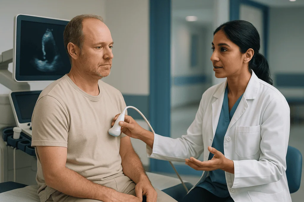
Echo is also the better tool for certain questions. Doppler, the part of the exam that measures blood velocity, gives a detailed read of valve leaks and narrowings, filling pressures and the way blood moves through the chambers in real time. For a first look at someone who is short of breath or has a new murmur, the NHS and Mayo Clinic both describe echocardiography as the usual starting point for suspected cardiomyopathy.
Think of the two tests as a general practitioner and a specialist. Echo asks the broad questions: Is the heart enlarged? Is it pumping well? Are the valves working? It answers them quickly and repeatably, which is why it is also the workhorse for routine follow-up. MRI is called in when the broad answers raise a narrower question, most often one of these:
- The echo images were incomplete or of poor quality.
- The heart is clearly abnormal but the cause is not obvious.
- A specific diagnosis is suspected that depends on tissue findings, such as inflammation or infiltration.
- A precise, reproducible measurement is needed before a decision about a device or a medicine.
Seen that way, the referral is not a sign that the first test failed. It is a sign that the first test did its job and found something worth understanding properly.
What is the gold standard for diagnosing cardiomyopathy?
People search this phrase constantly, and the honest answer disappoints anyone hoping for a single decisive test. Cardiomyopathy is not one disease. It is an umbrella term for conditions in which the heart muscle itself is abnormal in structure or function without a valve problem, high blood pressure or coronary disease fully explaining it. A diagnosis is assembled, not read off a screen.
The assembly usually includes a careful history (including family history, alcohol use, prior cancer treatment and pregnancy), a physical examination, an electrocardiogram, blood tests that may include markers of heart strain and screens for iron or abnormal proteins, and imaging. Genetic testing is increasingly part of the picture for inherited forms, according to Mayo Clinic’s overview of cardiomyopathy diagnosis. In selected cases a small sample of heart muscle, taken through a vein under X-ray guidance, is still needed.
Within that assembly, cardiac MRI holds a specific title. It is regarded as the reference standard for measuring how big the chambers are, how thick the walls are and how much blood the ventricles eject, because the same heart scanned twice produces almost the same numbers. That reproducibility matters when a decision hinges on whether function has fallen below a threshold. MRI is also the most informative non-invasive test for the tissue itself.
So the accurate phrasing is this: there is no single gold standard for cardiomyopathy, but cardiac MRI is the gold standard for cardiac measurement and the leading non-invasive tool for tissue characterization. Echo remains the first-line test. Biopsy remains the final arbiter for a handful of conditions. The treating team decides which pieces the puzzle needs, and a scan result is always read next to the person, never instead of them.
Late gadolinium enhancement meaning: why scar changes the conversation
The phrase that appears on many cardiac MRI reports is late gadolinium enhancement, usually shortened to LGE. Gadolinium is a metal-based contrast agent injected into an arm vein partway through the scan. In healthy, tightly packed muscle it washes in and out within minutes. Where cells have died and been replaced by fibrous scar, or where the spaces between cells have expanded because of inflammation or deposits, gadolinium lingers. Images taken roughly ten minutes after the injection show those regions as bright against the dark of normal muscle.

The location of the brightness is the clue. Scar after a heart attack sits under the inner lining of the ventricle, because that is the layer that runs out of oxygen first, and it follows the territory of a blocked artery. Scar in a non-ischemic cardiomyopathy tends to appear as a thin stripe in the middle of the wall or spread across the outer layer. Patchy, scattered enhancement raises the possibility of sarcoidosis or hypertrophic disease. A diffuse glow across the inner layer of both ventricles, with contrast behaving unusually in the blood pool, points toward amyloid.
Why does anyone care about a bright patch? Because scar is electrically unstable tissue. It can act as the anchor for abnormal rhythms. Current guidance from major cardiology societies treats the presence and extent of LGE as one factor, among several, when weighing the risk of dangerous arrhythmias and the possible role of an implantable defibrillator. The scan does not make that decision. It contributes a piece of evidence that the team weighs alongside symptoms, family history, rhythm monitoring and the person’s own priorities.
One caution belongs here. Enhancement is a finding, not a verdict. Small amounts of scar are common in many settings, and a report that mentions LGE is a prompt for a conversation, not a reason for alarm.
Cardiac MRI in dilated cardiomyopathy: finding the cause behind a weak pump
Dilated cardiomyopathy means the left ventricle has stretched and its walls have thinned, so it pumps less blood with each beat. It is the form most often found on an echo ordered for breathlessness or swelling, and it is also the form where the echo most often leaves the cause open. Cardiac MRI earns its place here by helping the team sort that cause into groups that are managed differently.
The first split is ischemic versus non-ischemic. A ventricle can dilate because a silent heart attack destroyed part of the wall years earlier, or because a disease of the muscle itself has weakened it. On MRI, an old infarct declares itself as scar under the inner lining in a coronary territory, with thinning of the wall above it. A muscle disease usually shows no such pattern, or shows the mid-wall stripe described above. That single distinction shapes whether the next steps focus on the coronary arteries or on the muscle.
The second split is within the non-ischemic group. MRI can show swelling consistent with recent or ongoing inflammation of the heart muscle, called myocarditis, which raises different questions about recent infections and immune disease. It can show fatty replacement of the wall. Mapping techniques can suggest diffuse fibrosis even when no discrete bright area appears. In someone treated with certain cancer therapies or with heavy alcohol use, MRI cannot name the toxin, but it can document the extent of injury and provide a precise baseline for follow-up.
The third contribution is measurement over time. Because volumes and ejection fraction are so reproducible on MRI, a change between scans is more likely to reflect a real change in the heart than a difference in image quality. When the team needs to know whether the ventricle is recovering under treatment, that reliability is what they are asking for.
Cardiac MRI in hypertrophic cardiomyopathy: seeing the whole thickened wall
Hypertrophic cardiomyopathy (HCM) is a usually inherited condition in which part of the heart wall thickens without a mechanical reason such as long-standing high blood pressure or a narrowed valve. It is often first suspected on an echo, but echo has a blind spot: the very tip of the ventricle and its lateral wall can be difficult to visualize, and thickening confined to those areas can be underestimated or missed altogether.
Cardiac MRI images every segment of the wall at the same clarity, so the maximal thickness can be measured wherever it happens to sit. That number is one of the inputs to risk models used in both European and American guidelines, which is why accuracy matters. MRI also shows features that are hard to catch on echo: a small bulge at the apex of the ventricle, abnormalities of the muscle bundles inside the chamber, and the papillary muscles that anchor the mitral valve, which are sometimes misplaced in HCM and contribute to obstruction.
Tissue findings add a second layer. Scar in HCM often appears as patchy enhancement within the thickest segments, and its extent is taken into account when the team discusses arrhythmia risk. Mapping helps separate true HCM from conditions that mimic it. Athletes can develop thicker walls through training, but the muscle looks healthy on mapping and the thickening is usually balanced across the ventricle. Cardiac amyloidosis and Fabry disease, a rare inherited storage disorder, can both thicken the wall while producing distinctive mapping signatures that point away from classical HCM.
For relatives of someone with HCM, MRI is sometimes used when an echo is borderline, since a clean, complete picture of the wall can settle an uncertain screening result. Whether and when to scan family members is a decision for the cardiology and genetics team, guided by the specific gene variant when one is known.
Restrictive and infiltrative disease: amyloid, sarcoid, iron and inflammation
Restrictive cardiomyopathy describes a heart that is stiff rather than weak or thick. The ventricles fill poorly, pressures back up, and the person is breathless with a pump that may look normal in size and only modestly reduced in function. Echo can suggest stiffness through filling patterns, but it usually cannot say why the muscle is stiff. This is where cardiac MRI’s ability to read the tissue is most decisive.
Cardiac amyloidosis occurs when misfolded proteins deposit between muscle cells. On MRI the walls are thickened, mapping values are markedly raised, and gadolinium behaves in a characteristic way: it clears from the blood unusually quickly and lights up the inner layer of both ventricles and often the atria. The pattern is distinctive enough that MRI frequently triggers the blood and nuclear tests that confirm the type of amyloid, a step that matters because the two main types are treated quite differently.
Cardiac sarcoidosis involves clumps of inflammatory cells scattered through the muscle. MRI shows patchy swelling on the water-sensitive sequences and patchy scar that does not follow an artery, often in the septum, which is the wall between the ventricles. Because sarcoid can disturb the heart’s electrical wiring, this finding frequently changes how closely rhythm is monitored.
Iron overload, seen in inherited anemias that need repeated transfusions and in hemochromatosis, is one of the clearest wins for MRI. A sequence called T2 star measures how quickly signal decays, and iron shortens that time in proportion to how much has accumulated. The technique is used to track iron in the heart without a biopsy.
Myocarditis, inflammation of the muscle, shows as swelling plus non-ischemic enhancement, assessed against internationally agreed imaging criteria. Arrhythmogenic cardiomyopathy, which mostly affects the right ventricle, can show wall thinning, bulging and fatty change. In each case MRI narrows the field so that the confirmatory tests can be chosen rather than guessed.
Cardiac MRI for cardiomyopathy: who is usually referred, and who is asked to wait
Referral patterns follow the logic above. People are usually sent for cardiac MRI when the echo was technically limited; when the ventricle is clearly abnormal but the cause is unexplained; when a tissue-dependent diagnosis such as amyloid, sarcoid, myocarditis or iron overload is on the table; when HCM is suspected or needs risk assessment; when precise function measurements will influence a decision about a device or a medicine; or when a baseline is needed before treatments known to affect the heart. Some centers also use it to screen close relatives of people with inherited cardiomyopathy when the echo is inconclusive.
Being asked to wait, or to have a different test, is common and rarely means the scan is off the table for good. Typical reasons include:
- Implanted devices. Many modern pacemakers and defibrillators are labeled MRI-conditional, meaning they can be scanned under specific settings with device checks before and after. Older or unknown devices need individual assessment, and some cannot be scanned. Retained lead fragments and certain other implants also require review.
- Kidney function. Gadolinium is cleared by the kidneys. In severe kidney disease the team weighs the value of contrast against a small risk, and may scan without contrast or choose another test.
- Pregnancy. MRI without contrast is generally considered acceptable when clinically needed, but gadolinium is usually avoided unless essential, so scans are often deferred until after delivery.
- Unable to lie flat or hold breath. Severe breathlessness, an unstable rhythm or acute illness can make a 45-minute scan impractical; stabilization comes first.
- Metal fragments. Shrapnel, some older aneurysm clips and metal in the eye must be ruled out or cleared.
Claustrophobia deserves its own line. It is one of the most common reasons scans are abandoned, and it is also one of the most manageable: shorter protocols, a companion in the room, music and, in some cases, mild sedation arranged by the team can make the difference. Raise it before the appointment rather than on the day.
Cardiac MRI vs echocardiogram: a side-by-side view
The two tests are often described as rivals. They are better understood as a pair with different strengths, which is why most people with cardiomyopathy end up having both. The table sets out the practical differences the way a cardiologist tends to think about them.
| Feature | Echocardiogram | Cardiac MRI |
|---|---|---|
| How it images | Sound waves through a chest window | Magnetic field and radio waves, any plane |
| Radiation | None | None |
| Typical duration | About 20–45 minutes | About 30–90 minutes |
| Chamber size and ejection fraction | Good, but depends on image quality | Reference standard, highly reproducible |
| Tissue (scar, inflammation, iron, deposits) | Cannot assess directly | Core strength, with and without contrast |
| Valves and blood flow | Excellent, real-time Doppler | Good, but less detailed for valve anatomy |
| Where it can be done | Bedside, clinic, emergency room | Dedicated scanner suite only |
| Contrast | Occasionally, microbubble agent | Usually gadolinium, unless kidneys or pregnancy dictate otherwise |
| Main limitations | Poor windows, operator dependence | Implants, claustrophobia, breath-holds, availability |
Two rows deserve emphasis. The tissue row is the reason the second scan exists; nothing in an echo report can tell you whether a wall is scarred or inflamed. The valve row cuts the other way, and it is why a normal cardiac MRI does not make the echo redundant. When a cardiomyopathy coexists with a leaking mitral valve, which is common in dilated hearts, echo remains the better tool for following that leak.
Duration figures are typical ranges reported by the NHS and Cleveland Clinic for standard scans. A focused scan without contrast may be shorter; a full protocol with mapping, contrast and stress imaging sits at the longer end.
What happens on the day, and what the following days usually look like
The appointment begins with paperwork that matters more than usual: a safety questionnaire about implants, surgery, metal fragments, kidney problems, allergies and pregnancy. Answer it thoroughly. Then jewelry, watches, hearing aids, hairpins and anything containing metal come off, cards with magnetic strips stay in the locker, and a gown replaces clothing with zips or underwire.
Inside the scanner room, small sticky electrodes go on the chest so the machine can time its pictures to the heartbeat. A thin plastic tube is placed in an arm vein for the contrast. The bed slides into a tunnel that is open at both ends, a light padded coil rests on the chest, and headphones or earplugs are offered because the scanner knocks and buzzes loudly as it works. A technologist watches and talks through an intercom the whole time.
The scan itself is a series of breath-holds. The voice asks for a breath in, a breath out, then a hold of roughly ten to fifteen seconds while one set of images is captured, repeated many times. Between holds there is time to breathe normally. Partway through, the contrast is injected; a cool feeling in the arm or a brief metallic taste is common and passes quickly. According to the NHS, a whole MRI appointment typically lasts between 15 and 90 minutes depending on the area and the number of images, and cardiac protocols sit toward the upper part of that range.
Afterwards there are no restrictions for most people. Eating, driving and returning to work the same day are normal unless a sedative was given, in which case someone else drives. The gadolinium leaves the body through the kidneys in urine, mostly within a day, per the NHS. Mild tiredness from the concentration and breath-holding is common and short-lived.
Results are not read in the room. A specialist analyzes hundreds of images and produces a report over the following days, and the ordering cardiologist explains it at a follow-up. Ask at the desk how and when the results will be communicated so that silence does not feel like bad news.
Cardiac MRI with contrast safety: implants, kidneys and gadolinium
The first reassurance is the simplest. MRI uses no ionizing radiation, so unlike CT scans and nuclear tests, repeated cardiac MRIs do not add to lifetime radiation exposure. That matters for younger people who may need serial imaging over decades, and for anyone tracking a condition that changes slowly.
Gadolinium is the part that prompts most questions. It is not the same as the iodine-based dye used for CT, so an iodine allergy does not predict a gadolinium reaction. Mild effects such as a cold sensation, brief nausea, headache or a metallic taste are the most common experiences, and serious allergic reactions are rare, according to MedlinePlus and Mayo Clinic. Staff are trained and equipped to treat a reaction if one occurs.
Two longer-term concerns have shaped current practice. Nephrogenic systemic fibrosis, a rare thickening of skin and tissues, was linked to some older gadolinium agents in people with severe kidney failure. Kidney function is now checked before contrast when there is any doubt, and the agents in routine use carry a much lower risk. Separately, trace gadolinium has been found retained in the brain and other tissues after repeated doses. Regulators have not identified harm from this retention, but they advise using contrast only when the diagnostic benefit is clear, which is exactly how cardiac protocols are designed.
Implants are handled by protocol rather than guesswork. Every device has a make and model, and the radiology team checks its MRI labeling. MRI-conditional pacemakers and defibrillators are reprogrammed before the scan and checked afterward. Coronary stents, sternal wires and most heart valves are routinely scanned. Cochlear implants, some neurostimulators and certain older clips are the ones that most often rule a scan out.
Finally, the magnet itself is on all the time, even between patients. Loose metal objects can become projectiles, which is why the screening feels repetitive. It is repetitive on purpose.
What people often get wrong about cardiac MRI
Does a cardiac MRI show all heart issues? No, and this is the most important correction. MRI is exceptional for the muscle and for chamber measurement, but it is not the best test for the coronary arteries, whose narrowings are usually assessed with CT angiography or catheter angiography. It cannot see the tiny vessels involved in microvascular disease. It does not record rhythm over time; a bright patch may explain a palpitation, but only a monitor can capture the rhythm itself. Valve detail is generally better on echo. A normal cardiac MRI is reassuring about structure and tissue; it is not a certificate that the heart is free of all disease.
MRI replaces echo. It does not. The two answer different questions, and echo remains the everyday tool for follow-up.
Scar on the report means something terrible is coming. Scar is a finding that feeds into risk assessment alongside many other factors. Small areas are common, and how much weight they carry depends on the underlying condition, symptoms and rhythm data. The team, not the report, translates it into a plan.
MRI involves radiation. It does not. The confusion usually comes from CT, which does.
The contrast is the same as CT dye. Gadolinium and iodine are unrelated compounds with different side-effect profiles.
A pacemaker means no MRI, ever. Many current devices are MRI-conditional. Whether a scan can proceed depends on the specific device and leads, so ask rather than assume.
Cardiac MRI is only for research or rare diseases. It has become part of standard guideline-based evaluation for unexplained cardiomyopathy, suspected myocarditis, HCM assessment and iron monitoring.
If I felt fine, the scan would be normal. Several cardiomyopathies produce tissue changes long before symptoms, which is precisely why MRI is sometimes used in family screening.
Questions to ask your care team before and after the scan
A good scan is preceded by a good conversation. The questions below are the ones cardiologists and radiographers say they wish more people asked, because each one either improves the scan or makes the result easier to act on. Write down the answers; reports arrive days later and the details blur.
Before the scan:
- What specific question is this scan meant to answer that the echo could not?
- Will contrast be used, and has my kidney function been checked recently?
- I have a device, stent, clip or metal from surgery. Has its MRI compatibility been confirmed, and will the device need reprogramming?
- I struggle in enclosed spaces. What options exist to make the scan manageable, and should I discuss sedation in advance?
- Can I take my usual medicines that morning, and should I eat beforehand? (Follow the specific instructions given; do not change medicines on your own.)
- How long should I expect to be in the department, and can someone come with me?
After the results:
- What did the scan show about the size and function of my heart, in plain language?
- Was there any scar, inflammation or infiltration, and what pattern did it show?
- Does the result change the working diagnosis, and if so, what is it now?
- Does it change the treatment plan, the follow-up interval or the need for further tests such as genetic testing or a biopsy?
- Should any of my relatives be offered assessment?
- Will I need another MRI, and roughly when would that be considered?
- Which symptoms should prompt me to call before the next appointment?
One more, worth asking gently: is there anything in the report that you are uncertain about? Imaging often produces findings of unclear significance, and clinicians appreciate the chance to say so plainly rather than have a phrase on a report become a source of private worry.
When to call your doctor
Cardiac MRI itself is a low-risk test, but the conditions it investigates are not trivial, and the period around a scan is a time when symptoms deserve prompt attention rather than patience. Treat the following as red flags that warrant emergency care, whether or not a scan is scheduled:
- Chest pain or pressure that lasts more than a few minutes, spreads to the arm, jaw or back, or comes with sweating or nausea.
- Fainting or near-fainting, especially during exertion or without warning.
- Sudden or rapidly worsening breathlessness, or waking at night unable to breathe.
- A racing, pounding or irregular heartbeat accompanied by dizziness, confusion or collapse.
- Rapid swelling of the legs or abdomen with weight gain over a few days.
In the hours after a scan with contrast, call the imaging department or seek urgent care for hives, facial or throat swelling, wheeze or difficulty breathing, since these can indicate an allergic reaction. Redness, pain or swelling spreading from the injection site over the following days should also be reported.
Less urgent, but still worth a call within a day or two: new breathlessness climbing stairs you managed last month, a persistent cough at night, palpitations that are new or more frequent, or a family member’s recent diagnosis of a heart muscle condition. The NHS lists breathlessness, swelling, palpitations and fainting among the symptoms that should prompt assessment for cardiomyopathy, and the point of raising them is not to self-diagnose but to let the team decide whether the plan needs to change.
Every decision described in this article, from whether to scan to what the findings mean and what follows, sits with the treating team who know the full picture. A report is one document in a much longer conversation, and the scan’s real value is measured in how well that conversation goes.
Frequently asked questions
What is the gold standard for diagnosing cardiomyopathy?
There is no single gold standard test; cardiomyopathy is diagnosed by combining history, examination, ECG, blood tests, imaging and sometimes genetic testing or biopsy. Within that workup, cardiac MRI is considered the reference standard for measuring heart size and function and the leading non-invasive method for assessing the muscle tissue. Echocardiography is usually the first test, and the treating team decides which further tests are needed.
What is the best test to diagnose cardiomyopathy?
The best first test is usually an echocardiogram, because it is quick, widely available and shows heart size, pumping and valves well. The best test for explaining why the muscle is abnormal is often cardiac MRI, which can show scar, inflammation, iron or protein deposits. Most people with suspected cardiomyopathy end up having both, along with an ECG and blood tests, and the combination matters more than any one result.
Does a cardiac MRI show all heart issues?
No. Cardiac MRI is excellent for the heart muscle, chamber measurements and tissue changes, but it is not the preferred test for coronary artery narrowings, small-vessel disease or valve detail, and it cannot record rhythm problems over time. A normal cardiac MRI is reassuring about structure and tissue, yet coronary CT or angiography, echo and rhythm monitors each answer questions MRI does not.
What does cardiac MRI show that other tests miss?
Cardiac MRI shows the composition of the heart muscle itself: areas of scar, swelling from inflammation, fatty change, iron loading and protein infiltration, using contrast enhancement and mapping techniques. It also images every part of the heart at equal clarity regardless of body shape or lung disease, so thickening at the apex or lateral wall that echo can miss becomes visible. These tissue findings often narrow the cause of a cardiomyopathy.
What are four signs of cardiomyopathy?
The NHS lists breathlessness, swelling of the legs or abdomen, palpitations and fainting or dizziness among the common symptoms, and some people have no symptoms at all. These features overlap with many other conditions, so they cannot be used to self-diagnose. Their value is as a prompt to see a doctor, who can arrange an ECG and echocardiogram and decide whether further imaging such as cardiac MRI is warranted.
How long does a cardiac MRI take?
A cardiac MRI typically takes about 30 to 90 minutes in the scanner, according to Cleveland Clinic and the NHS, with the total appointment longer once screening and preparation are included. Most of the scan is a series of short breath-holds of roughly ten to fifteen seconds, with time to breathe normally in between. Protocols that include contrast, mapping or stress imaging sit toward the longer end of that range.
Is the gadolinium contrast used in cardiac MRI safe?
For most people gadolinium contrast is well tolerated, with mild effects such as a cool sensation, metallic taste or brief nausea being the most common and serious allergic reactions rare. Kidney function is checked beforehand because gadolinium is cleared by the kidneys, and it is generally avoided in pregnancy unless essential. Trace retention in tissues has been reported without identified harm, which is why contrast is used only when it adds diagnostic value.
Can I have a cardiac MRI with a pacemaker or defibrillator?
Often yes, but it depends on the device. Many modern pacemakers and defibrillators are labeled MRI-conditional, meaning they can be scanned under specific settings with the device checked and reprogrammed before and after. Older devices, abandoned leads or unknown models need individual assessment, and some cannot be scanned. Bring the device identification card and tell the referring and imaging teams well before the appointment.
Will I need the cardiac MRI repeated?
Sometimes. Because MRI measurements are highly reproducible, a repeat scan is a reliable way to track whether heart size and function are changing under treatment, to monitor iron levels in overload conditions, or to reassess after myocarditis. Many people, however, have a single MRI to establish the diagnosis and are then followed with echocardiograms. Whether and when to repeat the scan is a decision for the cardiology team based on the specific condition.
Does a normal cardiac MRI rule out cardiomyopathy?
A normal cardiac MRI makes a significant structural cardiomyopathy much less likely, because it reliably measures size, thickness, function and tissue. It does not rule out everything: early inherited disease can precede visible change, some conditions are detected only on ECG, rhythm monitoring or genetic testing, and coronary or valve problems are better assessed by other tests. The team interprets a normal result in light of symptoms and family history.
References
This article is for general information only and is not a substitute for professional medical advice. Please consult a qualified doctor about your individual situation.

More from the Blog

Cardiomyopathy and Other Myocardial Diseases: How Heart Muscle Conditions Are Treated
Treatment for cardiomyopathy depends on its type. Dilated cardiomyopathy is usually managed with heart failure medicines that ease the heart's workload, hypertrophic cardiomyopathy with…

How Do Follow-Up Tests Change Pulmonary Hypertension Treatment Over Time?
Follow-up tests for pulmonary hypertension, usually an echocardiogram, a six-minute walk test, BNP blood tests and a functional class assessment, are repeated every few…

The Portfolio Diet: The Cholesterol-Lowering Eating Pattern With Trial Evidence
The portfolio diet is a plant-forward eating pattern that combines four cholesterol-lowering food groups, viscous fiber from oats, barley and psyllium, plant sterols, soy…

What Happens During Coronary CT Angiography, From Contrast Injection to the Scan Itself
During coronary CT angiography, you lie on a narrow table while a small cannula in an arm vein delivers iodine-based contrast that briefly feels…

Living With Cardiorenal Syndrome: Daily Weight, Fluid Limits and the Kidney-Friendly Plate
Living well with cardiorenal syndrome usually rests on three daily habits: weighing yourself each morning to catch fluid gain early, keeping to the fluid…

High Ferritin and Iron Overload: When Stored Iron Becomes a Problem
A high ferritin result means the body is storing more iron than usual or, just as often, that inflammation, liver strain, alcohol, obesity or…