

Why Did Your Doctor Order an Electromyography? When Numbness or Weakness Needs an EMG

Key Takeaways
- An EMG records electrical activity in muscle and is usually paired with a nerve conduction study that measures signal speed and strength along a nerve, so one appointment yields two complementary sets of data.
- Electromyogram and electromyography refer to the same test; the first is the recording and the second is the technique, and both are abbreviated EMG.
- Mayo Clinic puts a typical appointment at 30 to 60 minutes, with soreness at needle sites that generally settles within a few days.
- Skin lotions, caffeine, tobacco and cold limbs can all distort readings, which is why laboratories ask you to arrive with clean, warm skin and to avoid stimulants for a few hours beforehand.
- A normal EMG does not rule out small fiber neuropathy, central nervous system causes or intermittent problems, because standard studies examine large nerve fibers in the limbs only.
- After an acute nerve injury, the muscle changes a needle EMG detects take time to develop, so specialists often schedule the study a few weeks later rather than immediately.
Doctors usually order an electromyography (EMG) when numbness, tingling, weakness, cramping or muscle wasting could come from a nerve, a muscle or the connection between them, and a physical examination alone cannot tell which. The test records electrical activity in muscles, often alongside a nerve conduction study, to locate the problem and judge how severe it is. It guides diagnosis; it does not treat anything.
The referral letter arrives with a word you have to sound out twice: electromyography. Somewhere below it is a date, a clinic number, and a line about needles. If you are already lying awake wondering why did my doctor order an EMG, you are in good company. Most people who are sent for this test have been living with something vague for weeks: a hand that goes dead on the steering wheel, a foot that catches on stairs, a thumb that will not pinch the way it used to.
Here is the honest frame. An EMG is not a scan, and it is not a treatment. It is a listening device. It picks up the tiny electrical signals your muscles and nerves make, and it lets a specialist work out whether the trouble starts in the spine, along a nerve, in the muscle, or at the junction between the two.
That distinction changes everything that follows, which is exactly why your doctor wanted it answered before anyone talks about next steps.
Why did my doctor order an EMG? The short version
Numbness and weakness are among the least specific complaints in medicine. A pins-and-needles hand can come from a pinched nerve at the wrist, a compressed nerve root in the neck, a generalized nerve disorder linked to diabetes, or, less often, a disease of the muscle or of the motor neurons that drive it. On examination these can look alike. Reflexes may be slightly reduced, strength a little off, sensation patchy. The clinician needs a way to tell them apart, and that is the job an EMG does.
Electromyography records the electrical activity that muscle fibers produce when they rest and when they contract. Healthy muscle at rest is electrically quiet. Muscle that has lost its nerve supply, or muscle that is itself diseased, produces characteristic patterns the examiner can recognize on a screen and through a speaker. According to Mayo Clinic, doctors use the results to help evaluate muscle disorders, disorders of the nerve-muscle connection, peripheral nerve problems such as carpal tunnel syndrome, motor neuron diseases and nerve root conditions such as a herniated disk.
So the practical reasons behind the order usually fall into four buckets:
- To confirm or rule out a suspected diagnosis before treatment is planned.
- To find the exact level of a nerve problem, for example wrist versus neck.
- To judge severity, which affects whether watchful waiting or a procedure is discussed.
- To track change over time when a known condition is being monitored.
None of this is a sign that your doctor fears the worst. It is a sign that they want the right answer rather than a plausible one. The test cannot see pain itself, and it cannot read anxiety or fatigue. It measures one thing well: how nerves and muscles are talking to each other.
Is an electromyogram the same as electromyography?
Yes, for practical purposes. Electromyography is the technique; an electromyogram is the recording it produces, in the same way that electrocardiography produces an electrocardiogram. Clinicians, scheduling staff and appointment letters use the abbreviation EMG for both, and MedlinePlus treats the terms interchangeably on its patient page. If one document says electromyogram and another says electromyography, you have not been booked for two different tests.
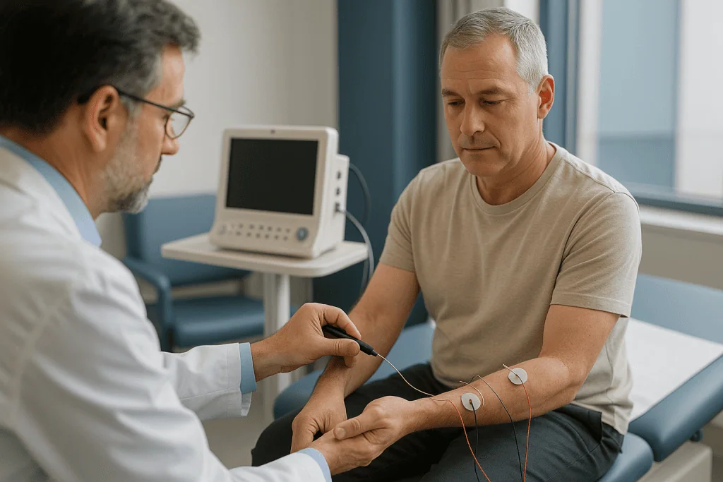
Where the vocabulary does matter is in the pairing you will probably hear next: EMG and NCS. A nerve conduction study, usually shortened to NCS, is a separate measurement that tests how fast and how strongly an electrical signal travels along a nerve. The two are often described together, booked together and reported together, which is why so many people assume they are one test. They are two tests that complement each other, and the next section explains how.
You may also hear surface EMG and needle EMG. Surface EMG uses stick-on electrodes on the skin and is common in research, rehabilitation and biofeedback. Needle EMG, the version used for diagnosing nerve and muscle disease, uses a fine electrode inserted into the muscle. When a neurologist or physiatrist orders an EMG for numbness or weakness, they almost always mean needle EMG, because surface electrodes cannot pick up the fine-grained activity needed to distinguish nerve from muscle problems.
One more term worth decoding: electrodiagnostic testing. This is the umbrella phrase covering EMG, nerve conduction studies and related techniques. If your referral says electrodiagnostic study, EDX or EMG/NCS, expect the combined appointment described below rather than a single quick measurement.
How an EMG actually works: what the needle hears
Every time you move, a motor neuron, the nerve cell that commands muscle, fires an electrical impulse down its fiber to a cluster of muscle cells it controls. That cluster, the neuron plus its muscle fibers, is called a motor unit. When the fibers contract they generate a small voltage, and that voltage is what the EMG electrode records.
The needle electrode is thin, closer to an acupuncture needle than a blood-draw needle, and it carries no injection. Once it sits inside a muscle, the examiner studies three things in turn.
First, the muscle at rest. A healthy resting muscle is almost silent. If fibers have lost their nerve supply, they begin to fire spontaneously, producing crackling signals called fibrillations and positive sharp waves. Their presence tells the examiner that nerve damage has reached the muscle, and roughly how long it has been there, because these changes take time to develop after an injury.
Second, a gentle contraction. You are asked to tense the muscle just slightly, and the examiner looks at the shape and size of individual motor unit potentials on the screen. Units that are unusually large and long suggest nerves have regrown to cover for lost neighbors, a sign of a chronic nerve problem. Units that are small and brief point toward disease of the muscle itself.
Third, a stronger contraction. The examiner watches how many units are recruited and how quickly. Nerve loss leaves fewer units working harder; muscle disease leaves many units firing early but weakly.
The signals are shown as waveforms and also played through a loudspeaker, which is why you will hear static, pops and a sound like rain on a tin roof during the test. Experienced examiners read the sound as much as the screen. Cleveland Clinic notes the test is interpreted by a specialist trained in electrodiagnostic medicine, and the interpretation, not the raw tracing, is what reaches your doctor.
EMG nerve conduction study: why most appointments include both
If the needle EMG listens to muscle, the nerve conduction study measures the wire. The examiner places small surface electrodes on the skin over a nerve and the muscle it supplies, then delivers a brief electrical pulse at one point and records when and how strongly the signal arrives further along. Dividing distance by time gives conduction velocity; the height of the response reflects how many nerve fibers are still working.
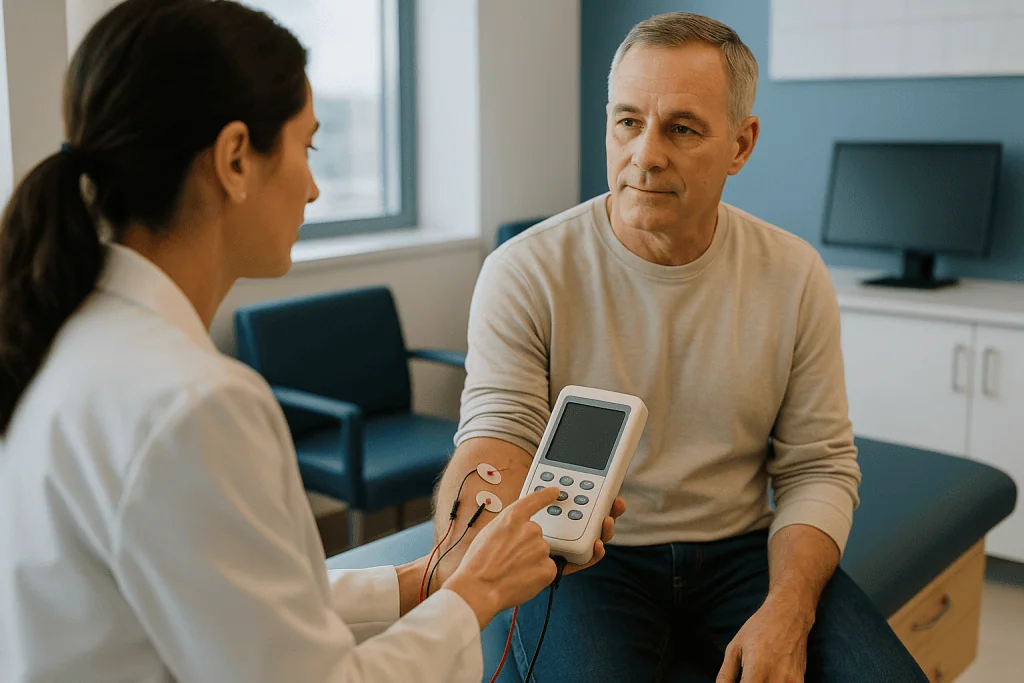
The two measurements answer different questions, and together they are far more informative than either alone.
- A slow signal with a preserved response suggests the nerve’s insulating sheath, the myelin, is damaged, as in carpal tunnel syndrome where the median nerve is compressed at the wrist.
- A normal speed with a small response suggests nerve fibers themselves have been lost, a pattern common in many diabetic and toxic neuropathies.
- Normal nerve conduction alongside abnormal needle findings points the examiner toward the nerve roots near the spine, or toward a muscle disease, because the nerve segments studied in the limb are intact.
A third technique, repetitive nerve stimulation, delivers a short train of pulses to see whether the muscle response fades. Fading suggests a problem at the neuromuscular junction, the tiny gap where nerve meets muscle, which is the mechanism behind myasthenia gravis. MedlinePlus lists this group of disorders among the conditions electrodiagnostic testing is used to evaluate.
In practice, the nerve conduction portion usually comes first because its results tell the examiner which muscles are worth sampling with the needle. An appointment ordered as an EMG for hand numbness may therefore involve several minutes of taps and pulses on the forearm before any needle appears. This is not padding; it is the map that keeps the needle portion short and targeted.
What does an EMG test show, and what can it not show?
An EMG is strongest at localization and characterization. It can tell a clinician where along the pathway a problem sits, whether it affects nerve fibers or their insulation, whether muscle has been denervated, whether the process looks recent or long-standing, and whether it is confined to one nerve or spread across many. Those answers steer the next decision: imaging of the neck, blood tests for a metabolic cause, a muscle biopsy, a referral for hand surgery discussion, or simply reassurance and a plan to re-examine.
It is equally important to know what the test does not do.
- It does not image the spine or the brain. A herniated disk can be suspected from an EMG pattern, but the disk itself is seen on MRI.
- It does not measure pain. Someone can have severe pain and a normal study, especially when small nerve fibers, which carry pain and temperature signals, are the ones affected. Standard nerve conduction studies test large fibers only.
- It does not diagnose multiple sclerosis, stroke or other conditions of the central nervous system, because it examines the peripheral nerves and muscles.
- It does not detect problems that are purely sensory in the spinal cord or brain.
A normal EMG is still useful information. It makes certain diagnoses much less likely and shifts attention elsewhere. The NHS, in its guidance on diagnosing peripheral neuropathy, describes nerve conduction and EMG studies as tests that help identify the type and extent of nerve damage, not as a universal screen for every cause of numbness.
Think of the report as one layer of a puzzle. Your history, the examination, blood work and imaging are the other layers, and the treating team reads them together. An EMG finding on its own rarely settles a diagnosis; combined with the rest, it often does.
EMG test for numbness and weakness: who is usually referred, and who is asked to wait
Referral patterns vary, but certain presentations reliably prompt the order. Numbness or tingling that follows the territory of a single nerve, such as the thumb, index and middle fingers in suspected carpal tunnel syndrome, is one. Weakness that is objective on examination rather than a feeling of heaviness is another. So is muscle wasting, visible twitching called fasciculations, or symptoms in both hands and both feet that suggest a generalized neuropathy. The NINDS notes that diabetes is the most common cause of peripheral neuropathy in the United States, and people with diabetes and new foot numbness are frequently tested.
Timing matters more than many patients realize. After a sudden nerve injury, the spontaneous muscle signals the needle looks for take time to appear. Specialists therefore often prefer to schedule the study a few weeks after an acute event rather than immediately, because an early test can look falsely reassuring. If your appointment seems slow to arrive, this may be the reason, and it is worth asking.
Some people are usually asked to wait, or to have a different test first:
- When symptoms are brief, intermittent and improving, since a normal result in between episodes may not reflect the problem.
- When a central cause such as a stroke is suspected, where brain imaging takes priority.
- When the leading question is structural, for instance a tumor or fracture, and imaging will answer it directly.
- When bleeding risk or a skin infection at the planned needle site needs to be addressed first.
Children and people who struggle with needles can still be tested, typically with a shortened protocol and a focus on the muscles most likely to give the answer. The decision about whether, when and how extensively to test belongs to the ordering clinician and the examiner, who adjust the study in real time to what the early findings show.
Conditions an EMG helps sort out, and what the pattern usually looks like
The table below summarizes the conditions most often behind an EMG referral and the broad electrodiagnostic patterns associated with them, drawn from the condition lists published by Mayo Clinic and MedlinePlus. Patterns are simplified; real reports contain far more nuance, and only the treating team can match them to your situation.
| Suspected condition | Where the problem sits | What the study typically examines |
|---|---|---|
| Carpal tunnel syndrome | Median nerve at the wrist | Slowed conduction across the wrist; needle changes in thumb muscles if severe |
| Cervical or lumbar radiculopathy | Nerve root near the spine | Normal limb nerve conduction; needle changes in muscles sharing one root |
| Peripheral neuropathy | Many nerves, usually longest first | Reduced or slowed responses in feet and hands, often symmetric |
| Ulnar neuropathy | Ulnar nerve at the elbow | Slowing across the elbow segment; ring and little finger muscles sampled |
| Myasthenia gravis | Neuromuscular junction | Fading response on repetitive stimulation; needle often normal |
| Myopathy (muscle disease) | Muscle fibers | Small, brief motor units recruiting early; nerve conduction normal |
| Motor neuron disease | Motor neurons in spinal cord and brainstem | Widespread denervation across several body regions with preserved sensory studies |
A few points stand out. Radiculopathy is one of the most common reasons for referral, and it is a good illustration of why both halves of the test are needed: the limb nerves conduct normally, so only the needle reveals the root-level problem. Myasthenia gravis is the reverse; the needle may be unremarkable while the repetitive stimulation test is diagnostic. Motor neuron disease sits at the serious end of the list, and it is the condition people most fear when they search the term. It is also among the least common reasons for a referral, and specialists apply strict criteria across multiple regions before they consider it.
What should you not do before an EMG?
Preparation is light, but a few habits can genuinely interfere with the recording.
- Skip lotions, oils and moisturizers on the limbs being tested on the day of the appointment. Mayo Clinic advises showering to remove skin oils and avoiding creams, because residue raises electrical resistance and weakens the signal the surface electrodes pick up.
- Avoid caffeine and tobacco in the hours beforehand. Johns Hopkins Medicine suggests not smoking for a few hours before the test and limiting caffeine, as both can alter muscle activity and make the tracing harder to read.
- Do not stop a prescribed medicine on your own. Blood thinners, in particular, matter because needle sampling can cause small bleeds. Tell the testing team what you take and let them decide whether any adjustment is needed; that decision sits with your prescribing clinician, not with you or the examiner alone.
- Do not wear tight clothing. The examiner needs access to the forearm, upper arm, lower leg and sometimes the back or neck. Loose sleeves and trousers, or a gown provided on the day, make this easier.
- Do not arrive cold. Cool limbs slow nerve conduction and can mimic disease. Many laboratories warm the hands or feet before testing; arriving with warm layers helps.
Also mention, when booking and again on arrival, if you have a pacemaker, implanted cardiac defibrillator, deep brain stimulator or any other electrical device. Mayo Clinic lists this as something to tell the team in advance, because the nerve conduction pulses may need to be adjusted. The same applies to a bleeding disorder, lymphedema in a limb, or a skin infection where the needle would go.
You can eat normally, and there is no need for fasting or a sedative for most adults. Bring a list of symptoms, when they started, what makes them better or worse, and any previous test results, since the examiner tailors the study to that story.
What happens during the appointment, step by step
Mayo Clinic puts the typical appointment at 30 to 60 minutes, though a study covering several limbs or a complex question can run longer. Here is the usual rhythm.
You will first meet the examiner, often a neurologist or a physical medicine and rehabilitation physician, sometimes working with a technologist. They will ask about your symptoms and examine the affected area briefly. This conversation matters; it determines which nerves and muscles are tested, and a good examiner will explain the plan before touching an electrode.
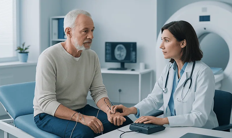
Nerve conduction comes next. Small adhesive electrodes are placed on the skin, the limb may be warmed, and a handheld stimulator delivers brief pulses at several points along a nerve. Each pulse feels like a sharp tap or a static shock and lasts a fraction of a second. The examiner may stimulate the same nerve a dozen times at different sites, measuring responses on the screen between pulses.
Then the needle portion. The skin is cleaned, a fine electrode is inserted into a chosen muscle, and you are asked to relax completely, then to tense slightly, then more firmly, while the examiner watches and listens. Each muscle takes a minute or two. A focused study might sample four or five muscles; a broad one may sample more across both sides of the body. The needle is withdrawn and the site pressed briefly before moving on.
Throughout, you can talk. Tell the examiner if any position is painful or if you need a pause. Examiners routinely adjust the order of muscles, skip ones that will not add information, or stop early once the question is answered.
At the end, you can get dressed and leave without a recovery period. Most people drive themselves home. The examiner may give a brief verbal impression, but the formal report is written afterward and goes to the referring clinician, who interprets it alongside everything else they know about you.
How painful is an EMG nerve test? An honest answer
Not painless, rarely severe, and shorter than most people expect. That is the fairest summary, and it is worth separating the two halves of the test because they feel different.
The nerve conduction pulses produce a sudden, brief jolt that makes the muscle twitch. People describe it as a rubber-band snap, a static shock from a doorknob, or a quick tap. The surprise is often worse than the sensation. By the third or fourth pulse most people have stopped flinching, though stimulation near the elbow, knee or ankle can be sharper than at the wrist.
The needle portion is a different sensation. Insertion feels like a pinprick, similar to or milder than a vaccination, because the electrode is thin and nothing is injected. The more noticeable discomfort comes while the needle sits in the muscle and you contract it, which produces a deep, pressure-like ache. Some muscles, such as those in the hand, the calf or along the spine, are more tender than others. Mayo Clinic describes the procedure as causing some discomfort and notes that the muscle may feel sore or slightly bruised for a few days afterward.
What makes it easier:
- Relaxing the muscle fully between contractions, since tense muscle hurts more and gives noisier signals.
- Breathing out slowly as the needle goes in.
- Telling the examiner immediately if a particular site is sharply painful; the needle can often be repositioned.
- Asking how many muscles are planned, so there is a visible end point.
Anxiety amplifies all of this. People who arrive expecting an ordeal tend to report more pain than those who understand the sequence. A brief, well-run study is more like a series of small, tolerable annoyances than a single painful procedure. If you have a known needle phobia or a chronic pain condition that could flare, tell the team when booking; shortened protocols and extra breaks are common accommodations.
Risks, side effects and who needs extra precautions
Electrodiagnostic testing is among the safer diagnostic procedures, and the risks that exist are small and mostly local. Mayo Clinic lists bleeding, infection and nerve injury at the needle sites as rare possibilities, with minor bruising and soreness as the common after-effects. There is no radiation, no contrast dye and no sedation for routine adult studies.
A few circumstances deserve explicit discussion with the team beforehand:
- Blood thinners and bleeding disorders. Needle sampling in deep muscles, such as those near the spine or in the chest wall, carries a slightly higher bleeding risk. Examiners often choose more superficial muscles or apply longer pressure. Never stop an anticoagulant on your own to prepare for the test; that decision belongs to the clinician who prescribed it.
- Implanted electrical devices. Pacemakers and defibrillators are generally compatible with nerve conduction studies, but the examiner may avoid stimulating near the device or adjust pulse settings. Tell them in advance.
- Lymphedema or a limb with prior lymph node surgery. Many laboratories avoid needles in that limb to reduce infection risk, and will discuss alternatives.
- Pregnancy. Routine limb studies are not known to pose a risk, but mention it so the examiner can avoid unnecessary stimulation near the abdomen.
- Skin infection or open wounds at a planned site, which usually means choosing another muscle or postponing.
Soreness is the most frequent complaint afterward and typically settles within a few days, according to Mayo Clinic. A cool pack and ordinary activity are usually all that is needed. Small bruises at needle sites are common and fade on their own.
What you should not expect: lasting weakness, numbness caused by the test itself, or any effect on a pacemaker’s function when the team has been informed. If something unexpected does occur, the section on when to call your doctor below lists the signs that warrant a prompt call.
The days and weeks after an EMG: results and next steps
The hours after the test are usually uneventful. You can return to work, drive, exercise and bathe as normal. Mild aching at needle sites is expected and, per Mayo Clinic, generally lasts a few days at most. Bruising, if it appears, follows the ordinary course of any small bruise.
The report is the part people wait for. The examiner writes up each nerve and muscle studied, states whether the findings are normal or abnormal, and offers an interpretation: for example, moderate median neuropathy at the wrist consistent with carpal tunnel syndrome, or chronic changes in muscles sharing a single nerve root. Some laboratories share a preliminary impression at the end of the visit; others send the complete report only to the referring clinician. Ask before you leave how and when you will hear, so you are not refreshing a patient portal for a document that will arrive by letter.
What happens next depends entirely on the pattern.
- A single compressed nerve may lead to a discussion of splinting, activity changes, injections or surgical options, weighed against severity on the study.
- A root-level pattern usually prompts spinal imaging if it has not already been done, to see what is pressing on the root.
- A generalized neuropathy pattern typically triggers blood tests to look for diabetes, vitamin deficiencies, thyroid disease or other causes that the NINDS lists as common contributors.
- A muscle or neuromuscular junction pattern may lead to antibody testing, a muscle biopsy or referral to a subspecialist.
- A normal study shifts attention to other explanations, including small fiber neuropathy, central causes or musculoskeletal problems, and may lead to different tests.
Repeat studies are sometimes scheduled months later to measure recovery after an injury or to track a progressive condition. Whether that is needed, and when, is a call for the treating team, who will weigh what a second study would actually change about your care.
What people often get wrong about EMG
Myths cluster around this test, partly because of its name and partly because most people have never watched one. A few deserve direct correction.
It is a scan. No. There is no magnet, no X-ray and no picture of your anatomy. An EMG produces waveforms and sounds that describe function, not structure. If you want to know what a disk looks like, that is MRI’s job.
The doctor suspects something terrible. The test is ordered far more often for carpal tunnel syndrome, pinched nerves in the neck or back, and diabetes-related neuropathy than for rare, serious diseases. Being referred reflects the need for precision, not a hidden fear.
A normal result means nothing is wrong. Standard studies examine large nerve fibers. Small fiber neuropathy, which causes burning pain and temperature changes, can produce an entirely normal EMG. So can early or intermittent conditions. A normal result narrows the search; it does not end it.
The shocks are dangerous. Nerve conduction pulses are brief, low-current and designed to be safe, including for most people with pacemakers when the team is informed. They are uncomfortable, not harmful.
You must stop your medicines first. Most medicines, including the drug classes commonly used for nerve pain, do not affect the test. Blood thinners need a conversation, not a unilateral pause. Only one class, the medicines used to treat myasthenia gravis by boosting signals at the neuromuscular junction, may sometimes be timed around a repetitive stimulation study, and that is a decision for the prescribing clinician.
The needle injects something. It records only. Nothing goes in except the electrode itself.
More muscles tested means a worse prognosis. A broader study often reflects a broader question, such as distinguishing a root problem from a generalized one, rather than worse disease. The number of muscles is a design choice, not a verdict.
The thread running through all of these is the same: the EMG describes, it does not decide. Your care team decides, using the description alongside everything else.
Questions to ask your care team
A good consultation before and after the test leaves you knowing why the study was ordered and what will change because of it. These questions tend to produce the most useful answers.
Before the test:
- What specific question are you hoping the EMG will answer, and what diagnoses are you trying to distinguish?
- Will the appointment include nerve conduction studies, needle EMG or both, and roughly which areas of the body?
- Is there a reason to wait before testing, for example recent injury, and if so how long?
- I take a blood thinner or have an implanted device; does anything need to change before the day, and who makes that decision?
- If the study is normal, what would the next step be?
On the day:
- How many muscles do you plan to sample, and can we pause if I need to?
- Will you be able to tell me a preliminary impression at the end?
- Is the limb warm enough for accurate readings?
After the result:
- Does the report point to the nerve, the nerve root, the muscle or the junction between them?
- How severe does the study suggest the problem is, and does that change the options you would discuss?
- Are the findings consistent with my symptoms, or is there a mismatch we should explore?
- Would a repeat study at some future point tell us anything new?
Bring a notebook or a companion. Electrodiagnostic reports use dense language, and it is easy to leave with the sense of having understood, only to lose the thread at home. Ask the team to write the one-line conclusion in plain words. Most are glad to, and it anchors every later conversation about treatment, which remains, at each step, a shared decision between you and the people responsible for your care.
When to call your doctor
Numbness and weakness are usually slow-moving problems, and an EMG appointment that is a few weeks away is typically appropriate. Some changes, however, should not wait for the test, and a few after-effects of the test itself warrant a call. Contact your doctor promptly, or seek urgent care, if you notice any of the following.
Before or while waiting for the EMG:
- Weakness that appears suddenly, especially on one side of the body, or sudden facial drooping, slurred speech or confusion. These can signal a stroke and need emergency assessment, not an outpatient nerve test.
- Weakness or numbness that spreads rapidly over hours to days, particularly if it climbs from the feet upward or affects breathing or swallowing.
- New difficulty controlling the bladder or bowel, or numbness in the inner thighs and genital area, which can indicate pressure on the lower spinal cord or nerve roots.
- Numbness following a significant injury to the neck, back or a limb.
- Fever, severe back pain and new leg weakness together.
- Rapid, unexplained wasting of muscle, or weakness that is clearly worsening week by week.
After the EMG:
- Increasing redness, warmth, swelling or pus at a needle site, or a fever, which could indicate infection.
- A rapidly enlarging, tense or very painful bruise, especially if you take a blood thinner.
- New weakness or numbness that was not present before the test and does not settle within a day.
- Any concern about an implanted device after the study.
For anything less dramatic, such as mild soreness, small bruises or lingering questions about the report, a routine message or the scheduled follow-up appointment is the right channel. The treating team can judge whether a symptom changes the plan, and that judgment, informed by the EMG but never replaced by it, is where every decision about your care belongs.
Frequently asked questions
Why did my doctor order an EMG for hand numbness?
Hand numbness can come from a nerve compressed at the wrist, at the elbow or at a root in the neck, and these can look alike on examination. An EMG with nerve conduction studies locates the problem along that pathway and grades its severity, which shapes whether splinting, imaging of the neck or a procedural discussion comes next. The result informs the treating team’s decision rather than making it.
Is an electromyogram the same as electromyography?
Yes. Electromyography is the technique and an electromyogram is the recording it produces, just as electrocardiography produces an electrocardiogram. Both are shortened to EMG in referral letters and appointment schedules. If you see both words on different documents, you have one test booked. The term you are more likely to need to clarify is nerve conduction study, which is a separate measurement commonly performed in the same visit.
What should you not do before an EMG?
Avoid lotions and oils on the skin of the limbs being tested, skip caffeine and tobacco for a few hours beforehand, and do not arrive cold, since all three interfere with the electrical readings. Do not stop any prescribed medicine on your own, particularly a blood thinner; tell the testing team what you take and let the prescribing clinician decide. Mention pacemakers or other implanted devices when booking.
What can an EMG tell you about numbness and weakness?
It can show whether the problem lies in a single nerve, a nerve root near the spine, many nerves at once, the muscle itself or the junction between nerve and muscle. It also indicates whether nerve fibers or their insulation are affected, roughly how severe the damage is, and whether changes look recent or long-standing. It cannot measure pain, image anatomy or assess the brain and spinal cord.
How painful is an EMG nerve test?
Uncomfortable rather than severely painful for most people. Nerve conduction pulses feel like brief static shocks that make the muscle twitch, and the needle portion feels like a pinprick followed by a deep ache while the muscle is contracted. Mayo Clinic describes some discomfort during the test and soreness for a few days afterward. Relaxing between contractions and telling the examiner about painful sites help considerably.
What does an EMG test show that an MRI cannot?
Function. MRI shows structure, such as a bulging disk or a narrowed canal, but it cannot tell whether that structure is actually disturbing a nerve’s signals. An EMG shows whether the nerve and muscle are working normally, and if not, where and how badly. Many people have imaging findings that cause no symptoms, so the two tests are often read together to decide which abnormality matters.
How long does an EMG nerve conduction study take?
Mayo Clinic gives a typical range of 30 to 60 minutes for the combined appointment, though a focused study of one wrist can be shorter and a broad study of several limbs can take longer. The nerve conduction portion usually comes first and guides which muscles are sampled with the needle. There is no recovery period, and most people leave and resume normal activities immediately.
Can I drive myself home after an EMG test?
Yes, in nearly all cases. The test involves no sedation, no anesthesia and no effect on coordination, so driving, working and exercising afterward are all fine. The only common after-effects are mild soreness and small bruises at needle sites. If you have a needle phobia and have arranged a sedative with your clinician, follow their specific guidance about transport.
Is an EMG safe with a pacemaker or on blood thinners?
Usually, with precautions. Nerve conduction studies are generally compatible with pacemakers and defibrillators when the team knows in advance and adjusts stimulation sites and settings. Blood thinners raise the small risk of bleeding at needle sites, so examiners may choose superficial muscles and apply longer pressure. Never stop an anticoagulant on your own; the prescribing clinician decides whether any change is appropriate.
What happens if my EMG is normal but I still have symptoms?
A normal study narrows the possibilities rather than dismissing them. Standard testing examines large nerve fibers, so small fiber neuropathy, early or intermittent conditions, and central causes in the brain or spinal cord can all produce normal results. Your doctor may order blood tests, imaging, specialized skin biopsy or a repeat study later. The finding is one piece of evidence the treating team weighs with everything else.
References
- MedlinePlus: Electromyography (EMG) and Nerve Conduction Studies
- Cleveland Clinic: EMG (Electromyography)
- NHS: Peripheral neuropathy, Diagnosis
- NIH NINDS: Peripheral Neuropathy
This article is for general information only and is not a substitute for professional medical advice. Please consult a qualified doctor about your individual situation.

More from the Blog

How to Prepare for an Ultrasound: Why Some Scans Need Fasting and Others a Full Bladder
Preparing for an ultrasound depends on the body part being scanned. Abdominal scans of the liver, gallbladder or pancreas usually require fasting for several…
What Doppler Ultrasound Adds: How Blood-Flow Imaging Changes a Vascular or Pregnancy Exam
Doppler ultrasound is a form of ultrasound that measures how blood moves through vessels, not just how tissue looks. It uses the Doppler effect:…

Why a Doctor Orders a Liver Ultrasound, and What It Can Find
A doctor usually orders a liver ultrasound to look for a physical explanation for abnormal liver blood tests, pain or fullness under the right…
What Happens During an Ultrasound Scan? The Gel, the Probe and How Long Each Exam Takes
During an ultrasound scan you lie on a couch while a sonographer spreads water-based gel on your skin and moves a handheld probe over…

Who Benefits From a Comprehensive Check-Up? How Age and Family History Shape the Test List
Most adults benefit from a periodic comprehensive check-up, but the right test list depends on age, sex, family history, and existing conditions rather than…
EMG vs Nerve Conduction Study vs MRI: Which Test Finds the Cause of Numbness and Weakness
EMG, nerve conduction studies and MRI answer different questions. Nerve conduction studies measure how fast and how strongly a nerve carries electrical signals; EMG…