

Why Does Volume Settle After Breast Fat Transfer? Results Surgeons Expect Over the Months

Key Takeaways
- Grafted fat cells survive only if new capillaries reach them within the first days, so cells placed in thick clumps or far from breast tissue are reabsorbed rather than retained.
- The breast is largest in the first week because swelling and non-surviving fat are both present; most volume loss occurs between roughly weeks two and month four before plateauing.
- Systematic reviews report retained volume that varies widely between studies and techniques, which is why no surgeon can reliably promise a personal survival percentage.
- Fat that survives past about six months is permanent tissue but shrinks and grows with the person's overall weight, unlike an implant.
- Oil cysts and calcifications from fat necrosis can appear on mammograms, so every future radiologist should be told that fat grafting was performed.
- Larger changes are usually staged because each session can only add what the existing tissue can nourish, and the second session is typically planned after the first has plateaued.
Volume settles after breast fat transfer because only part of the injected fat survives. Fat cells must gain a new blood supply within days; those that do not are reabsorbed over weeks to months, while surgical swelling also fades. Surgeons typically judge the lasting result at around six months, and published reviews report that retained volume varies widely between patients and techniques.
Three weeks after surgery, she stands sideways in front of the bathroom mirror and feels quietly pleased. The bruising along her hips has faded to a yellow tide line, and her chest looks fuller than the photographs she brought to her first consultation. Then she remembers the line her surgeon repeated twice: “What you see now is not what you keep.”
That sentence is the heart of understanding fat transfer breast augmentation results. Unlike an implant, which arrives at a fixed size, grafted fat is living tissue moved from one part of the body to another. Some of it settles in and stays for years. Some of it does not survive the journey. The breast a person sees in the early weeks is a mixture of both, plus swelling, and the proportions shift month by month.
This explainer follows that shift from operating room to the one-year review, using what mainstream surgical evidence actually shows rather than what social media promises.
What actually happens during fat transfer breast augmentation
Fat transfer breast augmentation, sometimes called autologous fat grafting, moves a person’s own fat from one body region to the breasts. “Autologous” simply means the tissue comes from the same person. The procedure has three stages, and each one influences how much volume eventually stays.
The first stage is harvest. A surgeon performs liposuction, a technique that removes fat through a thin hollow tube called a cannula, usually from the abdomen, flanks, or thighs. Fluid containing local anesthetic and a small amount of a blood-vessel-constricting medicine is often infused first to reduce bleeding and bruising; this is known as a tumescent technique. Mayo Clinic notes that liposuction itself is a surgical procedure with its own recovery and risks, not a minor add-on.
The second stage is processing. The harvested material is a slurry of fat cells, oil from ruptured cells, blood, and infused fluid. Surgeons separate the intact fat by letting it settle, spinning it, rinsing it, or filtering it. The aim is to inject fat cells that are as undamaged as possible, because damaged cells will not survive.
The third stage is placement. Using fine cannulas, the surgeon deposits the fat in many small threads across different depths of the breast, under and within the existing breast tissue and over the chest muscle. Spreading it thinly matters: a fat cell can only draw nutrients from tissue within a fraction of a millimeter, so a large single pocket of fat starves at its center.
The whole operation is usually done under general anesthesia or deep sedation and, depending on how much is harvested and placed, takes a few hours. Because the added volume is limited by how much fat the breast can safely accept in one sitting, a substantial size change is frequently planned as more than one session.
Why does volume settle after breast fat transfer? The biology of graft take
The settling that patients notice is not the fat “leaking” or being squeezed out. It is a biological sorting process that decides which transplanted cells become permanent residents.

A fat cell, or adipocyte, is essentially a storage bubble with a thin rim of living machinery. When it is moved, it loses the tiny blood vessels that fed it. For the first few days it survives by soaking up nutrients from the surrounding tissue fluid, a phase called imbibition. That is a stopgap, not a solution.
Over the following days, new capillaries grow toward the graft from the surrounding breast tissue, a process called neovascularization. Cells that sit close enough to be reached by those vessels resume normal life. Cells buried deeper inside a clump, or damaged during harvest and processing, die. Their contents are broken down and cleared by the body’s scavenger cells, and the volume they occupied disappears.
Some of the dead cells do not clear cleanly. Their oil can pool into an oil cyst, a small fluid-filled pocket, or the area can scar and, over time, pick up calcium deposits. This is fat necrosis, meaning fat tissue that has died, and it is discussed later because it has implications for breast imaging.
Layered on top of this cellular story is ordinary surgical swelling. Every injected thread of fat comes with a small amount of fluid and triggers inflammation, and the breast tissue itself swells in response to hundreds of cannula passes. Swelling inflates the early result, then drains over several weeks.
Put those two timelines together and the pattern becomes predictable: an early peak, a steady decline through the first three to four months, and then a plateau once surviving cells have a stable blood supply and swelling is gone.
Fat transfer breast augmentation results: what the first weeks and months usually look like
Timelines below are typical ranges drawn from general surgical recovery guidance published by Mayo Clinic and MedlinePlus, not promises, and an individual’s course belongs to their surgical team to interpret.
| Stage | Breasts | Donor areas | What surgeons are watching for |
|---|---|---|---|
| First week | Firm, swollen, tender; largest they will appear | Bruising, soreness, fluid drainage; compression garment usually worn | Infection, bleeding, wound problems |
| Weeks 2 to 6 | Swelling eases; volume visibly decreasing | Bruising fades; numbness or firm patches common | Early lumps, uneven settling |
| Months 2 to 4 | Softening; most reabsorption happens here | Contour changes become clearer | Fat necrosis, asymmetry |
| Months 6 to 12 | Stable volume; final assessment usually made | Residual swelling gone; skin retraction settled | Need for a further session |
Mayo Clinic describes liposuction swelling and bruising that can take several weeks to subside, with final contour visible only after several months, and MedlinePlus gives similar recovery guidance for breast surgery. The breast follows a parallel arc because it is recovering from the same kind of tissue trauma.
Two practical points flow from that table. Judging the outcome before month three is judging swelling, not fat. And the donor site deserves as much attention as the breast: liposuction leaves its own contour that also changes for months, and complications such as fluid collections (seromas) or irregularities occur there, not in the chest.
How much fat survives? What the evidence actually shows
Patients ask for one number, and honesty requires refusing to give one. Systematic reviews of autologous fat grafting to the breast indexed on PubMed describe retained volume that varies considerably from study to study, with many reports clustering somewhere around half to two-thirds of the injected amount at follow-up of six months or longer, and individual studies falling well outside that band in both directions.
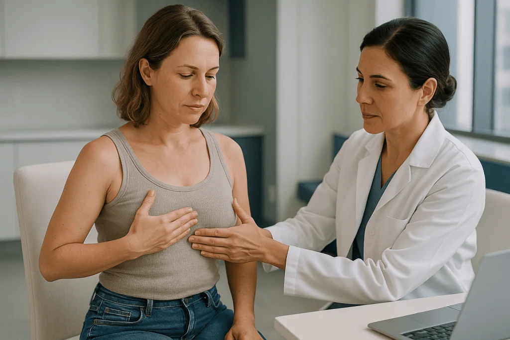
Why so wide? Several reasons stand out in the literature.
Measurement differs. Some studies estimate volume by tape measure or photographs; others use three-dimensional surface scanning or MRI. The same breast can produce different retention figures depending on the tool.
Technique differs. Harvest pressure, processing method, cannula size, injection speed, and how thinly the fat is layered all affect how many cells survive, and few of these steps are standardized between surgeons or trials.
Recipient tissue differs. A breast with more space and better blood supply relative to the volume placed tends to hold more. Overfilling a tight breast produces higher early volume and lower late volume, because the excess cannot be fed.
Patients differ. Body weight stability, smoking status, age, and underlying health all influence healing and blood-vessel growth.
The responsible takeaway is that a surgeon who quotes a precise personal survival percentage in advance is speculating. What experienced teams do instead is plan for loss: they place a calculated surplus, accept that the early appearance will exceed the target, and set expectations for a review at around six months when the retained volume can be assessed and a further session considered if the person wants more.
Who is fat transfer breast augmentation usually for, and who is asked to wait
Suitability is a clinical judgment, but the pattern in mainstream guidance is consistent.
The procedure is most often considered for people seeking a modest increase, typically described as up to about one cup size per session, who want to avoid an implant, or who want to soften the edges of an existing implant or correct minor asymmetry. It is also widely used in breast reconstruction after cancer surgery to refine shape, a setting where the same biology applies.
A person also needs enough fat to harvest. Someone very lean may not have adequate donor tissue for a meaningful change, and taking too aggressively from a small reserve raises the risk of contour irregularities where the fat was removed.
Several groups are commonly asked to pause or reconsider. People who smoke or vape nicotine are usually asked to stop for a period before and after surgery, because nicotine narrows blood vessels and impairs the new capillary growth on which graft survival depends. People actively losing or gaining weight are often asked to reach a stable weight first, for reasons covered below. Anyone with uncontrolled diabetes, bleeding disorders, or conditions that impair wound healing needs those addressed with their physicians beforehand. And people with a personal or strong family history of breast cancer, or with prior abnormal imaging, warrant a careful discussion about how grafted fat may affect future mammograms.
The NHS advises anyone considering cosmetic surgery to check that the surgeon is appropriately registered and to expect a cooling-off period of about two weeks between consultation and consent. That pause exists precisely because expectations about outcome, including settling, are often clearer after a second conversation than a first.
Fat transfer vs breast implants: how the results behave differently
The most common decision people face is between grafted fat and an implant, and the two behave so differently after surgery that comparing them side by side clarifies what “settling” really means.
| Feature | Fat transfer | Implants |
|---|---|---|
| Size change per operation | Modest; larger changes usually staged | Wide range chosen before surgery |
| Early appearance | Larger than final; volume declines for months | Sits high and firm at first; drops into position over weeks |
| Final volume | Uncertain until roughly six months | Predictable; matches the device |
| Response to weight change | Grafted fat gains and loses with the rest of the body | Unchanged |
| Feel | Natural breast tissue | Depends on device and coverage |
| Incisions | Small entry points on breast and donor areas | Breast incision, typically longer |
| Long-term considerations | Fat necrosis, oil cysts, imaging changes | Rupture, capsular contracture, likely revision or replacement over time |
| Additional procedure | Liposuction with its own recovery | None |
Mayo Clinic and the NHS both stress that implants are not lifetime devices and that further surgery is likely at some point. Fat carries no device to fail, but it carries the biological uncertainty described throughout this article, plus a second surgical site.
Neither approach is superior in the abstract. Someone who wants a large, guaranteed increase in one step is generally steered toward an implant. Someone who wants a subtle change, has fat to spare, and accepts a staged plan may be steered toward grafting. Many people ultimately combine both. The choice belongs in a consultation, not a comparison table.
Why one breast may settle differently than the other
Asymmetry after fat transfer surprises people who started symmetrical, and it is more common than the glossy photographs suggest.
Most breasts are slightly uneven before surgery, and surgeons often place different volumes on each side to compensate. Because retention is proportional rather than absolute, the side that received more fat also loses more in absolute terms, and the two sides may not shrink in step.
Blood supply is not identical on both sides either. Prior surgery, a previous biopsy, radiation treatment, or simply anatomical variation can leave one breast better able to nourish new tissue. Scar tissue from earlier operations is particularly poor at growing new vessels, which is why grafting into previously operated or irradiated breasts is often planned more conservatively.
Sleeping position and early compression can play a small role too. Sustained pressure on a fresh graft in the first days may reduce blood flow to that region, which is one reason many surgeons advise sleeping on the back and avoiding tight underwire bras for a period; the exact instructions are the surgical team’s to give.
Then there is measurement. A difference that is invisible clothed can be obvious in a mirror, and the human eye is very good at detecting small side-to-side differences in a paired structure.
Reasonable expectations help here. Minor asymmetry after the first session is not a failure; it is information. It tells the surgeon which side holds fat better, and a second, smaller session can be planned to even things out once the first has fully settled. Chasing symmetry before month six risks adding fat to a breast that is still shrinking.
Fat necrosis after fat transfer: lumps, oil cysts, and what they mean for mammograms
A new lump in a breast is frightening in any context, and it is one of the more common findings after fat grafting. Knowing the likely explanation matters, but so does knowing that a lump still needs to be assessed rather than assumed.
Fat necrosis is the medical term for fat tissue that has died. After grafting it happens when a cluster of injected cells sits too far from a blood supply. The body walls off the dead tissue. Sometimes the liquefied contents collect into an oil cyst, a smooth, sometimes tender nodule that can appear weeks to months after surgery. Sometimes the area scars and, over time, calcifies, meaning calcium salts deposit within it.
These changes matter for screening because mammograms look for exactly those features, calcifications and masses, as possible signs of cancer. Reviews of fat grafting to the breast indexed on PubMed note that the imaging changes it produces are generally distinguishable from malignant patterns by experienced radiologists, but that additional views, ultrasound, or occasionally biopsy may be needed to be sure. That is not a reason to avoid the procedure; it is a reason to make sure every future radiologist knows it was done.
Practical steps that mainstream guidance supports: tell any breast imaging service about prior fat grafting, including approximate dates; keep operative records; and ask the surgical team whether a baseline mammogram or ultrasound before or after surgery is advisable for your age and risk profile. Screening intervals themselves do not change because of grafting; they remain a decision for the person’s primary care physician or breast specialist.
Small, stable oil cysts often need no treatment. Larger, painful, or enlarging lumps are sometimes drained or removed. The decision sits with the treating team after imaging.
Does breast fat transfer last? How weight change affects the result
Once a grafted fat cell has survived the first few months and secured its blood supply, it behaves like any other fat cell in the body. That single fact answers most questions about longevity.
Surviving cells are considered permanent in the sense that they do not spontaneously disappear later. They do, however, respond to the body’s overall energy balance. If a person gains weight, those cells store more fat and the breasts enlarge; if a person loses weight, the cells shrink and so does the breast volume, just as the abdomen or thighs would.
There is a nuance surgeons sometimes mention. Fat cells appear to retain some memory of where they came from. Fat harvested from the abdomen and placed in the breast may respond to weight change more like abdominal fat than like native breast fat. The evidence for how strongly this occurs in humans is limited, so it is better described as a possibility to keep in mind than a rule.
This is why a stable weight before surgery is more than a preference. A person who is mid-way through significant weight loss will see the grafted breast shrink along with everything else and may conclude the surgery failed when the fat in fact survived. Conversely, someone who gains weight after surgery may see the breasts become larger than planned, sometimes unevenly.
Pregnancy and breastfeeding add another variable. Native breast tissue enlarges and then involutes, and grafted fat within it follows the person’s overall weight pattern. Hormonal changes at menopause alter breast composition too. Grafted fat is durable, but it lives in a body that changes, and the honest way to describe fat transfer breast augmentation results over years is “lasting, within the limits of your own body’s changes.”
Why some people need a second session, and when it is usually planned
Being told a second session may be needed can feel like being told the first one did not work. In reality, staging is often part of the plan from the start.
The limiting factor is not how much fat can be harvested but how much the breast can nourish. Every square millimeter of recipient tissue can support only so many new cells. Inject beyond that capacity and the surplus simply dies, adding to fat necrosis without adding to lasting volume. Surgeons therefore place what the tissue can plausibly feed and stop.
Once that first layer has survived and become vascularized, it is itself living tissue with blood vessels. The breast is now a slightly larger, better-supplied recipient site than it was before, and it can accept another layer. This is why sequential sessions can build more total volume than a single oversized one.
Timing follows the biology described earlier. Most surgeons wait until the first graft has clearly plateaued, commonly around six months, before assessing whether more is wanted and how much. Operating earlier means grafting into tissue that is still inflamed and still losing volume, which makes both planning and judging the outcome unreliable.
A second session also means a second round of liposuction. Donor sites that were already treated may have less accessible fat and more scar tissue, so a different area is sometimes used. The recovery is broadly similar to the first, and the same settling curve applies.
Not everyone wants or needs a second round. Some people are content with a modest change. Others, after seeing how their body holds fat, decide an implant would suit their goal better. Both are legitimate outcomes of a first session done well.
What people often get wrong about fat transfer breast augmentation results
Myths around this procedure tend to come from compressing a months-long biological process into a single “after” photograph. A few deserve direct correction.
“Massage or special bras help the fat take.” There is no mainstream evidence that massaging grafted fat improves survival, and pressure on a fresh graft may reduce blood flow. Surgeons typically advise the opposite: protect the area and avoid compression on the breast in the early weeks.
“Eating more fat feeds the graft.” Newly grafted cells depend on capillary growth, not dietary fat. Weight gain after surgery enlarges all fat cells, including the grafted ones, but it also enlarges everywhere else and cannot be targeted.
“What I see at two weeks is my result.” The early appearance is the combination of surviving fat, fat that will be reabsorbed, and swelling. Mayo Clinic’s guidance on liposuction recovery describes final contour emerging over several months, and the breast follows the same course.
“Fat transfer causes or hides breast cancer.” Systematic reviews indexed on PubMed have not shown that grafting increases breast cancer risk in people without a prior diagnosis, and imaging changes such as oil cysts and calcifications can generally be distinguished by radiologists who know grafting occurred. The evidence in people with a prior cancer is still evolving, which is why that group needs individualized advice.
“A good surgeon gets near-total survival.” Retention varies with technique, but no method reliably achieves it. Anyone who guarantees a specific percentage is stepping beyond what the literature supports.
“Stem cell enrichment makes it permanent.” Techniques that concentrate cells from fat before grafting remain under study; mainstream evidence does not establish them as superior, and they should be treated as investigational unless a surgeon can point to published data.
Risks and alternatives in plain language
Every operation carries risk, and fat transfer carries two sets: one for the breast and one for the donor areas. Neutral description helps people weigh them.
At the breast, the main issues are fat necrosis and oil cysts, described earlier; infection, which is uncommon but possible at any surgical site; bleeding or bruising; asymmetry; loss of more volume than expected; and temporary changes in nipple sensation. Very rarely, injected fat can enter a blood vessel and travel elsewhere, a fat embolism, which is why placement technique and cannula design receive so much attention in training.
At the donor areas, MedlinePlus and Mayo Clinic list contour irregularities such as dents or waviness, fluid collections called seromas, prolonged numbness, skin discoloration, and, uncommonly, injury to deeper structures. Because compression garments and positioning matter here, following the team’s instructions closely is part of the treatment.
Systemic risks apply as with any surgery under anesthesia: reactions to anesthetic agents and blood clots in the legs or lungs. Early walking after surgery is the standard clot-prevention measure, and people are typically asked to avoid prolonged immobility in the first days.
Alternatives depend on the goal. Implants offer a larger, more predictable size change with different long-term trade-offs. A breast lift without added volume reshapes rather than enlarges. Combining a small implant with fat to soften its edges is common. And doing nothing is a legitimate choice; the NHS encourages people considering cosmetic surgery to take time, discuss motivations, and seek a second opinion if anything feels rushed.
None of these options is right for everyone. The comparison is between a specific person’s anatomy, goals, and health and what each approach can realistically deliver, and that conversation belongs with the treating surgeon.
Questions to ask your care team before fat transfer to the breast
A consultation is more useful when the questions are specific to how volume behaves over time. These are the ones that tend to change decisions.
- How much fat do you plan to place in each breast, and how much do you expect to remain at six months for someone with my tissue and health profile?
- Will you overfill to allow for loss, and what will the early appearance look like compared with the target?
- Are you planning one session or staging the result, and what would prompt a second?
- Which donor areas would you use, and how might those areas look afterward?
- How do you process the harvested fat, and why do you use that method?
- What is your approach if one side holds fat better than the other?
- How should I sleep, dress, and move in the first weeks to protect the graft?
- Should I have baseline breast imaging before surgery given my age and family history?
- How will future mammograms be affected, and what should I tell a radiologist?
- What lumps or changes should I report, and which are expected?
- What are your registration and training credentials, and who covers emergencies after hours?
- If I lose or gain weight later, how would that change the result?
Write down the answers. The NHS recommends a cooling-off period of around two weeks before consenting to cosmetic surgery, and a second appointment to revisit these points is time well spent. A team that welcomes detailed questions about settling and retention, rather than deflecting them with photographs, is demonstrating the realism that the procedure requires.
When to call your doctor after fat transfer breast augmentation
Most of the changes described in this article are expected: swelling that eases, volume that declines, bruising at the donor sites that fades over weeks. Some changes are not, and they need the surgical team the same day or emergency care immediately.
Contact your surgical team promptly if you notice a breast or donor area becoming increasingly red, hot, swollen, or hard rather than gradually improving; fever or chills; spreading redness or pus from any incision; a rapidly enlarging, tense, or painful area that may indicate bleeding under the skin; a new lump that grows quickly or becomes very tender; sudden loss of volume on one side; or numbness or skin color change that worsens after the first days.
Seek emergency care without waiting for a callback if you develop shortness of breath, chest pain, coughing up blood, sudden confusion, a rapid heartbeat, fainting, or pain and swelling in one calf or thigh. These can indicate a blood clot in the lung or leg or, very rarely, fat that has entered the circulation, and all are treated as emergencies after any surgery.
Between those extremes sit changes that are worth a phone call rather than a search engine: a lump you cannot explain, asymmetry that seems to be increasing rather than stabilizing, or persistent pain beyond what the team predicted. Nobody on a surgical team minds hearing about something that turns out to be an oil cyst; every team would rather know about an infection at day three than day ten.
Beyond the recovery period, keep ordinary breast awareness. Report any new lump, skin change, or nipple discharge to your primary care physician or breast specialist, tell them you have had fat grafting, and keep routine screening on the schedule they recommend. Grafted fat does not exempt a breast from the same care as any other.
Frequently asked questions
Why do my breasts look smaller a month after fat transfer than they did in the first week?
Because swelling is going down and fat cells that did not gain a blood supply are being reabsorbed. Both processes peak in the first weeks and continue for several months. The first-week size includes fluid and inflammation that were never going to stay. Surgeons generally assess the true retained volume at around six months, and comparing yourself with week one will always look like loss.
Does breast fat transfer last, or does the fat go away completely?
Fat that survives the first few months and becomes vascularized is lasting tissue and does not spontaneously disappear later. It does behave like fat elsewhere in the body, enlarging with weight gain and shrinking with weight loss. Total disappearance is not the expected course; partial loss in the early months followed by a stable plateau is.
Fat transfer vs breast implants: which settles more predictably?
Implants settle more predictably in terms of volume, because the size is fixed before surgery and only position changes afterward. Fat transfer settles in volume as well as position, and the final amount cannot be known until months later. Implants carry their own long-term considerations, including likely future surgery, so predictability is one factor among several rather than a deciding one.
What is fat necrosis after fat transfer, and is it dangerous?
Fat necrosis is fat tissue that has died because it did not receive a blood supply. It can form a smooth oil cyst or a firm, sometimes calcified nodule. It is not cancer and is usually harmless, but any new breast lump should be examined and imaged rather than assumed benign. Larger or painful areas are sometimes drained or removed by the surgical team.
How long is fat transfer breast recovery time before I see the final result?
Most surgeons consider the result stable at around six months, with some continued softening up to a year. Physical recovery is faster: Mayo Clinic describes liposuction bruising and swelling that ease over several weeks, and most people return to desk work within one to two weeks and unrestricted activity within about six. Seeing the final volume simply takes longer than feeling recovered.
Can I do anything to help the fat survive after surgery?
The measures with a plausible basis are avoiding nicotine in all forms, keeping weight stable, following the team’s advice on sleeping position and avoiding compression of the breasts early on, and attending follow-up. Massage, special diets, and supplements have no mainstream evidence of improving graft survival. Most of what determines retention is decided by technique and tissue before the person wakes up.
Will fat transfer affect my mammograms?
It can. Oil cysts and calcifications from fat necrosis are visible on imaging and may prompt additional views or an ultrasound. Radiologists who know grafting was performed can usually distinguish these from suspicious findings. Always tell the imaging service about prior fat grafting, keep your operative records, and continue screening on the schedule your physician recommends.
Why is my surgeon suggesting a second session?
Usually because the amount of fat a breast can nourish in one operation is limited, so a larger change is built in layers. Once the first graft has survived and developed blood vessels, it becomes better tissue for accepting more. The second session is typically planned after the first has plateaued, commonly around six months, and only if the person wants additional volume.
Does losing weight after fat transfer undo the results?
It reduces them in proportion to the weight lost, because grafted fat cells shrink along with fat elsewhere. The cells themselves remain. This is why surgeons prefer a stable weight before surgery and why someone planning significant weight loss is often asked to reach their goal first. Regaining weight enlarges the cells again, though not always evenly.
Is fat transfer to the breast safe for people with a history of breast cancer?
It is widely used in breast reconstruction after cancer surgery, and systematic reviews indexed on PubMed have not demonstrated an increased recurrence risk in most groups studied, though the evidence continues to evolve and follow-up in many studies is limited. Anyone with a personal cancer history needs individualized advice from their oncology and surgical teams before deciding.
References
- MedlinePlus Medical Encyclopedia: Breast augmentation surgery
- NHS: Breast enlargement (breast implants)
- NHS: Liposuction
- PubMed: systematic reviews of autologous fat grafting to the breast and volume retention
This article is for general information only and is not a substitute for professional medical advice. Please consult a qualified doctor about your individual situation.

More from the Blog
How Gynecomastia Surgery Is Performed: Gland Excision, Liposuction and the Incision Placement
A gynecomastia surgery procedure removes enlarged male breast tissue using two techniques that are often combined: liposuction, which suctions out excess fat through small…
When Breast Prosthesis Removal Is Considered: Rupture, Discomfort and Changing Preferences
Breast implant removal is usually considered for one of three broad reasons: a problem with the device itself, such as rupture or capsular contracture;…
Gynaecomastia Explained: Hormonal and Medical Causes and How Male Breast Reduction Is Planned
Gynaecomastia is a benign enlargement of glandular breast tissue in men and boys, caused by a shift in the balance between estrogen and testosterone.…
Breast Augmentation Incision Options: Under the Fold, Around the Areola or in the Armpit
Breast augmentation incision options are the inframammary incision in the fold beneath the breast, the periareolar incision along the edge of the areola, and…
Can You Breastfeed After Nipple Aesthetics Procedures? Sensation, Ducts and Planning Ahead
Many people can breastfeed after nipple surgery, but how much milk they make depends on which structures the operation touched: the milk ducts running…
Can Breast Reduction Relieve Neck and Back Strain? How the Decision Is Made With Your Surgeon
Breast reduction surgery can ease neck, shoulder and upper back strain for many people whose symptoms are linked to heavy breast tissue, and major…