

Why Leukemia and Lymphoma Plans Are Built by Hematology and Oncology Together

Key Takeaways
- Most US physicians who treat leukemia and lymphoma completed a combined hematology and oncology fellowship, so the two specialties are usually one clinician backed by a multidisciplinary team.
- Diagnosis rests on a blood smear and bone marrow or lymph node biopsy read with flow cytometry, chromosome analysis and gene testing, and the plan is normally finalized only after all three return.
- Chronic lymphocytic leukemia and indolent lymphomas are often monitored rather than treated at diagnosis, because trials have not shown that earlier chemotherapy helps people live longer.
- Acute lymphoblastic leukemia treatment runs through induction, consolidation and maintenance, with the NHS describing maintenance as commonly lasting about two years, largely at home.
- After a stem cell transplant the NHS describes a hospital stay of several weeks and a recovery period that can extend to a year or more, with infection and graft-versus-host disease the main risks.
- Transfusions and preventive antimicrobials during low-count weeks are planned parts of therapy, not signs of failure, and a fever during that window is treated as an emergency.
Leukemia and lymphoma begin in blood-forming and immune cells, so the specialists who read blood counts and bone marrow (hematology) and those who plan cancer therapy (oncology) work as one team, often the same physician. Together they confirm the exact subtype, assess genetic risk, choose whether to treat now or monitor, and manage the blood and infection complications that treatment itself creates.
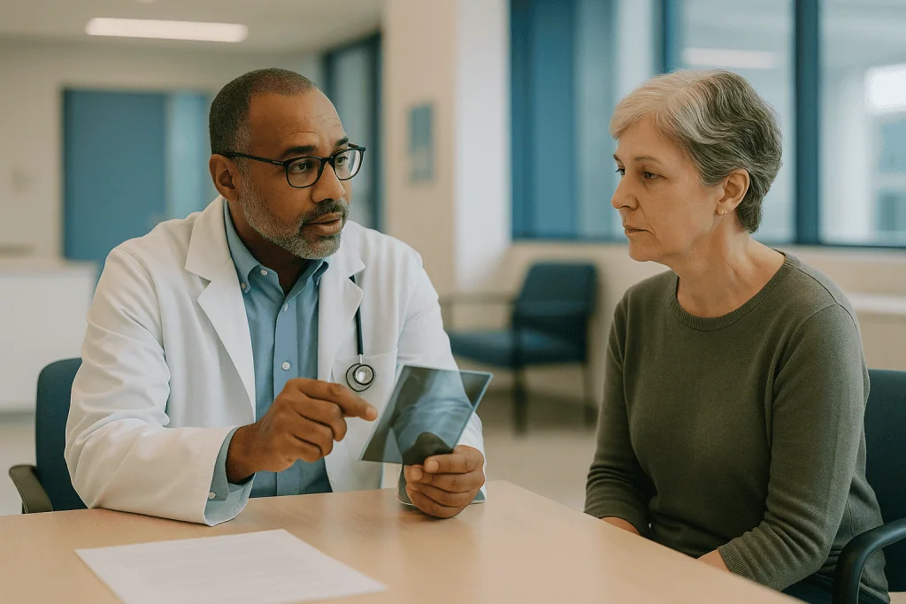
The first appointment usually ends with a question nobody expected: “Which doctor is actually in charge?” A woman in her fifties, referred after a routine blood test came back with white cells nobody could explain, sits with a folder of results and the name of a specialist she has never heard of. The badge reads hematology and oncology, two words that sound like two departments. Her son wants to know why there is not simply a cancer doctor.
The honest answer is that the hematology oncology team leukemia lymphoma patients meet is not a committee bolted together for convenience. It reflects where these cancers live. Leukemia starts in the marrow that manufactures blood. Lymphoma starts in the immune cells that patrol it. Treating either one means understanding blood the way a mechanic understands an engine, while also planning chemotherapy, immunotherapy or transplant the way any cancer specialist would.
What follows explains how that shared plan comes together, what happens in the first weeks, and which questions are worth asking before anyone signs a consent form.
What is a hematologist oncologist, and why do blood cancers need both skills?
A hematologist is a physician trained in diseases of the blood and bone marrow: anemia, clotting disorders, and the cancers that arise from blood-forming cells. An oncologist is a physician trained to treat cancer with medicines, and to coordinate surgery and radiation when they are needed. In the United States, most doctors who treat leukemia and lymphoma complete a combined fellowship in both, which is why a single name tag carries two specialties.
The overlap is not a bureaucratic accident. Leukemia and lymphoma are cancers of the very cells a hematologist studies. The diagnosis rests on counting and identifying blood cells under a microscope and with laboratory markers, not on locating a lump on a scan. Treatment then relies on chemotherapy, antibody therapies and sometimes stem cell transplant, which sit squarely in oncology.
There is a second, quieter reason the two disciplines cannot be separated. Almost every treatment for a blood cancer temporarily damages normal blood production. Red cells fall and cause fatigue and breathlessness. Platelets fall and raise bleeding risk. Infection-fighting white cells fall and turn an ordinary fever into an emergency. The same physician who chose the therapy must also read the daily blood counts, decide when a transfusion is needed, and recognize when the marrow is recovering on schedule. Splitting those decisions between two people who rarely speak would be unsafe.
So when patients ask about a hematologist vs oncologist, the practical answer is that for leukemia and lymphoma they are usually the same clinician, supported by a wider team. Where they are separate people, as in some centers that keep benign hematology and malignant hematology apart, they meet formally to build one plan. MedlinePlus describes both cancer groups as beginning in cells of the blood and immune system, and that shared origin is the whole logic of the pairing (see references).
How does the hematology oncology team diagnose leukemia and lymphoma?
Diagnosis starts with the least dramatic test in medicine: a complete blood count, the standard lab panel that measures red cells, white cells, platelets and hemoglobin. A number that is unexpectedly high or low sends the sample to a hematologist, who examines a thin smear of blood under the microscope looking for blasts, the immature cells that should stay inside the marrow and not circulate.
If blasts or other abnormal cells appear, the next step is usually a bone marrow biopsy, in which a needle draws liquid marrow and a small core of bone from the back of the pelvis under local anesthetic. According to Mayo Clinic, the sampling itself typically takes around ten minutes, though the visit is longer with preparation and a short rest afterward. Patients often describe a deep pressure sensation rather than sharp pain.
For lymphoma, the key tissue usually comes from a lymph node, the small bean-shaped immune filter that has become enlarged. A needle sample is sometimes enough, but pathologists generally prefer an excisional biopsy, removing the whole node, because the architecture of the node helps distinguish Hodgkin lymphoma from the many kinds of non-Hodgkin lymphoma.
The samples then pass through three laboratories. Flow cytometry attaches fluorescent tags to proteins on the cell surface and sorts millions of cells, identifying exactly which cell type has gone wrong. Cytogenetics looks at the chromosomes for breaks and swaps, such as the Philadelphia chromosome that defines most chronic myeloid leukemia. Molecular testing hunts for specific gene mutations that change risk and, increasingly, change which drug classes are appropriate.
Imaging plays a supporting role in leukemia but a central one in lymphoma, where a PET-CT scan maps which nodes and organs are involved. Each result adds a line to the plan. The team does not usually finalize therapy until the full picture, cell type, chromosomes and mutations together, has come back from the lab.
Leukemia vs lymphoma: what actually differs, and why one team handles both
Patients frequently hear the two words used together and assume they are near-synonyms. They are cousins rather than twins. Leukemia is a cancer of blood-forming cells in the bone marrow; the abnormal cells spill into the bloodstream and crowd out normal production. Lymphoma is a cancer of lymphocytes, the immune cells that live in lymph nodes, spleen and lymphatic vessels; it usually forms solid masses in those tissues, though some types also appear in blood and marrow. The dividing line is genuinely blurry: chronic lymphocytic leukemia and small lymphocytic lymphoma are the same disease named by where it is found.
| Feature | Leukemia | Lymphoma |
|---|---|---|
| Cell of origin | Immature blood-forming cells in marrow (myeloid or lymphoid) | Mature or maturing lymphocytes (B cells or T cells) |
| Where it shows up | Blood counts, marrow; sometimes spleen, liver, brain lining | Enlarged nodes, spleen, sometimes marrow or organs |
| Main types | Acute lymphoblastic, acute myeloid, chronic lymphocytic, chronic myeloid | Hodgkin lymphoma; many non-Hodgkin subtypes |
| Key diagnostic test | Blood smear plus bone marrow biopsy | Lymph node biopsy plus PET-CT staging |
| How urgency is judged | Acute vs chronic; blast count; genetic risk group | Aggressive vs indolent; stage I to IV; symptoms |
| Typical treatment tools | Chemotherapy phases, targeted agents, transplant | Chemo-immunotherapy, antibodies, radiation, sometimes transplant |
The columns look different, yet the skills to read them are identical. Both diseases are defined by the immune and blood cells a hematologist is trained to recognize, and both are treated with systemic therapy an oncologist is trained to deliver. The NHS and MedlinePlus pages on these conditions describe them side by side for exactly this reason. One team, one laboratory pathway, one set of supportive-care rules for low blood counts.
Who needs treatment right away, and who is usually asked to wait?
Nothing surprises families more than hearing a cancer diagnosis followed by the words “we are going to watch this for now.” Yet in blood cancers the decision to wait is a real treatment decision, made by the same team that would otherwise be admitting the patient that afternoon.

Acute leukemias, both acute lymphoblastic and acute myeloid, tend to progress quickly. Blasts multiply in the marrow within days to weeks, normal blood production collapses, and the risk of severe infection or bleeding rises. The NHS describes acute lymphoblastic leukemia as a condition where treatment usually begins within days of diagnosis. For these patients, the plan is drafted fast, sometimes before every genetic result is back, and refined as data arrive.
Aggressive lymphomas, such as diffuse large B-cell lymphoma, also call for prompt therapy, though there is usually time for full staging scans and a fertility discussion first.
The other end of the spectrum looks quite different. Chronic lymphocytic leukemia and many indolent, or slow-growing, lymphomas can sit quietly for years without causing harm. For these, guidelines from major cancer bodies support active monitoring: regular blood counts, examinations and sometimes scans, with treatment reserved for when symptoms appear, counts fall, or nodes grow. Starting chemotherapy earlier has not been shown in trials to help people live longer with these conditions, and it exposes them to side effects they may not otherwise face for a long time.
Chronic myeloid leukemia occupies a middle ground. It usually needs treatment at diagnosis, but with a daily targeted pill class rather than hospital chemotherapy, and its pace allows for careful planning.
Who is asked to wait, then, is not decided by age or how frightening the word sounds. It is decided by the biology of the specific subtype, the blood counts, and whether the disease is actually causing problems. That judgment rests with the treating team, who will explain which category applies and what would prompt a change of course.
What happens at the lymphoma multidisciplinary team meeting, or tumor board?
Most people picture their doctor deciding alone at a desk. In practice, blood cancer plans are usually finalized in a scheduled meeting where the case is presented to a room of specialists, often called a tumor board or multidisciplinary team meeting. The patient is not present, but the plan that emerges is what they will hear at their next visit.
Around the table sit the hematologist-oncologist, a hematopathologist who reviewed the marrow or node under the microscope, a radiologist who read the scans, and a radiation oncologist, since some lymphomas are treated partly or entirely with radiation. A transplant physician joins when a stem cell transplant is a possibility. Specialist nurses and a pharmacist are often there, along with a clinical trials coordinator who can flag studies the patient might be eligible for.
The discussion has a structure. The pathologist confirms the exact subtype and reports the genetic findings. The radiologist walks through the staging, using the four-stage system for lymphoma that runs from a single node region to widespread organ involvement. The treating physician summarizes the patient’s other health conditions, kidney and heart function, and what the patient has said about their own priorities. Only then does the room debate the plan: which regimen, how many cycles, whether radiation should follow, whether a transplant should be planned early or held in reserve.
This format matters for two reasons. First, blood cancers include dozens of subtypes, and even experienced physicians benefit from a second opinion on a rare one. Second, the plan is written down and becomes the reference document for everyone who cares for the patient, including the emergency department at two in the morning.
Patients can ask when their case is scheduled for review and what was concluded. Many teams provide a written summary. Asking for it is reasonable and increasingly routine.
What are the leukemia treatment phases, and why is the plan built in stages?
Acute leukemia is treated in named phases rather than as one continuous course, and understanding the names helps the calendar make sense. The NHS describes acute lymphoblastic leukemia treatment as running through induction, consolidation and maintenance, and acute myeloid leukemia follows a broadly similar logic with induction and consolidation.
Induction, sometimes called remission induction, is the intense opening phase. Its goal is to clear the marrow of visible leukemia cells so that normal blood production can restart. It usually means a hospital stay because blood counts fall to their lowest during this period and infections need immediate treatment. Remission, in this context, means that fewer than a set small percentage of marrow cells are blasts and counts are recovering; it does not mean every cancer cell is gone.
Consolidation, also called post-remission therapy, aims at the cells too few to see. This phase may be delivered in cycles with breaks between them, sometimes partly as an outpatient, and it is here that the genetic results gathered at diagnosis have their biggest influence. Higher-risk features may lead the team to recommend a stem cell transplant as consolidation instead of further chemotherapy.
Maintenance, used in acute lymphoblastic leukemia rather than most acute myeloid leukemia, is a long, lower-intensity phase. According to the NHS, it commonly continues for about two years, largely at home with regular clinic checks. Life often looks close to normal during maintenance, though blood counts still need watching.
Running alongside all three phases in acute lymphoblastic leukemia is treatment directed at the fluid around the brain and spinal cord, because standard chemotherapy reaches that space poorly and leukemia cells can hide there.
Each phase has a decision point. The team reviews marrow results, sometimes with highly sensitive tests for residual disease, before moving on. The plan is a map, not a contract, and it is redrawn as results come in.
How is a lymphoma treatment plan put together?
Lymphoma planning starts with two questions the team must answer before anything else: which subtype, and how much of the body is involved. The subtype comes from the pathologist. The extent comes from staging, using a PET-CT scan that combines a metabolic tracer with anatomical imaging, plus in some cases a bone marrow biopsy.
Hodgkin lymphoma, defined by a distinctive abnormal cell type visible under the microscope, is usually treated with combination chemotherapy delivered in cycles, sometimes followed by radiation to areas that were bulky at diagnosis. The NHS notes that it most often affects people in their twenties and thirties and again in later life, which shapes conversations about fertility and long-term heart and lung health.
Non-Hodgkin lymphoma is not one disease but a large family. Aggressive B-cell lymphomas are commonly treated with chemo-immunotherapy, meaning a chemotherapy combination plus a monoclonal antibody, a laboratory-made protein that locks onto a marker on the surface of B cells and flags them for destruction. Indolent lymphomas may be monitored, treated with antibody therapy alone, or treated with radiation when disease is confined to one region. T-cell lymphomas follow their own protocols and are rarer.
A distinctive feature of lymphoma planning is the interim scan. After a set number of cycles, often two or three, the team may repeat imaging to see how the disease is responding and adjust intensity accordingly. Where the plan includes radiation, the radiation oncologist maps the treatment field to spare as much normal tissue as possible.
Everything is written as a regimen with a fixed number of cycles and clear stopping rules, which is why patients receive a schedule at the outset. Nurses and pharmacists check each cycle against blood counts and kidney and liver tests before it is given, and any deviation is discussed with the treating physician rather than simply skipped or delayed.
Where do targeted therapy and immunotherapy fit in blood cancer plans?
Blood cancers were among the first places where medicine moved from indiscriminate chemotherapy toward drugs aimed at a specific fault, and the hematology background of the team is a large part of why. Because these cells are so easy to sample and sequence, their molecular defects were mapped early.
Targeted therapy refers to medicines designed to block a particular protein that the cancer depends on. The classic example is chronic myeloid leukemia, where a chromosomal swap creates an abnormal enzyme that drives uncontrolled growth. A class of oral medicines known as tyrosine kinase inhibitors sits in that enzyme’s active site and switches it off. Similar principles now apply to certain mutations in acute myeloid leukemia and to signaling proteins in chronic lymphocytic leukemia and some lymphomas. These medicines are usually taken long term, and the team monitors blood-level markers of the disease at regular intervals to confirm the response is holding.
Immunotherapy uses the immune system itself. Monoclonal antibodies, already mentioned for lymphoma, are the oldest form. A newer approach engineers a patient’s own T cells outside the body to recognize a marker on the cancer, then infuses them back; this is called CAR T-cell therapy and is reserved for specific situations, typically after other treatments have not held. Bispecific antibodies, which grip a cancer cell with one arm and a T cell with the other, are another emerging class.
None of these is a universal replacement for chemotherapy, and their side effects differ rather than disappear. Targeted pills can affect the heart, liver or fluid balance. Immune therapies can trigger a surge of inflammatory signals that needs hospital monitoring. Whether any of them belongs in a particular plan depends on the mutations found at diagnosis, the subtype, prior treatment and overall health, all of which the treating team weighs together. Patients can ask whether their molecular results open any of these options, including through clinical trials.
When is a stem cell transplant part of the plan, and what does it involve?
A stem cell transplant, also called a bone marrow transplant, replaces a patient’s blood-forming system with new stem cells after high-dose treatment. It is one of the clearest examples of hematology and oncology working as a single discipline, because it is simultaneously a cancer treatment and a rebuilding of the blood itself.
Two broad types exist. An autologous transplant uses the patient’s own stem cells, collected beforehand and returned after intensive chemotherapy; it is used in some lymphomas and in myeloma. An allogeneic transplant uses cells from a donor, either a matched relative or an unrelated volunteer from a registry, and it is the type more often used in acute leukemia because the donor immune cells can also attack remaining leukemia.
The team recommends transplant based on risk. Higher-risk genetics at diagnosis, disease that has come back, or an incomplete response to initial therapy all push the discussion toward transplant. Age, organ function and the availability of a suitable donor pull in the other direction. Fitness, rather than a birthday, is the deciding factor, and many people in their sixties and seventies are considered.
The process itself has a rhythm. Conditioning treatment, high-dose chemotherapy with or without radiation, clears the marrow. The stem cells are infused through a vein like a transfusion. Then comes the waiting period while the new cells settle and begin producing blood. According to the NHS, patients usually remain in the hospital for several weeks, and full recovery can take a year or more.
Risks are real and are discussed frankly. Infections during the low-count period, organ strain from conditioning, and, after donor transplants, graft-versus-host disease, in which the new immune system attacks the patient’s own tissues, are the main concerns. Alternatives, including continued chemotherapy, targeted agents or clinical trials, are laid out alongside. The recommendation, and the timing, remain with the transplant and treating teams together.
What do the first weeks of leukemia or lymphoma treatment usually look like?
The opening weeks differ sharply depending on which disease and which phase, so it helps to sketch both ends.
For acute leukemia, the first weeks are typically spent as an inpatient. A central venous line, a soft tube placed into a large vein in the chest or arm, is inserted so chemotherapy, fluids, transfusions and blood draws do not require repeated needle sticks. Blood counts are checked daily. Around a week to ten days into induction, white cells reach their lowest point, and the ward becomes a careful place: hand hygiene for every visitor, cooked rather than raw foods in some centers, and a low threshold for starting antibiotics at the first fever. Transfusions of red cells and platelets are common and are not a sign that treatment is failing; they are expected consequences of the marrow being cleared. Toward the end of induction, counts begin to climb, and a repeat marrow test tells the team whether remission has been reached.
For lymphoma treated as an outpatient, the pattern is cyclical. A treatment day in the infusion suite, often several hours long, is followed by a few days of fatigue and possibly nausea, then a low-count window roughly one to two weeks later, then recovery before the next cycle. Nurses give a number to call and a thermometer-based rule for when to use it.
Across both, certain supports start immediately. Anti-nausea medicine is given before chemotherapy rather than after. Medicines to protect the kidneys from the breakdown products of dying cancer cells are used in the first days of high-burden disease. Growth factor injections that speed white cell recovery may be scheduled. Fertility preservation, where wanted and where time allows, happens before the first dose.
Patients often say the second cycle feels more manageable than the first, simply because the pattern is known. The team will ask at every visit how the previous cycle went and adjust supportive care accordingly.
Supportive care: the hematology half of the plan that keeps treatment safe
If chemotherapy and immunotherapy are the visible part of a blood cancer plan, supportive care is the scaffolding that lets it stand. Much of it is pure hematology.
Transfusion is the most frequent intervention. Red cell transfusions relieve the breathlessness and exhaustion of severe anemia; platelet transfusions reduce bleeding risk when counts fall low. Both are matched to the patient’s blood type, and people who may later need a donor transplant sometimes receive specially treated blood products to avoid complications down the line. The team sets thresholds for when to transfuse based on counts and symptoms rather than on a fixed calendar.
Infection prevention is the second pillar. Because treatment lowers neutrophils, the white cells that fight bacteria, many teams prescribe preventive antimicrobials during the lowest-count weeks. Vaccination status is reviewed, though live vaccines are generally avoided during treatment. Patients are taught to check their temperature and given a clear number at which to call, commonly a reading at or above 100.4°F (38°C), though the team’s own written instructions take precedence.
Bleeding and clotting both need attention. Some leukemias disturb the clotting system directly in the first days, requiring close laboratory monitoring. Later, central lines and reduced mobility raise clot risk, and the team weighs that against low platelets.
Beyond blood, the plan usually includes a dietitian, since appetite and taste change; a pharmacist who screens every new medicine, including supplements, for interactions; and access to psychological support. Harvard Health and Mayo Clinic both emphasize that anxiety and depression are common during cancer treatment and respond to treatment themselves. Fertility counseling before therapy, and a conversation about long-term heart and lung monitoring after certain drug classes and radiation, round out the picture.
None of this is optional garnish. Trials of intensive leukemia therapy succeed or fail partly on how well these supports are delivered, which is why the same team owns both halves.
What people often get wrong about blood cancer care
Several beliefs surface in nearly every first consultation. Correcting them early spares needless fear and, occasionally, poor decisions.
“Leukemia is a children’s disease.” Acute lymphoblastic leukemia is the most common childhood cancer, which is where the impression comes from, but MedlinePlus notes that leukemia overall is diagnosed more often in adults than in children, and acute myeloid leukemia and chronic lymphocytic leukemia are largely diseases of later life.
“If it is cancer, waiting is always dangerous.” For chronic lymphocytic leukemia and indolent lymphomas, monitoring is the evidence-based standard, and early treatment has not been shown to extend life. Waiting is a decision, not neglect.
“A bone marrow biopsy is a major operation.” It is done under local anesthetic in a clinic room, usually taking around ten minutes for the sampling itself, according to Mayo Clinic. Soreness for a day or two is typical.
“Needing transfusions means treatment is not working.” Transfusions are an expected part of induction because the therapy is designed to empty the marrow. Their frequency usually falls as normal cells return.
“Lymphoma always means chemotherapy.” Some early-stage lymphomas are treated with radiation alone, some indolent types with antibody therapy alone, and some are monitored. The subtype decides.
“Remission means it is gone.” Remission is a laboratory and clinical definition meaning the disease is below a detection threshold and counts have recovered. It is a milestone, and the team will explain what monitoring follows it.
“Supplements or special diets can replace or boost treatment.” No dietary regimen has been shown to treat leukemia or lymphoma, and some supplements interact with chemotherapy or affect bleeding. The pharmacist should know about everything taken, including herbal products.
“The hematologist and oncologist might disagree and leave me stuck.” In blood cancers they are typically the same physician, and where they are not, the multidisciplinary meeting exists precisely to produce one agreed plan.
Questions to ask your hematology oncology team about a leukemia or lymphoma plan
A good plan survives questioning; a rushed one does not. These are the questions experienced nurses say they wish more patients asked, ideally written down and brought to the visit with a second pair of ears.
- What is the exact subtype, and what did the genetic and molecular tests show? Can I have a copy of the pathology report?
- Is this a disease that needs treatment now, or one that can be safely monitored? What would prompt a change from watching to treating?
- What is the goal of this first phase of treatment, and how will you know whether it has worked?
- How many cycles or phases are planned, roughly how long is each, and where will they take place, hospital or clinic?
- Which side effects should I expect, which are emergencies, and what is the number to call at night and on weekends?
- Will this treatment affect my fertility, and is there time to see a fertility specialist before we start?
- Is a stem cell transplant part of the plan now, held in reserve, or not relevant to my situation?
- Are there clinical trials I might be eligible for, and how would joining one change my schedule?
- Do any of my current medicines or supplements need to stop or change, and who reviews that?
- Who is my main point of contact, and how does information reach my primary care doctor?
- What long-term monitoring will I need after treatment ends, for the disease and for late effects on the heart, lungs or bones?
- When did the multidisciplinary team review my case, and can I have the written summary?
Answers may come from different members of the team, the physician, the nurse coordinator, the pharmacist, and that is normal. What matters is that every question has an owner. If an answer is “we do not know yet,” the right follow-up is when it will be known and what test will settle it. Decisions about whether and how to proceed remain with you and the treating team; the questions simply make sure the decision is an informed one.
When to call your doctor: red-flag signs during leukemia and lymphoma care
Blood cancer treatment turns some ordinary symptoms into urgent ones, because the body’s usual defenses are temporarily down. The team will hand over its own written thresholds, and those override anything general. As a guide, the following warrant a call to the treatment line or emergency care the same day, not the next appointment.
- A temperature at or above the threshold your team gave you, often 100.4°F (38°C), or shaking chills even without a recorded fever. During low white cell counts this can signal a bloodstream infection that needs antibiotics within the hour.
- Bleeding that does not stop with pressure, blood in urine or stool, black stools, coughing or vomiting blood, or a sudden spread of pinpoint red spots or bruises on the skin.
- New breathlessness, chest pain, a racing heartbeat, or fainting, which may reflect severe anemia, infection, a clot or a heart effect of treatment.
- Redness, swelling, pain or discharge around a central line, or a line that will not flush.
- Severe headache, confusion, drowsiness, new weakness on one side, or vision change.
- Persistent vomiting or diarrhea that prevents keeping fluids down, or signs of dehydration such as very dark urine and dizziness on standing.
- A painful, swollen or red leg or arm, which may indicate a clot.
- Sudden severe abdominal pain, or a rash that spreads quickly or blisters.
Between treatments and after therapy ends, the list changes shape but does not disappear. Report a new or growing lump, night sweats that soak clothing, unexplained weight loss, fatigue that worsens rather than eases, or infections that keep returning. These do not automatically mean the disease is back, and only the team can interpret them, but they should never be saved for the next scheduled visit.
One practical habit helps more than any list: keep the treatment line number in your phone and by the bed, and when in doubt, call. Nurses on those lines would far rather hear about a symptom that turns out to be nothing than learn about one that was waited out.
Frequently asked questions
What is a hematologist oncologist, and do I need both a hematologist and an oncologist for leukemia?
A hematologist oncologist is a physician trained in both blood disorders and cancer treatment, and for leukemia or lymphoma this single specialist usually leads your care. You typically do not need two separate doctors. Where a center divides the roles, the two meet in a multidisciplinary review to produce one plan. Either way, a wider team of pathologists, radiologists, nurses and pharmacists contributes to the decisions.
Hematologist vs oncologist: who reads my blood counts and who chooses the chemotherapy?
In blood cancer care the same physician generally does both, which is the point of the combined specialty. Blood counts guide when treatment can proceed, when transfusions are needed and when infection risk peaks, so the person choosing the chemotherapy needs to read them daily. Nurses and pharmacists check each cycle against those counts before any dose is given.
What are the leukemia treatment phases and why are there several?
Acute leukemia is treated in phases called induction, consolidation and, for acute lymphoblastic leukemia, maintenance. Induction aims to clear visible leukemia from the marrow, consolidation targets cells too few to detect, and maintenance uses lower-intensity treatment over a longer period. The NHS describes maintenance as commonly running about two years. Each phase ends with a marrow check before the team decides the next step.
What does a lymphoma multidisciplinary team meeting decide?
The meeting confirms the exact subtype, reviews the staging scans, and agrees on a regimen, the number of cycles, whether radiation should follow, and whether a transplant or clinical trial should be considered. Attendees include the treating physician, a hematopathologist, a radiologist, a radiation oncologist and often a transplant specialist and nurses. Patients can ask for the written summary of what was concluded.
Why would a cancer team tell me to wait instead of treating my leukemia or lymphoma?
Because for chronic lymphocytic leukemia and slow-growing lymphomas, starting treatment before symptoms or falling blood counts has not been shown to help people live longer, while it does expose them to side effects. Active monitoring with regular blood tests and examinations is the guideline-supported approach. Your team will explain which changes would prompt a move to treatment.
Does a bone marrow biopsy hurt, and how long does it take?
Most people describe deep pressure and brief aching rather than sharp pain, because the skin and bone surface are numbed with local anesthetic. According to Mayo Clinic, the sampling itself usually takes around ten minutes, with additional time for preparation and a short rest afterward. Soreness at the site for a day or two is common and typically eases with simple measures your team suggests.
Are transfusions during leukemia treatment a sign that the treatment is failing?
No. Induction chemotherapy is designed to empty the marrow of leukemia cells, and normal red cells and platelets fall alongside them, so transfusions are an expected part of the plan. Their frequency usually decreases as healthy marrow recovers. The team sets transfusion thresholds based on your counts and symptoms, and a repeat marrow test, not the number of transfusions, tells them how the disease is responding.
When is a stem cell transplant recommended for leukemia or lymphoma?
Transplant is usually considered when genetic features at diagnosis indicate higher risk, when disease returns after initial treatment, or when the response to first-line therapy is incomplete. Fitness and organ function matter more than age. The NHS describes a hospital stay of several weeks and recovery that can take a year or more. The transplant and treating teams weigh risks, alternatives and donor availability together with you.
What is the difference between remission and being disease-free?
Remission is a defined milestone meaning the disease has fallen below a set detection threshold and blood counts have recovered; it does not guarantee every cancer cell is gone. Highly sensitive tests for residual disease may be used to refine the picture. After remission, the team schedules monitoring blood tests and sometimes marrow or imaging checks, and will explain what the results mean for further treatment decisions.
Which symptoms during blood cancer treatment should make me call the team immediately?
A temperature at or above the threshold your team gave you, shaking chills, bleeding that will not stop, black stools, new breathlessness or chest pain, confusion, a swollen painful limb, or redness around a central line all need a same-day call. During low white cell counts a fever can signal a bloodstream infection that requires antibiotics within the hour. Use the treatment line number rather than waiting for the next appointment.
References
- MedlinePlus: Leukemia
- MedlinePlus: Lymphoma
- NHS: Acute lymphoblastic leukaemia
- NHS: Stem cell and bone marrow transplants
This article is for general information only and is not a substitute for professional medical advice. Please consult a qualified doctor about your individual situation.

More from the Blog

Mouth Sores and Taste Changes During Chemotherapy: Daily Care That Makes Eating Easier
Mouth sores during chemotherapy (oral mucositis) happen because chemotherapy damages the fast-dividing cells lining the mouth. They usually appear 5 to 10 days after…

After Ureteral Tumor Surgery: Stents, Catheters and the Return to Normal Activity
After surgery for a ureteral tumor, a temporary ureteral stent (a soft tube keeping the ureter open) and a bladder catheter are common. The…

Who Is Assessed for a Stem Cell Transplant in Multiple Myeloma, and When Does It Happen?
In multiple myeloma, a stem cell transplant is usually considered for people whose overall fitness, organ function and other health conditions suggest they can…
What a CyberKnife Session Feels Like: No Incision, No Frame, Just Lying Still
Most people describe a CyberKnife session as uneventful: you lie still on a padded table, often in a soft mesh mask or body cradle,…

Preparing for Brachytherapy: Fasting, Bowel Preparation and the Anesthesia Check
To prepare for brachytherapy, most people are asked to stop eating for roughly six hours and clear fluids for about two hours before anesthesia,…

When Is a Stem Cell Transplant Part of Acute Lymphocytic Leukemia Treatment?
A stem cell transplant is usually considered for acute lymphocytic leukemia when chemotherapy alone is unlikely to keep the disease away: high-risk genetic features,…