

Staging Stomach Cancer: What Endoscopy, CT and Laparoscopy Each Contribute
Key Takeaways
- Stomach cancer staging answers three separate questions, depth through the wall (T), lymph node involvement (N) and distant spread (M), and no single test answers all three.
- Endoscopic ultrasound is the most precise test for how deeply a tumor has grown because it images the individual layers of the stomach wall from inside.
- Contrast CT of the chest, abdomen and pelvis is the main test for liver, lung and distant node spread but commonly misses peritoneal deposits only millimeters across.
- Staging laparoscopy with peritoneal washings is used before planned surgery for deeper tumors because free cancer cells in abdominal fluid count as stage IV even when nothing is visible.
- PET-CT is used selectively in stomach cancer because diffuse and signet-ring cell types often do not take up the tracer, so a negative result is not reassurance.
- The stage given before treatment is a clinical estimate; the definitive stage comes from pathology after surgery, which is why a stage can legitimately change.
Stomach cancer staging tests work as a set. Endoscopy with biopsy confirms the diagnosis and, with endoscopic ultrasound, measures how deeply the tumor has grown and whether nearby lymph nodes look involved. A contrast CT scan of the chest, abdomen and pelvis looks for spread to distant organs. Staging laparoscopy lets surgeons inspect the abdominal lining directly for small deposits that scans can miss before major surgery is planned.
The envelope from the endoscopy unit says the biopsy showed cancer. The next line says more tests are needed before anyone can talk about treatment. For most people that sentence lands as a contradiction: you already looked inside my stomach, you already have a name for this, so why is the plan still blank?
The honest answer is that a biopsy tells the team what the cells are, not where the cancer has reached. Stomach cancer staging tests answer the second question, and they do it in layers. Each test sees something the others cannot: the camera sees the inner lining, ultrasound through the endoscope sees the wall, CT sees the organs and lymph nodes around it, and a laparoscope sees the surface of the abdominal cavity itself.
Knowing what each one contributes makes the waiting easier to bear, and it makes the conversation with your team sharper. This article walks through all of them, including what they routinely miss.
What stomach cancer staging tests are actually asking
Staging is the process of mapping how far a cancer has spread, and it is built around three questions. How deeply has the tumor grown into the stomach wall? Have cancer cells reached nearby lymph nodes, the small bean-shaped filters along the stomach’s blood vessels? Has the cancer traveled to distant sites such as the liver, lungs or the peritoneum, the thin membrane that lines the abdominal cavity and covers its organs?
No single test answers all three. The stomach wall has distinct layers, roughly comparable to the layers of a garden hose: an inner lining, a muscular middle and an outer skin. Tumor depth matters because cancers that stay in the inner layers behave very differently from those that have pushed through the outer surface. A camera can show a lump on the inside but cannot see how far down it goes. CT can show enlarged lymph nodes and liver spots but cannot reliably tell a node swollen by inflammation from one containing cancer. Laparoscopy, a keyhole operation with a small camera passed into the abdomen, can see tiny peritoneal deposits that are simply below the resolution of scans.
That is why guideline pathways from bodies such as the NHS and the National Cancer Institute describe staging as a sequence rather than a single appointment. Each result narrows the options and decides whether the next test is needed at all. A cancer that CT shows has already spread to the liver may not need laparoscopy, because the treatment plan would not change. A cancer that looks confined to the stomach on CT may need laparoscopy precisely because surgery is being considered and the team wants to be sure the peritoneum is clear.
The stage that emerges shapes everything that follows, from whether chemotherapy comes before surgery to whether surgery is offered at all. Those decisions always rest with the treating team, but understanding the map behind them is worth the effort.
Stomach cancer stages explained: the TNM system in plain language
Doctors describe stomach cancer using the TNM system, where T stands for tumor depth, N for lymph nodes and M for metastasis, meaning spread to distant parts of the body. Each letter receives a number, and the combination is then grouped into an overall stage from 0 to IV.
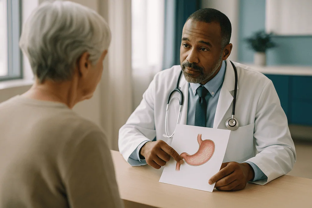
T describes the layer the tumor has reached. T1 cancers are limited to the inner lining or the layer just beneath it. T2 tumors have grown into the muscle layer. T3 tumors have reached the connective tissue beneath the outer surface, and T4 tumors have broken through that surface or invaded neighboring structures such as the pancreas, spleen or colon.
N is scored by the number of regional lymph nodes containing cancer, from N0 (none) up to N3 (many). This number is only truly known after surgery, when a pathologist examines the removed nodes under a microscope. Before surgery, N is an estimate based on how nodes look on ultrasound and CT.
M is simpler: M0 means no distant spread has been found, M1 means it has. Peritoneal deposits, cancer cells found in fluid washed from the abdomen during laparoscopy, and spread to the liver or lungs all count as M1, and any M1 finding places the cancer in stage IV regardless of T or N.
Two details often surprise people. First, the same stage can be reached by different combinations: a deep tumor with no nodes and a shallow tumor with several positive nodes may land in the same group. Second, there are different stage tables for different moments. A clinical stage (cTNM) is assigned from imaging before any treatment; a pathological stage (pTNM) is assigned after surgery; and a post-treatment stage (ypTNM) is used when chemotherapy was given first. Your letter may carry one of these prefixes, and asking which one applies is a fair question.
Endoscopy and biopsy: where staging begins
An upper endoscopy, sometimes called a gastroscopy, passes a thin flexible tube with a camera through the mouth, down the esophagus and into the stomach. According to Mayo Clinic, the procedure itself usually takes about 15 to 30 minutes, and most people receive a throat spray, sedation, or both. It is the test that found the cancer in the first place, and it still has staging work to do.
The endoscopist records where the tumor sits: near the junction with the esophagus, in the body of the stomach, or near the outlet into the small intestine. Location changes the surgical options, because a tumor at the top of the stomach may need part of the esophagus removed, while one near the outlet may allow the upper stomach to be kept. The scope also shows how much of the circumference the tumor occupies and whether the stomach wall looks stiff and poorly expanding, a pattern that can suggest a diffuse type of cancer that spreads within the wall rather than forming a single lump.
Biopsies, small pinches of tissue, are taken from several points. Pathologists confirm the cancer type, most often adenocarcinoma, which arises from the gland cells of the lining. In many pathways those same samples are tested for biomarkers, including HER2 protein, mismatch repair status and PD-L1, which may influence later treatment choices. These are laboratory findings on the tissue, not treatment decisions, and your oncologist will explain what, if anything, they mean for you.
What endoscopy cannot do is see beneath the surface. A tumor that looks small on camera may already have grown through the wall, and a bulky one may still be shallow. For depth, the scope needs a different sensor, and that is the subject of the next section.
Endoscopic ultrasound for stomach cancer: measuring the wall
Endoscopic ultrasound, or EUS, uses an endoscope fitted with a tiny ultrasound probe at its tip. Placed directly against the stomach lining, it produces a cross-sectional image of the wall in which the individual layers appear as alternating bright and dark bands. A tumor shows up as a disruption of those bands, and the depth of that disruption gives the T stage more precisely than any external scan.
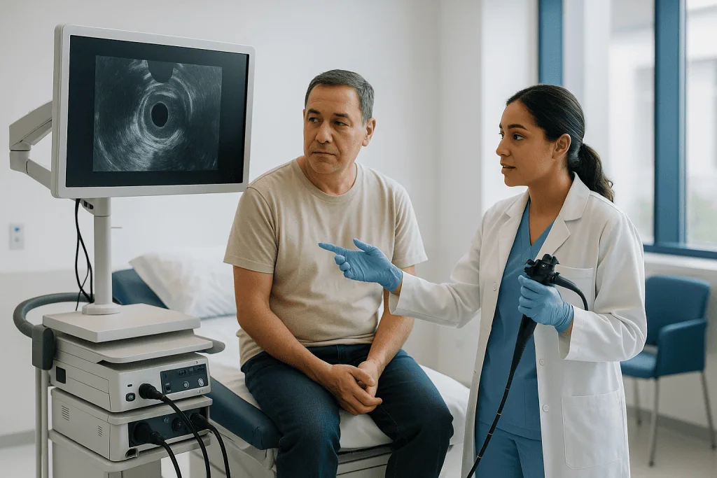
EUS also sees the lymph nodes immediately around the stomach. Nodes that are round, dark, sharply edged and larger than a few millimeters raise suspicion. When a node looks worrying, a fine needle can be passed through the scope into it to collect cells, a technique called fine-needle aspiration. A positive sample upgrades the clinical N stage with real tissue evidence rather than an impression from a picture.
The Mayo Clinic and National Cancer Institute both list EUS among the standard staging tests, and its main value lies at two ends of the spectrum. For very early tumors, confirming that the cancer is limited to the inner lining may make a patient a candidate for endoscopic removal, where the tumor is lifted off from inside the stomach without open surgery. For more advanced tumors, showing invasion through the wall or into nearby nodes helps the team decide whether chemotherapy before surgery is appropriate.
Its limits are real. EUS is operator dependent, meaning accuracy varies with the experience of the person holding the scope. It can overstage inflammation as tumor and understage microscopic invasion. It cannot see the liver surface or the far reaches of the peritoneum. In other words, EUS answers the depth and nearby node questions well and leaves the distant spread question to CT and laparoscopy.
CT scan for stomach cancer: the wide-angle view
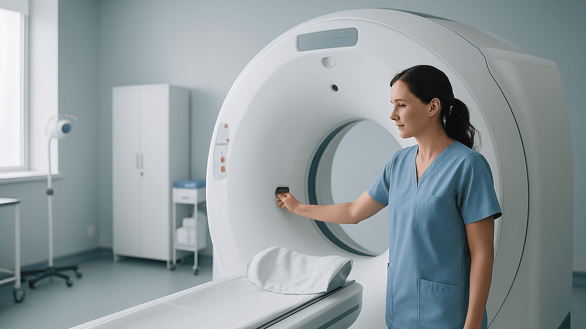
A computed tomography scan, or CT, uses X-rays taken from many angles to build detailed cross-sectional images of the body. For stomach cancer, the scan usually covers the chest, abdomen and pelvis in one session, with contrast dye injected into a vein so that blood vessels, liver and lymph nodes stand out. Patients are often asked to drink water or a contrast liquid shortly beforehand so the stomach is distended, which makes the wall easier to assess. The NHS notes the scan itself typically takes only a matter of minutes, though preparation and waiting add to the visit.
CT answers the M question first. It looks for deposits in the liver, nodules in the lungs, enlarged nodes away from the stomach, fluid in the abdomen and thickening or nodularity of the peritoneum. It also gives a second opinion on T stage by showing whether the fat outside the stomach wall looks clean or streaky and whether the tumor abuts the pancreas or other organs.
Where CT struggles is in the small details. Peritoneal deposits only a few millimeters across are commonly invisible. Lymph nodes are judged mainly by size, and size is an imperfect proxy: small nodes can harbor cancer and large ones can be reactive. Flat, diffuse tumors that thicken the wall without forming a mass can be hard to see at all.
Radiologists therefore report CT findings in graded language such as suspicious, indeterminate or consistent with, and the multidisciplinary team reads them alongside the endoscopy and EUS reports. A clean CT is reassuring but not final, which is exactly why guidelines add laparoscopy for tumors that have grown through the wall.
Staging laparoscopy for gastric cancer: looking where scans cannot
Staging laparoscopy is a short keyhole operation under general anesthesia in which a surgeon makes one or more small cuts in the abdominal wall, inflates the cavity with carbon dioxide gas and inserts a camera to inspect the organs directly. The National Cancer Institute lists it among the standard stomach cancer staging tests, and its purpose is narrow but important: to find peritoneal disease that CT missed before someone is committed to a major operation.
During the procedure the surgeon examines the surface of the liver, the lining of the abdominal wall, the diaphragm, the fatty apron called the omentum and the pelvis. Tiny white or gray nodules on these surfaces are biopsied. Sterile fluid is then washed over the surfaces and suctioned back, a step called peritoneal lavage or washings. The fluid is sent to the laboratory to search for free-floating cancer cells. A positive washing counts as distant spread in the TNM system even when no visible deposit is found, because it signals that cells have already escaped the stomach.
This matters because peritoneal spread changes the plan. Surgery to remove the stomach is a large operation with real risks, and guidelines reserve it for situations where it can realistically clear the disease. Finding occult peritoneal deposits at laparoscopy spares a person that operation and redirects treatment toward systemic therapy. Some teams also use the same session to place a feeding tube if nutrition is a concern.
Laparoscopy is not needed for everyone. Early tumors confined to the inner wall have little chance of peritoneal spread, and cancers already shown by CT to have reached the liver do not need confirmation of a stage they already have. It is typically a day-case or overnight procedure; your surgeon will explain what applies to you.
PET-CT, MRI and blood tests: the supporting cast
Beyond the three core tests, several others appear in some pathways, and it helps to know why they are sometimes used and often not.
PET-CT combines CT with a scan that tracks a radioactive sugar taken up by metabolically active cells. Many cancers light up brightly, which can reveal distant spread that CT alone showed only vaguely. Stomach cancers are an uneven case. Intestinal-type tumors often take up the tracer well, while diffuse and mucinous types, including signet-ring cell cancers, frequently do not, so a dark PET scan in those types is not reassurance. For this reason PET-CT is used selectively in stomach cancer rather than routinely, and a negative result does not replace laparoscopy.
MRI, which builds images from magnetic fields rather than X-rays, is occasionally used to characterize a liver lesion that CT could not classify, or when contrast dye for CT is unsuitable because of kidney function or allergy. It is not a standard first-line stomach cancer staging test.
Blood tests do not stage cancer, but they support the process. A full blood count can reveal anemia from slow bleeding; liver and kidney function tests guide the safe use of contrast and later treatment; nutritional markers such as albumin flag whether a person is losing weight and muscle, something the team will want to address before any surgery. Tumor markers in the blood are sometimes measured, though MedlinePlus and Mayo Clinic are clear that they are neither specific enough nor sensitive enough to diagnose or stage stomach cancer on their own.
Finally, the biomarker tests already mentioned on biopsy tissue sit alongside staging. They describe the biology of the cancer rather than its extent, and together the two sets of information frame the treatment discussion.
Who usually needs each stomach cancer staging test, and who is asked to wait
Everyone diagnosed with stomach cancer has had an endoscopy with biopsy, and nearly everyone proceeds to a contrast CT of the chest, abdomen and pelvis. From there the pathway branches according to what the first two tests show, and it is worth seeing the branches clearly.
Endoscopic ultrasound is most useful when the answer would change the plan: a tumor that looks early on camera and clean on CT, where confirming a shallow depth could open the door to endoscopic removal; or a tumor whose depth and nodal status decide whether chemotherapy should come before surgery. It is less helpful once CT has already shown distant spread, because the T stage is then secondary.
Staging laparoscopy is generally offered when surgery with curative intent is being considered and the tumor appears to have grown into or through the outer wall (T3 or T4 on EUS or CT), or when there is fluid in the abdomen, a diffuse-type cancer or anything suspicious on the peritoneum. People with very early tumors are usually spared it, as are people whose CT already demonstrates liver or lung metastases.
Some people are asked to wait for reasons unrelated to the cancer itself. Someone recovering from a recent heart attack or stroke, or with an uncontrolled lung condition, may need optimization before a general anesthetic for laparoscopy. Someone with impaired kidney function may need a different contrast strategy before CT. People with a pacemaker or certain metal implants may need the MRI question handled separately. Pregnancy changes the sequencing of X-ray-based imaging.
None of these are refusals; they are sequencing decisions, and a good team will explain the reason for any pause. If a test has been deferred and no one has told you why, ask.
Side by side: what each stomach cancer staging test contributes
A map of the tests, read across, shows how little overlap there is. Each column answers a question the others cannot, and the gaps in one row are filled by the row beneath.
| Test | Best at | Commonly misses | What it involves |
|---|---|---|---|
| Endoscopy with biopsy | Confirming the cancer type, locating the tumor, obtaining tissue for biomarkers | Depth of invasion, lymph nodes, anything outside the stomach | Flexible camera through the mouth; sedation or throat spray; about 15–30 minutes (Mayo Clinic) |
| Endoscopic ultrasound (EUS) | T stage (depth through the wall), nearby lymph nodes, needle sampling of nodes | Distant organs, small peritoneal deposits; accuracy varies with operator | Same route as endoscopy with an ultrasound tip; sedation |
| Contrast CT chest, abdomen, pelvis | Liver, lung and distant node metastases, large peritoneal disease, ascites, local invasion of neighboring organs | Peritoneal nodules only millimeters across, cancer in normal-sized nodes, flat diffuse tumors | Intravenous contrast, drinking water or oral contrast; scan time measured in minutes (NHS) |
| Staging laparoscopy with washings | Small peritoneal deposits, free cancer cells in abdominal fluid, liver surface nodules | Disease deep inside organs, lymph node status, lung spread | Keyhole surgery under general anesthesia; usually day-case or overnight |
| PET-CT | Detecting active distant disease in tumor types that take up tracer | Diffuse, mucinous and signet-ring cell cancers that often do not light up | Injection of radioactive sugar, rest period, then scan |
Two patterns stand out. The tests that are best at local detail (endoscopy, EUS) are weakest at distant spread, and the test best at distant spread (CT) is weakest at the small-scale surface disease that most often derails surgery. Laparoscopy exists to close that last gap. When a team orders all four, it is not over-investigation; it is the only way to answer all three TNM questions before committing to a plan.
What the days and weeks of staging usually look like
The staging period has a rhythm, and knowing it reduces the sense that nothing is happening.
The first days after a cancer biopsy are often spent waiting for pathology, including biomarker tests, which require extra laboratory steps. During that window the CT scan is usually booked. You will be asked about kidney function, allergies and any diabetes medicines that interact with contrast dye, and to arrive with an empty stomach. The scan visit itself is short; the radiology report follows within days.
Endoscopic ultrasound, when needed, is scheduled as a second endoscopy. The preparation mirrors the first: nothing to eat for several hours beforehand, someone to take you home if sedation is used, and a sore throat or bloating for a day afterward. If a needle sample is taken, that result adds another short wait.
Staging laparoscopy sits last in the sequence because it depends on everything before it. It needs a pre-anesthetic assessment, a general anesthetic and a small operating slot. Most people go home the same day or the next morning, with a few small dressings and shoulder-tip discomfort from the gas, which settles over a day or two. Washings take a few days to be reported.
The results then go to a multidisciplinary team meeting, where surgeons, oncologists, radiologists and pathologists review the complete picture together. Only after that meeting is a treatment plan presented to you. NHS guidance describes this team review as a standard step, and it is the reason the full staging journey from biopsy to plan commonly spans a few weeks rather than days.
Nutrition matters throughout. If you are losing weight or struggling to eat, say so early; dietitians are part of the pathway and can begin support before any treatment starts.
Clinical stage, pathological stage and restaging after chemotherapy
The stage you are given at the first planning meeting is a clinical stage, built from pictures and small samples. It is the best available estimate, and it is sometimes wrong in either direction. The definitive stage comes from the pathologist’s examination of the removed stomach and lymph nodes after surgery, which is why some people are told a different stage weeks later. That is not an error; it is the system working as designed.
For cancers that have grown into or through the wall or involve nodes, many guideline pathways, including those summarized by the National Cancer Institute, describe chemotherapy before surgery, sometimes called neoadjuvant or perioperative treatment. The aim is to shrink the tumor and treat cells that may already have escaped. After this phase, restaging with CT, and sometimes a repeat endoscopy, checks whether the cancer has responded and whether surgery remains appropriate. A repeat laparoscopy is occasionally used if the first one was borderline or if new symptoms suggest peritoneal disease.
After surgery, the pathology report assigns a ypTNM stage. The prefix y signals that treatment was given beforehand, and tumor regression grading describes how much of the cancer was destroyed by chemotherapy. These findings guide the decision about further treatment afterward, which again sits with the oncology team.
For cancers already found to have spread at diagnosis, staging does not stop either. Scans are repeated at intervals to measure how treatment is working. The vocabulary changes from stage to response: stable disease, partial response, progression. Ask your team which scan is the comparison baseline, because the question being answered at each visit is different from the one asked at the start.
Can stage 4 stomach cancer cause fluid buildup in the abdomen?
Yes. Fluid accumulation in the abdominal cavity is called ascites, and in stomach cancer it most often signals peritoneal spread, which by definition places the cancer in stage IV. The peritoneum normally produces and reabsorbs a small amount of lubricating fluid every day. When cancer cells seed the membrane, they irritate it and block the tiny channels that drain fluid away, so it collects. The result is a gradually swelling abdomen, tight clothing, early fullness when eating and sometimes breathlessness as the diaphragm is pushed upward.
Ascites is also a staging clue in its own right. Fluid seen on CT in someone with a newly diagnosed stomach cancer raises the probability of peritoneal disease even when no nodules are visible, and it is one of the findings that pushes a team toward staging laparoscopy or toward sampling the fluid directly. A sample drawn with a fine needle under ultrasound guidance can be examined for cancer cells, which can confirm M1 disease without an operation.
Not all abdominal fluid in a person with cancer comes from the cancer. Liver disease, heart failure, low protein levels from poor nutrition and some infections also cause ascites, and the fluid analysis helps tell them apart.
Management is symptom-focused and decided by the treating team. Draining fluid through a temporary catheter can relieve pressure and breathlessness; the fluid often returns, and the interval between drains varies widely between people. Some are offered a longer-term drainage tube to manage at home. Systemic treatment that controls the underlying cancer can slow reaccumulation. None of this is a decision to make alone; it belongs in a conversation with your oncology and palliative care teams, who can balance relief against the small risks of each procedure.
What people often get wrong about stomach cancer staging
Myths cluster around staging because the process is slow and the vocabulary is unfamiliar. Some of the most common deserve correcting.
Myth: the stage from the first scan is final. It is a clinical estimate, revised by EUS, by laparoscopy and ultimately by pathology after surgery. Being told a different stage later usually means more information arrived, not that someone was careless.
Myth: a clear CT means the cancer has not spread. CT is excellent for liver and lung disease and poor for millimeter-sized peritoneal deposits. That blind spot is the entire reason staging laparoscopy exists for deeper tumors.
Myth: more tests mean the team suspects something terrible. Laparoscopy is ordered precisely when surgery is a realistic option and the team wants to be certain before offering it. It is a sign of a curative-intent plan being checked, not abandoned.
Myth: PET-CT is the most powerful scan, so everyone should have one. For diffuse and signet-ring cell stomach cancers, PET frequently shows nothing even when disease is present. A negative PET in those types can be falsely reassuring.
Myth: the size of the tumor sets the stage. Depth through the wall, not width across the lining, drives the T stage. A broad shallow tumor can carry a lower T stage than a small one that has punched through the outer surface.
Myth: biomarker results like HER2 or mismatch repair status are part of the stage. They describe the biology of the cancer and may shape treatment choices, but they sit alongside TNM rather than inside it.
Myth: you cannot eat during the staging weeks. Apart from fasting before each procedure, maintaining nutrition is one of the most useful things you can do, and a dietitian can help if eating has become difficult.
Questions to ask your care team about stomach cancer staging tests
Multidisciplinary meetings generate a lot of information, and the person receiving it is rarely at the table. Bringing specific questions turns a one-way summary into a conversation. These are the ones that tend to matter most.
- Which stage have I been given, and is it a clinical, pathological or post-treatment stage?
- What is my T, N and M, and which test contributed each letter?
- Did the CT show anything described as indeterminate, and how is the team interpreting it?
- Was endoscopic ultrasound done, and if not, why was it not needed in my case?
- Is staging laparoscopy part of my pathway, and what would a positive washing change?
- Were biomarkers tested on my biopsy, and are the results back?
- If chemotherapy is planned before surgery, when and how will I be restaged?
- What symptoms during the staging weeks should prompt me to call rather than wait?
- Who is my named contact, and how do I reach the team between appointments?
- Can I see a dietitian now rather than after treatment begins?
A few practical habits help. Ask for a written copy of the staging summary, including the TNM letters, so you can refer back to it. If a second opinion is something you want, say so plainly; it is a normal request in cancer care and teams are used to arranging it. Bring someone to appointments where results are discussed, because two sets of ears catch more than one.
Keep in mind that the team may not be able to answer every question until a test is complete. A straight answer of ‘we will know after the laparoscopy’ is more useful than a guess, and it is a reasonable thing to be told.
When to call your doctor during or after stomach cancer staging tests
Staging procedures are generally safe, but each carries a short list of warning signs, and the cancer itself can cause problems while you wait. Knowing when to call is part of the plan, not an afterthought. Use the contact number your team gave you; if you cannot reach them and any of the following are severe, use emergency services.
After endoscopy or endoscopic ultrasound, Mayo Clinic advises seeking care for chest pain, fever, breathing difficulty, severe or worsening abdominal pain, vomiting blood or material that looks like coffee grounds, or black tarry stools. A sore throat and bloating for a day are expected; pain that escalates is not.
After CT with contrast, a mild warm flush and metallic taste are normal. Hives, facial or throat swelling, wheezing or feeling faint during or shortly after the injection need immediate attention.
After staging laparoscopy, call for a temperature above 38°C (100.4°F), redness, discharge or spreading pain at any incision, abdominal pain that worsens rather than eases, persistent vomiting, inability to pass urine, or calf pain and swelling, which can signal a blood clot.
At any point during the staging weeks, contact your team for vomiting that prevents you keeping fluids down, inability to swallow solids, rapid abdominal swelling, new breathlessness, sudden severe abdominal pain, or signs of bleeding such as dizziness with dark stools. Unintentional weight loss that is accelerating also warrants a call, because nutrition support works best when started early.
None of these signs means the plan has failed. They mean something needs assessing today rather than at the next scheduled appointment, and your team would rather hear from you than not.
Frequently asked questions
How long does it take to recover from stomach cancer surgery?
Recovery after a gastrectomy is measured in weeks for the wound and months for eating. NHS guidance describes a hospital stay of roughly one to two weeks, with a gradual return to normal activity over the following weeks. Adjusting to a smaller or absent stomach takes longer: small frequent meals, dietitian follow-up and sometimes vitamin supplementation continue for months. Your surgical team will give a timeline specific to the operation you had.
Can stage 4 cancer cause fluid buildup in the stomach?
Yes, although the fluid collects in the abdominal cavity around the organs rather than inside the stomach itself. This is called ascites, and in stomach cancer it usually indicates spread to the peritoneum, the lining of the abdomen, which defines stage IV. It causes swelling, early fullness and sometimes breathlessness. Draining the fluid can relieve symptoms, and the oncology or palliative care team decides the safest approach.
How long can you live with stomach cancer without treatment?
There is no honest single answer, because it depends on the stage, the cancer’s biology, a person’s general health and nutrition. Early cancers confined to the lining can progress slowly over years, while advanced disease can change within months. Mainstream sources such as the National Cancer Institute present outlook by stage rather than as one figure. If you are weighing whether to have treatment, ask your team to talk you through your specific situation.
What should I avoid with stomach cancer?
During staging, avoid eating or drinking in the fasting window before each procedure and avoid stopping any prescribed medicine without checking with the team, especially blood thinners and diabetes drugs that interact with contrast. Beyond that, guidance from the NHS and Mayo Clinic emphasizes not smoking, limiting alcohol and maintaining nutrition rather than restrictive diets. Unproven supplements and alternative therapies are best discussed with your oncologist before use.
What are the stomach cancer stages explained simply?
Stage 0 is cancer confined to the surface cells. Stage I has grown a little into the wall with no or few nodes involved. Stages II and III describe deeper growth into or through the wall and increasing numbers of affected lymph nodes. Stage IV means the cancer has reached distant sites such as the liver, lungs or the peritoneum, including cancer cells found in abdominal washings. The exact grouping comes from TNM letters.
Is endoscopic ultrasound for stomach cancer painful?
It is not usually painful. EUS is performed like a standard endoscopy, with sedation or throat spray, and most people remember little of it. A sore throat, bloating and mild cramping for a day are common afterward. If a needle sample is taken from a lymph node, there is a small risk of bleeding or infection, and you will be told which symptoms should prompt a call to the unit.
Why do I need a staging laparoscopy for gastric cancer if my CT scan was clear?
Because CT cannot reliably see peritoneal deposits that are only millimeters across, and those deposits change the treatment plan entirely. Laparoscopy lets a surgeon look directly at the abdominal lining and wash the cavity to search for free cancer cells. It is offered when surgery is realistically being considered and the tumor has grown deep into the wall, precisely so that a major operation is not performed when it would not help.
What does a CT scan for stomach cancer show?
A contrast CT of the chest, abdomen and pelvis shows whether the cancer has spread to the liver, lungs or distant lymph nodes, whether it is invading neighboring organs such as the pancreas, and whether there is fluid or thickening on the peritoneum. It gives an approximate T stage from the appearance of the stomach wall. It is weakest for tiny peritoneal nodules and for cancer in normal-sized nodes.
Can the stage of my stomach cancer change after surgery?
Yes, and this is expected. The stage before surgery is a clinical estimate from scans and small samples. After surgery, the pathologist examines the whole removed stomach and every lymph node under a microscope, producing the pathological stage. It may be higher or lower than the clinical one. If chemotherapy was given beforehand, the post-treatment stage carries a y prefix and includes a measure of how much the tumor responded.
How long does stomach cancer staging take from biopsy to treatment plan?
Commonly a few weeks. Pathology and biomarker results take days, CT is usually scheduled promptly, EUS and laparoscopy each add an appointment and a short wait for results, and the findings are then reviewed at a multidisciplinary team meeting before a plan is presented, a step NHS guidance describes as standard. The exact span varies with local scheduling and with whether every test is needed in your case.
References
This article is for general information only and is not a substitute for professional medical advice. Please consult a qualified doctor about your individual situation.

More from the Blog
Is Radiation Needed After Endometrial Cancer Surgery? How Risk Factors Guide the Answer
Radiation is not automatically needed after hysterectomy for endometrial cancer. Many people with early, low-risk disease are treated with surgery alone and monitored. Radiation…

Fiducial Markers Before SBRT: Why Some Tumors Need Them and How They Are Placed
Fiducial markers before SBRT are tiny implanted objects, usually gold, placed in or near a tumor so the radiation team can see exactly where…
How CT, MRI and PET-CT Stage Kidney Cancer Before a Treatment Plan Is Written
Kidney cancer is staged mainly with a contrast-enhanced CT scan of the abdomen and chest, which measures the tumor, checks whether it has grown…

Monitoring Response in Lymphocytic Leukemia: What Blood Counts Show Between Visits
CLL treatment response monitoring uses regular complete blood counts, a physical examination of lymph nodes, spleen and liver, and sometimes imaging or a bone…
Why Chemotherapy and Radiation Often Come Before Esophageal Cancer Surgery
Chemotherapy and radiation are often given before esophageal cancer surgery because the combination can shrink the tumor, treat cancer cells that may have spread…
Transfusions and Infection Precautions During Acute Myeloid Leukemia Treatment
During acute myeloid leukemia treatment, chemotherapy drives red cells, platelets and infection-fighting white cells to very low levels for weeks. Red cell and platelet…