

Why Repeat X-Rays Matter During Scoliosis Bracing: Checking Fit, Growth and Curve Control
Key Takeaways
- Scoliosis is defined by a Cobb angle of 10 degrees or more, so a 7-degree reading is spinal asymmetry that is usually rechecked rather than treated.
- Bracing is generally considered for curves of roughly 25–40 degrees in a child with meaningful growth remaining, and surgery is usually discussed from about 45–50 degrees.
- During active growth, standing X-rays are typically repeated about every 4–6 months, with the interval shortened around the peak growth spurt and lengthened near maturity.
- The in-brace X-ray checks fit rather than destiny: it shows whether the pressure pad sits at the apex and how much the curve reduces inside the brace.
- The Risser sign, a 0–5 grade of how much the pelvic rim has turned to bone, tells the team how much growth is left and when brace weaning can begin.
- In the BRAIST randomized trial, 72 percent of braced adolescents reached maturity with curves under 50 degrees versus 48 percent of those observed, and the benefit rose with hours worn.
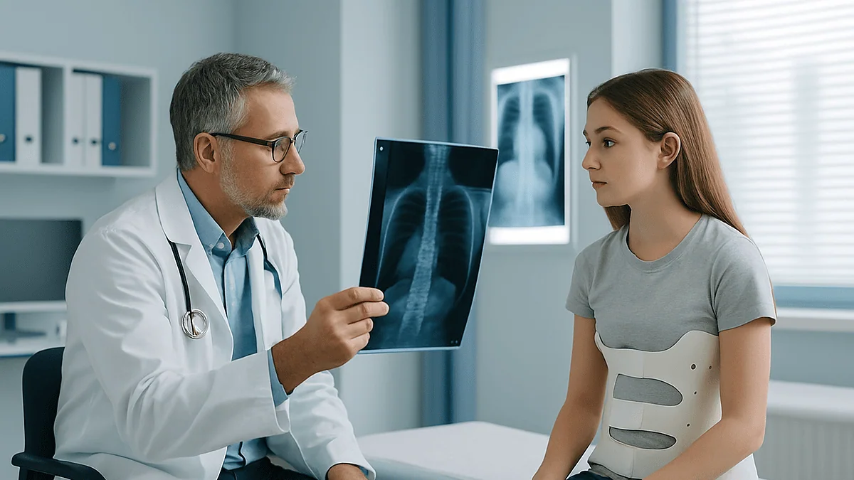
Repeat X-rays during scoliosis bracing let the care team measure the Cobb angle, check how well the brace is holding the curve, and track how much growth remains, usually through the Risser sign. Because curves change fastest during growth spurts, standing X-rays are typically repeated about every four to six months while the brace is worn, with an in-brace image after fitting to confirm the pads sit where they should.
The fitting room smells faintly of warm plastic. A 12-year-old turns sideways in the mirror, testing how the new brace sits under a hoodie, while a parent listens to the orthotist say, almost in passing, “We’ll get another picture in a few weeks, then again in four months.” The parent’s face asks the question before their voice does: another one? Didn’t we just do that?
It is a fair challenge. Scoliosis brace X-rays are the one part of treatment that families rarely hear explained, even though they drive every adjustment, every reassurance and every hard conversation that follows. The curve was measured once to decide on a brace. Why keep measuring?
Because a brace is not a cast. It works on a spine that is still growing, which means the target moves every few months. The repeat film is how the team checks whether the plastic is pushing in the right place, how much growing is left, and whether the plan is holding.
What scoliosis brace X-rays actually measure: the Cobb angle
Scoliosis is a sideways curve of the spine, and on a scoliosis brace X-ray that curve is turned into a number. The number is the Cobb angle: the angle between a line drawn along the top of the most tilted vertebra above the curve and a line along the bottom of the most tilted vertebra below it. A curve of 10 degrees or more meets the usual definition of scoliosis, and that one figure anchors nearly every decision that follows.
It helps to picture what the radiograph can and cannot show. It records the shape of the spine at the instant the image was taken, standing, in a particular posture, on a particular afternoon. Ten minutes of slouching, a long day on the feet, or a growth spurt since the last visit can all shift the reading by a few degrees. That is why spine teams do not react to a single film in isolation; they look for a trend across several images, and most only call a change progression once it clearly exceeds the ordinary wobble of measurement.
Repeat imaging is therefore doing three jobs at once. It tracks whether the curve is holding, growing or settling. It checks the brace itself, because an orthosis (the clinical word for a brace) is only useful if its pads are pushing on the right vertebrae. And it reads the growth plates of the pelvis to estimate how much growing is left, which is the single biggest predictor of whether a curve will worsen. A brace works by guiding growth rather than by straightening bone directly, so knowing where a child sits on that growth timeline is as important as the angle itself.
What actually happens when scoliosis X-rays are taken during bracing
The appointment is quicker than most families expect. The usual image is a standing, full-length view of the whole spine, often from pelvis to neck on one film, because scoliosis is a problem of the entire column and not just the few vertebrae at the apex (the point of the curve farthest from the midline). Standing matters: gravity loads the spine the way it is loaded all day, and lying down flattens the curve and gives a falsely reassuring number.

Most centers take the image from back to front, which radiology calls a posteroanterior projection. The spine looks the same either way, but entering from the back means the beam reaches breast and thyroid tissue after it has already passed through the body and been partly absorbed. A side view is sometimes added to assess the spine’s forward and backward curves. Depending on the equipment, the child stands against a plate or inside a low-dose scanning booth that captures front and side views in one sweep.
Preparation is simple. Hair is tied up, and necklaces, bras with metal clasps and belts come off, since they cast shadows. The team will say whether the brace should be on or off for that specific image; the two give different information, and turning up in the wrong state can mean a wasted exposure. Adolescents who could be pregnant are asked, routinely and without judgment. The exposure itself lasts a fraction of a second. Afterward, the clinician measures the Cobb angle, notes the Risser sign, checks vertebral rotation, and compares all three with the previous films before anyone talks about next steps.
In-brace X-ray scoliosis check: what it tells the orthotist about fit
An orthotist is the specialist who makes and adjusts braces, and the in-brace X-ray is their most honest feedback. It is taken once the brace has been worn for a few weeks and the skin has adapted, with the brace fastened exactly as it is worn at home. The question it answers is blunt: with the pads applying pressure, where does the spine actually sit?
Several things are read off that image. The first is in-brace correction, the difference between the Cobb angle with and without the brace. The second is position: the main pressure pad should sit just below the apex of the curve, pushing the spine toward the midline, with a relief area on the opposite side to give the trunk somewhere to move. If the pad lands a vertebra too high or too low, the brace can squeeze without correcting, and the only way to see that is to image through the plastic. The third is the pelvis and shoulders, which should stay level rather than being tipped by the brace.
What the in-brace number means for the long run is a fair question, and the honest answer is that it is a sign of good fit rather than a forecast. A larger reduction inside the brace generally suggests the design is doing mechanical work, and clinicians use it to decide whether to tighten straps, move a pad or remake the shell. It is not a promise about the final curve, because some of that correction is lost whenever the brace comes off, and growth has its own plans. The team reads it alongside wear time, comfort and the out-of-brace trend.
How often are scoliosis X-rays needed during bracing?
There is no single schedule that fits every spine, but a pattern recurs across guidelines and major centers. During active growth, follow-up visits with a standing X-ray typically happen every 4–6 months, the interval Mayo Clinic describes for growing children whose curves are being watched. Teams often shorten that during the peak growth spurt, when curves can move quickest, and lengthen it as the Risser sign climbs toward maturity. One image per visit is the norm; two is unusual and should come with a reason.
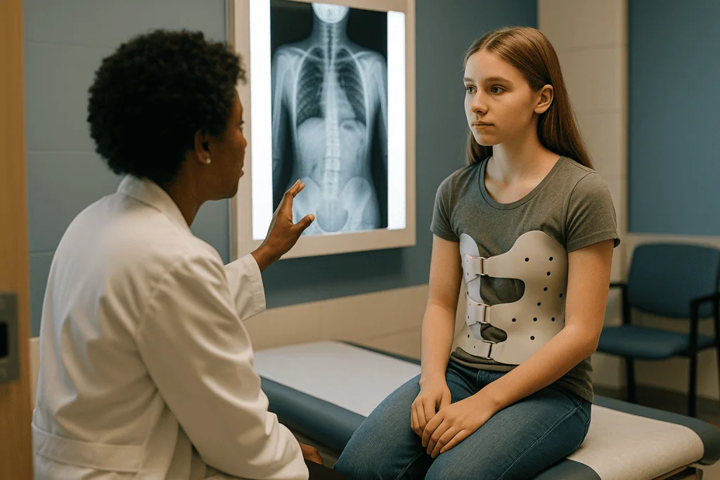
Some teams ask for the brace to be left off for a set number of hours before certain follow-ups so the image shows the unsupported spine. Others image in the brace for routine checks and reserve out-of-brace films for decision points. Both approaches are legitimate; what matters is that the same method is used each time so the trend is comparable.
| Image | Usual timing | What the team looks for |
|---|---|---|
| Baseline standing X-ray, out of brace | Before the brace is prescribed | Cobb angle, curve pattern, Risser sign, vertebral rotation |
| In-brace check | A few weeks after fitting | Pad position, in-brace correction, level pelvis and shoulders |
| Routine follow-up | About every 4–6 months while growing | Trend in Cobb angle, growth progress, need for brace adjustment |
| Out-of-brace check | At decision points, after a short break from the brace | True unsupported curve, how much correction is retained |
| Skeletal maturity image | When growth is judged complete | Final curve, readiness to wean the brace |
The last row is where many families relax, and it is also where imaging usually slows or stops: once bone growth has finished, a stable curve below the surgical range generally does not need regular repeat films, though the care team sets the exact plan.
Risser sign scoliosis grading: how X-rays track remaining growth
The Risser sign is a grade from 0 to 5 that describes how much of the growth plate along the top rim of the pelvis has turned to bone. It appears on the same film as the spine, which is why the full-length image is framed to include the hips. Risser 0 means the rim has not started to ossify and plenty of growth remains; Risser 4 or 5 means the rim is nearly or fully fused and trunk growth is close to finished. Clinicians read it with a glance, but it changes the entire conversation.
Growth is the engine of curve progression. The same 30-degree curve carries a very different outlook in a child at Risser 0 than in a teenager at Risser 4, and the brace is prescribed to steer the growing spine during that window rather than to straighten a finished one. This is also why bracing is typically continued until bones stop growing, as Mayo Clinic and the NIH both describe, and why the Risser grade on each repeat X-ray is tracked as closely as the Cobb angle.
Risser is not the only clue. Height measured at each visit shows growth velocity, the time since a first period is a strong marker in girls, and some teams add a hand and wrist X-ray to estimate bone age when the pelvis is ambiguous. None of these replaces the spine film, but together they let the team answer the question every family asks at every visit: how much longer? The answer shifts from “a while” to “months” to “we can start weaning” as the pelvic rim fills in, and that progression is one of the most reassuring things a repeat X-ray can show.
Who is usually offered a scoliosis brace, and who is usually asked to wait
Bracing is an intervention with a specific target: a growing child whose curve is big enough to be at risk but not so big that it has already crossed into surgical territory. Johns Hopkins and Mayo Clinic describe the usual pattern. Curves between 10 and about 25 degrees are generally observed with periodic X-rays rather than braced. Curves in the roughly 25–40 degree range in a child with meaningful growth remaining, usually Risser 0 to 2, are the classic bracing candidates. Curves around 45–50 degrees or more, or curves that keep progressing despite a well-worn brace, prompt a conversation about surgery.
Who is usually asked to wait? A child with a 15-degree curve, even one that worries their parents a great deal, is typically rechecked rather than braced, because most mild curves do not progress to a size that needs treatment and a brace carries a real daily burden. A near-mature teenager whose pelvis reads Risser 4 or 5 has little growth left for a brace to guide, so bracing is rarely started at that stage. Adults with scoliosis are not braced to control progression; their curves behave differently and are managed according to symptoms.
Waiting is not the same as doing nothing. It means measured intervals, a consistent imaging technique, attention to the growth spurt, and a plan for what happens if the next film shows a jump. Some families find observation harder than bracing because it feels passive, and it is worth saying so to the team. Equally, some curves fall between the categories, and there the decision rests on the child’s age, curve pattern, family preference and the clinician’s judgment, not on a threshold alone.
What does mild scoliosis look like on an X-ray, and is 7 degrees scoliosis?
On a standing film, a mild curve looks like a gentle lean rather than a bend: a shallow C in the lower back or mid-chest, or a soft S if there are two curves balancing each other. The vertebrae along it may show slight rotation, visible as the pedicles (the small bony columns at the back of each vertebra) shifting toward the midline on one side. Rib spacing may be a little uneven. To an untrained eye the spine often looks nearly straight, and that is accurate; mild scoliosis is mild.
So is 7 degrees scoliosis? By the standard definition used by Mayo Clinic and MedlinePlus, no. Scoliosis is defined as a Cobb angle of 10 degrees or more. A 7-degree reading is usually described as spinal asymmetry, and in a growing child it may simply be rechecked at the next visit. Because measurement itself carries a few degrees of variation, a 7 on one film and a 10 on the next does not necessarily mean the curve grew; it may mean the ruler moved.
Mild scoliosis matters less for what it is now than for what it might become, and that depends almost entirely on growth. A 20-degree curve in a child at Risser 0 earns closer watching than the same curve in a teenager a year past their growth spurt. The radiologist’s report will usually note the Cobb angle, the direction and level of the curve, the Risser grade and any vertebral abnormalities. Most children with scoliosis have mild curves and never need a brace or surgery, as Mayo Clinic notes, which is the context in which that first number should be heard.
What the first weeks of scoliosis bracing usually look like
The first fortnight is mostly about skin and sleep. Braces are introduced gradually, a few hours at a time, with the wearing period lengthened over days as the trunk adapts. Red marks that fade within about half an hour of removal are expected; marks that stay, blister or break the skin are not, and the orthotist wants to hear about them early because a small pad shift usually solves the problem. A thin, seamless cotton layer under the brace helps.
Wear time is the variable the team cares about most. Mayo Clinic describes most braces as worn between 13 and 16 hours a day, with the brace removed for sport and bathing, while some nighttime designs are built to be worn only during sleep. A randomized trial published in the New England Journal of Medicine found that the protective effect rose with hours worn, so many clinics ask families to keep a simple log, and some braces include a small temperature sensor that records wear objectively. Honest logging helps more than perfect logging.
Somewhere in the first few weeks comes the in-brace X-ray, after which the orthotist may heat and reshape the shell, move a pad or add a strap. The first routine follow-up then falls a few months later. School, exams and most activities continue, and nearly all sports are allowed with the brace off. Expect questions from friends and a period of self-consciousness; many teenagers find it fades once routine sets in, and the team can connect families with peer support. By the second or third month, most families describe the brace as part of the furniture rather than the center of the day.
Radiation from repeat scoliosis X-rays: how teams keep the dose low
Every repeat film adds a small dose of ionizing radiation, and a child braced from age 11 to 16 may accumulate a dozen or more spine images. That deserves a straight answer rather than reassurance. Studies of women imaged for scoliosis decades ago, with far older equipment and techniques, raised concern about long-term breast cancer risk, and that history is the reason modern practice has changed so much. Doses from today’s full-spine techniques are a fraction of what they once were.
Several habits keep them low. The posteroanterior projection, taken from the back, lowers breast and thyroid dose substantially compared with imaging from the front. Collimation, which means narrowing the beam to the spine and pelvis only, cuts scatter to surrounding tissue. Low-dose biplanar systems capture front and side views in one standing sweep at very low exposure, and many pediatric centers use them precisely for scoliosis follow-up. Shielding is used in some centers for selected views, although its value with modern equipment is debated and it is never placed where it would hide the pelvis.
The biggest dose saving, though, is the one families can influence: only image when the result would change a decision. It is reasonable to ask whether a film is needed at a given visit, whether a recent image from elsewhere can be reused instead of repeated, and whether a side view adds anything today. Keep a simple record of the dates of every spine X-ray so that any new clinician knows the history. None of this should push a family to refuse an image the team genuinely needs; a missed progression carries its own cost. The aim is enough imaging to steer the brace well, and no more.
Can you correct scoliosis? What the evidence says bracing does and does not do
“Can you correct scoliosis?” is usually asking two different things. If the question means whether a curve can be straightened permanently without surgery, the honest answer from Mayo Clinic is that braces do not reverse a curve; they aim to stop it getting worse while the spine finishes growing. Some in-brace correction may persist after weaning, but the realistic goal written into every bracing plan is control, not reversal.
If the question means whether bracing actually works, the evidence improved sharply after a randomized controlled trial known as BRAIST, published in the New England Journal of Medicine and indexed in PubMed. It enrolled adolescents with curves of 20–40 degrees and substantial growth remaining. Treatment success was defined as reaching skeletal maturity with a curve under 50 degrees, the point at which surgery is commonly discussed. Success was recorded in 72 percent of braced adolescents compared with 48 percent of those observed, and the benefit grew with the number of hours the brace was worn each day. The trial was stopped early because the difference was clear.
Two cautions keep that result in proportion. First, the figures describe groups, not individuals; roughly a quarter of braced teenagers in the trial still progressed, and roughly half of unbraced ones did fine. Second, “success” in a trial means avoiding the surgical threshold, not a straight spine. The Cochrane systematic review of braces for adolescent idiopathic scoliosis reached a similar, more guarded conclusion: bracing appears to reduce progression, but the certainty of the evidence was rated low. Repeat X-rays are how that population-level finding is checked against one particular spine, visit by visit.
When scoliosis bracing doesn't work: what the X-rays show and what comes next
Sometimes the films tell a story nobody wanted. The curve climbs five degrees, then five more, despite good wear. The first response is not surgery but a search for a reason. Was the brace worn as prescribed, and does the sensor or log agree? Has the child grown out of it, so the pads now sit below the apex? Did the growth spurt arrive faster than the fitting schedule? Many “failures” are fit problems, and a remake or adjustment followed by a fresh in-brace X-ray settles the question.
If the curve keeps progressing with a well-fitting, well-worn brace, or reaches the 45–50 degree range that Johns Hopkins and Mayo Clinic describe as the usual threshold for surgical discussion, the conversation changes. The standard operation is spinal fusion, in which the curved segment is straightened with rods and screws and the vertebrae encouraged to grow into a single bone. Newer growth-guiding techniques, such as tethering the front of the spine with a flexible cord, aim to use remaining growth rather than fuse it; they are offered in selected centers and their long-term evidence is still being gathered.
Alternatives sit on the other side too. A curve that stalls in the high 30s near maturity may simply be observed. Scoliosis-specific physiotherapy programs are used in some countries alongside bracing; the evidence for them is limited and they are not a substitute for a prescribed brace. Whatever the path, the X-ray series is the shared document. It shows the pattern honestly, it dates each step, and it lets a surgeon, an orthotist and a family look at the same curve and decide together. The decision stays with the treating team and the family, not with a threshold.
What people often get wrong about scoliosis brace X-rays
Good intentions produce a surprising number of misunderstandings about scoliosis imaging. The most common deserve a clear correction.
- “A bigger in-brace correction means a better outcome.” It means a better fit. Some of that correction is lost once the brace comes off, and the final curve depends more on growth and wear time than on any single in-brace number.
- “The curve should be exactly the same on each X-ray.” Measurement varies by a few degrees between films and between readers. Spine teams look at the trend over several images before calling a change real.
- “An X-ray is needed at every appointment.” Not always. Skin checks, strap adjustments and height measurements do not need imaging, and a sensible team images only when the result might change the plan.
- “Seven degrees is scoliosis.” The usual definition is a Cobb angle of 10 degrees or more. Smaller asymmetries are common and are generally rechecked rather than treated.
- “The brace will straighten the spine for good.” Bracing is designed to control progression during growth. It does not reverse an established curve, and surgery is the only treatment that reliably reduces a large one.
- “Bad posture or heavy backpacks caused this.” The cause of adolescent idiopathic scoliosis is unknown, as Mayo Clinic and the NIH state; posture, backpacks and sports do not cause it, and no family is to blame.
- “Once the curve is under 25 degrees, imaging can stop.” While growth remains, even a mild curve is usually rechecked at intervals. Imaging stops when growth does and the curve is stable.
None of these myths is foolish; each comes from wanting a clearer answer than the biology offers. The repeat X-ray is the closest thing to that answer, read in context, by people who know the previous films.
Questions to ask your care team about scoliosis X-rays and bracing
Appointments are short, and the questions that matter tend to surface in the car afterward. Writing a few down beforehand turns the X-ray from something done to a family into something explained to them.
- What is the Cobb angle today, what was it last time, and do you consider that change real or within measurement variation?
- What Risser grade do you see, and roughly how much growth do you think remains?
- Was this image taken in or out of the brace, and will the next one use the same method so the trend is comparable?
- On the in-brace film, is the pad sitting at the right level, and is there anything about the fit you would like to change?
- How many hours a day is this brace designed for, and how are we tracking wear?
- At what Cobb angle, or what rate of change, would you start to discuss a different plan?
- Do you use a low-dose imaging system, and is a side view needed today?
- Could a recent X-ray from another clinician be reused instead of repeated?
- What signs between visits should prompt us to call rather than wait for the next appointment?
- When do you expect to begin weaning the brace, and what does that process look like?
Ask, too, who holds the full set of images and whether the family can keep copies. Scoliosis care often spans several years and sometimes more than one clinician, and a complete, dated series of films is the most valuable record a family can carry into any new appointment. A team that welcomes these questions is doing something right.
When to call your doctor
Most of scoliosis bracing is uneventful, and the routine X-ray schedule exists precisely so that problems are caught at planned visits. A few signs, though, should prompt a call to the care team before the next appointment rather than after it.
- New numbness, tingling or weakness in the legs or feet, or a change in the way the child walks.
- Any new difficulty with bladder or bowel control.
- Back pain that is severe, wakes the child at night, or comes with fever, unexplained weight loss or a general feeling of being unwell. Idiopathic scoliosis is usually not painful in children, and pain of this kind needs assessment in its own right.
- Shortness of breath or chest tightness, particularly with a large curve in the chest region.
- Skin under the brace that is broken, blistered, discolored beyond the usual fading redness, or increasingly painful.
- A brace that suddenly no longer closes or fits, a visible change in shoulder height, waist crease or rib prominence over a few weeks, or a growth spurt that has outpaced the planned review.
- A fall or injury while wearing the brace, with new pain afterward.
These are the situations in which an earlier X-ray, a brace adjustment or a different kind of examination may be needed, and the team would rather hear sooner than later. Everything in this article describes typical practice; the actual plan, the interval between images and every treatment decision belong to the treating clinicians who know the child, the films and the family. If you are unsure whether something warrants a call, that uncertainty is itself a good enough reason to make one.
Frequently asked questions
What does mild scoliosis look like on an X-ray?
A mild curve appears as a shallow C or soft S on a standing full-spine film, often close to straight at first glance. The Cobb angle measures between 10 and about 25 degrees, the vertebrae may show slight rotation, and rib spacing can be a little uneven. Most mild curves are observed with periodic imaging rather than braced, with the interval set by how much growth remains.
At what point do you need a brace for scoliosis?
Bracing is usually considered when a growing child has a curve in the roughly 25–40 degree range, or a smaller curve that is clearly progressing, with meaningful growth left on the Risser scale. Curves under about 25 degrees are generally watched, and curves of around 45–50 degrees or more lead to a surgical discussion. The treating team weighs age, curve pattern and family preference alongside the number.
Can you correct scoliosis without surgery?
Bracing does not reverse an established curve; its goal is to control progression while the spine finishes growing. Some correction seen inside the brace may be partly retained after weaning, but a brace is not designed to straighten the spine permanently. Surgery is the only treatment that reliably reduces a large curve, and the decision about whether it is needed rests with the treating team.
Is 7 degrees considered scoliosis?
No. Scoliosis is defined as a Cobb angle of 10 degrees or more, so a 7-degree reading is generally described as spinal asymmetry. In a growing child it may be rechecked at a later visit, because small differences between films can reflect measurement variation or posture rather than real change. Bracing is not used for curves of this size.
How often are scoliosis X-rays done during bracing?
During active growth, standing X-rays are typically repeated about every 4–6 months, the interval Mayo Clinic describes for monitoring children whose curves are being followed. Teams often image more frequently around the peak growth spurt and less often as the Risser sign shows maturity approaching. An additional in-brace image is usually taken a few weeks after fitting, and imaging generally stops once growth is complete and the curve is stable.
What is an in-brace X-ray, and should the brace be on or off for other films?
An in-brace X-ray is taken with the brace fastened as worn at home, usually a few weeks after fitting, to show whether the pressure pads sit at the right level and how much the curve reduces inside the brace. Other follow-up films may be in or out of the brace depending on the team’s protocol; the key is to use the same method each time, so always ask before the appointment.
What does the Risser sign mean on a scoliosis X-ray?
The Risser sign grades from 0 to 5 how much of the growth plate on the top rim of the pelvis has turned to bone, which estimates how much trunk growth remains. Risser 0 means a great deal of growth ahead and a higher risk of curve progression; Risser 4 or 5 means growth is nearly complete, which is when teams usually begin weaning the brace.
What is the Cobb angle on a scoliosis X-ray, and how accurate is it?
The Cobb angle is the angle between lines drawn along the most tilted vertebra above the curve and the most tilted vertebra below it, measured on a standing film. It is the standard measure of curve size, but readings vary by a few degrees between films and between readers because of posture and technique. That is why clinicians judge progression by the trend over several images rather than a single number.
Are repeat scoliosis X-rays safe for children?
Each film carries a small radiation dose, and modern techniques keep it low: imaging from back to front protects breast and thyroid tissue, the beam is narrowed to the spine and pelvis, and many centers use low-dose standing systems. Teams limit imaging to visits where the result could change the plan. Families can reasonably ask whether a film is needed and whether a recent image can be reused.
When do scoliosis X-rays stop?
Routine imaging usually winds down once the Risser sign and other growth markers show that bone growth has finished and the curve has stayed stable through weaning. A final maturity film documents the end point. Curves in the surgical range or those still changing may need continued follow-up, and adults with larger curves are sometimes imaged at longer intervals, as decided by the treating team.
References
This article is for general information only and is not a substitute for professional medical advice. Please consult a qualified doctor about your individual situation.

More from the Blog

After Herniated Disc Surgery: Wound Leaks, Fever and New Weakness That Need a Prompt Call
After herniated disc surgery, the signs that need a prompt call are fluid leaking from the wound, a temperature of 100.4°F (38°C) or higher,…

When Is Scoliosis Surgery Recommended? Curve Size, Progression and Symptoms That Matter
Scoliosis surgery is usually recommended when a curve measures about 45 to 50 degrees or more on the Cobb angle, is still progressing, and…

Who Is a Candidate for Nucleoplasty? Contained Disc Herniations and Who Needs Another Option
A nucleoplasty candidate is typically an adult with leg-dominant pain from a small, contained lumbar disc herniation, confirmed on MRI, whose symptoms have not…

Questions to Ask Before Spinal Stenosis Surgery: Levels, Fusion and the Recovery Plan
Before spinal stenosis surgery, ask your surgeon which spinal levels will be treated and why, whether a fusion is planned or decompression alone is…

Schroth Method Results: What Consistent Practice Can Realistically Change and What It Cannot
Consistent Schroth practice most reliably changes posture, trunk symmetry, breathing mechanics, muscle endurance, and how a person feels about their back. Small randomized trials…

When Does a Herniated Disc Need Surgery? Weakness, Persistent Sciatica and Time Limits
A herniated disc usually needs surgery only when a nerve is being damaged or when pain has outlasted a fair trial of nonsurgical care.…