

Why Ultrasound or Mammography Comes Before Accessory Breast Surgery: Confirming the Tissue

Key Takeaways
- Accessory breast tissue is real breast gland left over from the embryonic milk line, most often in the armpit, and it responds to the same hormones as the ordinary breast.
- Ultrasound is the first test because it distinguishes glandular tissue from a lipoma, lymph node, cyst or tumor and maps how far the tissue extends beneath the visible bulge.
- A mammogram is added mainly by screening age or when ultrasound shows a discrete finding, because it detects microcalcifications that ultrasound often misses.
- Standard mammographic views frequently do not include tissue high in the armpit, so a clear routine mammogram does not by itself evaluate an axillary lump.
- Radiologists grade findings with the BI-RADS scale, and a low category is the reassurance that lets a surgeon plan a cosmetic excision rather than a diagnostic one.
- Liposuction removes fat well and gland poorly, so imaging that shows mostly glandular tissue usually points toward direct excision for a lasting result.
Ultrasound, and sometimes mammography, comes before accessory breast surgery because a lump in the armpit or along the chest wall is not always breast tissue. Imaging confirms that the swelling is glandular breast tissue rather than a lymph node, fatty lump, cyst or tumor, checks it for suspicious features, and maps how far it extends so the surgical team can plan safely and decide whether a biopsy is needed first.
The swelling only shows up in certain clothes. A sleeveless top, a swimsuit, the way a bra strap sits: there is a soft mound just in front of the armpit that gets tender before a period and, for some people, once changed noticeably during pregnancy. A clinician has said the words accessory breast tissue and mentioned that surgery is possible. Then comes the sentence that stops most people short: before we discuss removing it, we need an accessory breast tissue ultrasound.
That request can feel like a delay, or even like a hint that something is wrong. Usually it is neither. It is the same discipline any careful team applies to any lump anywhere: look inside before you operate. The armpit is a crowded neighborhood of lymph nodes, fat, sweat glands, nerves and vessels, and several very different things can produce the same soft bulge under the skin.
This explainer walks through what the scan is really checking, when a mammogram is added, what the images can and cannot tell you, and how the results shape the decision that you and your treating team make together.
What is accessory breast tissue, and why does it need confirming?
Accessory breast tissue is breast gland that developed somewhere other than the two usual places, most often in the armpit or just below the normal breast. In early fetal life, a ridge of potential breast-forming tissue runs from the armpit down toward the groin on each side. Normally everything along that line regresses except one spot on each side of the chest. When a fragment persists elsewhere, it can grow into functioning glandular tissue, sometimes with a small nipple or areola, sometimes as nothing more than a soft fullness that swells with hormonal changes.
The catch is that a soft swelling in the armpit has a long list of possible explanations, and only some of them are breast tissue. Fat that has gathered into a lipoma, a benign fatty lump, feels almost identical to the touch. An enlarged lymph node can sit in exactly the same spot. A fluid-filled cyst, an inflamed sweat gland, a blocked hair follicle, or a rare tumor of the fat or connective tissue can all present as a lump that a person notices in the mirror.
Surgery removes what is under the skin, but it does not tell you beforehand what that is. Imaging does. On an ultrasound, true accessory breast tissue has the same layered, fibrous and glandular texture as the normal breast a few centimeters away, which is what a radiologist looks for when confirming the diagnosis. A lipoma, a lymph node and a cyst each look distinctly different. Confirming the tissue is therefore not a formality; it is the step that decides whether the planned operation is the right operation at all.
MedlinePlus describes breast lumps as any swelling, growth or mass in or near the breast, and notes that most are not cancer but should be evaluated rather than assumed. Accessory tissue in the armpit sits squarely inside that advice.
What causes accessory breast tissue?
The cause is developmental, not something a person did or ate. During roughly the sixth week of embryonic development, two thickened lines of skin tissue form on the front of the body, running from each armpit toward the groin. These are often called the milk lines. In most people, every part of that line disappears except a single site on each side of the chest, which becomes the ordinary breast. If a portion of the line fails to regress, the leftover cells can develop into breast tissue wherever they happen to sit.

Because the line begins at the armpit, that is by far the commonest location for leftover tissue. Below the normal breast is the next most frequent site, and rarer locations along the same track have been described. The tissue can appear on one side or both, and the amount varies enormously, from a nearly invisible patch to a distinct mound with its own nipple.
Accessory tissue often goes unnoticed for years because immature glandular tissue is quiet. It tends to announce itself when hormones surge: at puberty, before menstrual periods, during pregnancy and while breastfeeding. Some people first notice it when the tissue swells and aches in the second or third trimester, or when it fills with milk after delivery. In that situation a blocked duct in accessory tissue can form a galactocele, a milk-filled cyst, which is one of the specific things an ultrasound can identify.
A family tendency is sometimes reported, and accessory tissue occasionally accompanies other minor developmental variations, but for most people it is an isolated finding. It is not caused by weight gain, bra fit, deodorant, shaving or any lifestyle factor. Understanding the origin matters clinically for one reason above all: because this is real breast gland, it behaves like breast gland, responding to hormones and, rarely, developing the same diseases the ordinary breast can develop.
How an accessory breast tissue ultrasound actually works
Ultrasound uses high-frequency sound waves, far above human hearing, sent from a handheld probe into the body. The sound bounces off boundaries between tissues of different density and returns to the probe, which converts the echoes into a live grayscale picture. There is no radiation, nothing is injected for a standard study, and the scan can be repeated as often as needed. Johns Hopkins Medicine describes it as the usual first tool for evaluating a specific breast lump, particularly in younger people whose tissue is dense.
What happens on the day is simple. You lie on your back with the arm on the affected side raised behind your head, which flattens the armpit and spreads its contents. Warm gel goes on the skin so the probe makes airtight contact. The sonographer or radiologist slides the probe slowly across the swelling and the surrounding area, pausing to freeze and measure images. Most breast and axillary ultrasounds take roughly 15–30 minutes according to Johns Hopkins, longer if both sides are being mapped or if the operator wants to compare the finding with the normal breast.
The examiner is answering a series of questions in order. Is this solid tissue or fluid? If solid, does its texture match the glandular breast a few centimeters away, with the same interlacing bright fibrous strands and darker fatty lobules? Where are its edges, and how deep does it go toward the muscle? Is there a duct system? Are there any discrete masses within it, and do the nearby lymph nodes look normal? Color Doppler, a setting that shows blood flow, may be switched on to check whether any nodule has an unusual blood supply.
The result is a report that either confirms accessory breast tissue and documents its extent, or identifies something else entirely. Either outcome is useful. A surgeon planning to remove accessory tissue wants to know its full footprint, since tissue often extends further under the skin than the visible bulge suggests.
Accessory breast tissue mammogram: when is one added, and what does it show?
A mammogram is a low-dose X-ray of the breast taken while the tissue is gently compressed between two plates. It is not always needed before accessory breast surgery, and for many younger people ultrasound alone answers the question. There are, however, several situations in which a mammogram is added, and it helps to know why.
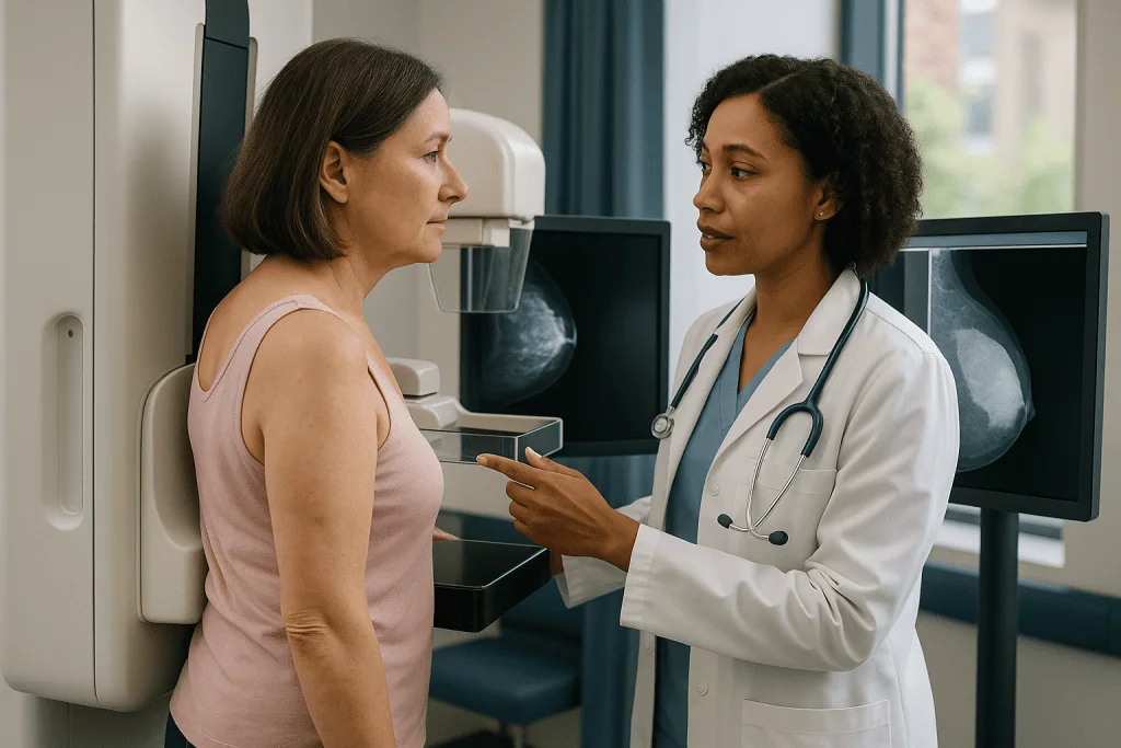
The first is age. Mayo Clinic notes that mammography is the standard screening tool for the general breast population, and if you are in the age range where routine screening is recommended, your team may want an up-to-date mammogram of both normal breasts as part of the overall assessment before any breast-related operation. The accessory tissue itself is often positioned too high or too far to the side to be captured well, but a specialized view that angles into the armpit can sometimes include it.
The second reason is a specific ultrasound finding. If the scan shows a discrete nodule, a cluster of tiny bright spots, or an area that is not clearly benign, mammography can look for microcalcifications, tiny calcium deposits that ultrasound often misses and that sometimes accompany early abnormal change. The third reason is density. Cleveland Clinic explains that mammograms show dense glandular tissue as white, the same shade as many masses, which is precisely why ultrasound leads in dense tissue and mammography adds value where fat predominates.
A mammogram itself typically takes around 20 minutes according to Mayo Clinic, with the actual compression lasting only seconds per image. The pressure is uncomfortable for some people, more so before a period, and the technologist can adjust positioning to capture tissue near the armpit.
Neither test replaces the other. Ultrasound characterizes what a lump is made of and defines its edges; mammography surveys the whole breast field and catches calcifications. Your team chooses the combination based on your age, your scan findings and your history.
Ultrasound vs mammogram vs MRI before accessory breast surgery
People often ask why they are not simply sent for the most detailed scan available. The answer is that each test has a different job, and the right sequence is usually the least invasive test that answers the question, followed by others only when needed. The table below summarizes how the three main imaging methods compare in this specific setting.
| Question | Ultrasound | Mammogram | MRI |
|---|---|---|---|
| Radiation | None | Low-dose X-ray | None (magnetic field, often with contrast injection) |
| Best at | Telling solid from fluid, matching texture to breast gland, guiding a needle | Surveying the whole breast, finding microcalcifications | Mapping extent in complex or unclear cases |
| Handles armpit location well? | Yes, probe goes exactly where the lump is | Partly, with angled views | Yes |
| Typical role here | First test for almost everyone | Added by age or after a suspicious ultrasound finding | Reserved for unresolved questions |
| Typical duration | About 15–30 minutes (Johns Hopkins) | About 20 minutes (Mayo Clinic) | Longer, often 30–60 minutes |
MRI, magnetic resonance imaging, builds pictures from the way hydrogen atoms respond to a strong magnet. It is exquisitely sensitive but also flags many harmless areas of enhancement, which can lead to extra biopsies. For an uncomplicated soft mound in the armpit with a textbook ultrasound appearance, MRI adds little and is rarely requested. It earns its place when ultrasound and mammography disagree, when the tissue is unusually extensive, or when there is a personal history that raises the stakes.
The practical point is that the sequence is deliberate. Starting with ultrasound is not a shortcut; it is the test most likely to settle the diagnosis of accessory breast tissue in a single sitting.
Why operate on a diagnosis, not a guess: the look-alikes in the armpit
The strongest argument for imaging first is the list of things that impersonate accessory breast tissue. Each has a different treatment, and some have no need for treatment at all.
- Lipoma. A soft, slow-growing lump of fat. On ultrasound it appears as a smooth, oval, fatty mass without the fibrous and glandular layering of breast tissue. Removing it involves a different, usually smaller, operation than excising a sheet of glandular tissue.
- Enlarged lymph node. Nodes swell with infection, inflammation and, occasionally, blood or lymphatic cancers or spread from a cancer elsewhere. On ultrasound a normal node has a characteristic kidney-bean shape with a bright fatty center. Nodes should never be casually excised as if they were cosmetic tissue.
- Cyst or galactocele. Fluid or milk in a sac. Ultrasound shows fluid as black with a sharp back wall. Aspiration with a needle may resolve it entirely; surgery is often unnecessary.
- Hidradenitis suppurativa. Chronic inflammation of sweat gland areas that produces tender nodules and tracts in the armpit. It is managed medically and surgically in a completely different way.
- Fibroadenoma or other mass within accessory tissue. Because accessory tissue is breast, it can host the same benign and malignant growths as the ordinary breast. A discrete mass changes the plan from cosmetic removal to diagnostic work-up.
- Soft-tissue tumors. Rare growths of fat, muscle or connective tissue can sit in the same location and require a specialist surgical approach.
The NHS makes the general point plainly: most breast lumps are harmless, but a clinician should check any new lump because it is not possible to tell the cause by touch alone. That principle does not stop at the edge of the breast. An operation planned for accessory tissue that turns out to be a lymph node or an unexpected tumor is a problem for both patient and surgeon, and it is exactly the scenario a pre-operative scan is designed to prevent.
Can accessory breast tissue be cancerous?
Yes, in the sense that any breast tissue can develop breast cancer, and accessory tissue is genuinely breast tissue. Case series in the medical literature describe carcinoma arising in axillary accessory breasts, and it is one of the reasons radiologists examine the tissue rather than simply confirming that it exists. That said, the honest framing matters: cancer in accessory tissue is uncommon, the overwhelming majority of people with an armpit mound have benign glandular tissue, and the evidence does not show that accessory tissue carries a higher risk per gram than the normal breast.
Why, then, does it come up so often? Three reasons. First, accessory tissue sits outside the areas covered by standard screening mammography, so any change in it is more likely to be found by touch than by routine imaging. Second, people often attribute a new firmness in the armpit to their known accessory tissue and wait, which can delay evaluation. Third, the armpit contains lymph nodes, and a firm node may be mistaken for accessory tissue or the other way around.
The practical safeguard is the same one applied to the ordinary breast. Soft, symmetrical tissue that swells and settles with the menstrual cycle and has a normal, layered ultrasound texture is reassuring. A new, distinct, firm lump within that tissue, skin changes over it, a nipple discharge from an accessory nipple, or a change on one side only deserves prompt imaging, and sometimes a biopsy, before anyone talks about cosmetic removal.
Mayo Clinic notes that a breast biopsy, the removal of a small sample of tissue for laboratory analysis, is the only way to confirm whether a suspicious area is cancer. Imaging narrows the question; pathology answers it. If your ultrasound report is entirely benign, the team can plan surgery with confidence. If it is not, the correct next step is diagnostic, and your team will explain what that involves and why the cosmetic plan must wait.
What does suspicious tissue look like on an ultrasound?
Radiologists do not rely on a single feature. They weigh a pattern, and they report their overall impression using a standardized scale called BI-RADS, the Breast Imaging Reporting and Data System, which grades findings from clearly benign to highly suspicious and tells the referring clinician what should happen next.
Normal accessory breast tissue looks like the ordinary breast: an irregular mix of bright fibrous strands and darker fatty lobules, without a discrete mass, blending gradually into the surrounding fat. A simple cyst is round or oval, completely black inside, with a thin wall and a bright echo behind it because sound passes straight through fluid. A typical fibroadenoma is oval, wider than it is tall, with smooth edges and a uniform gray interior.
Features that raise concern include a mass that is taller than it is wide, as if it is growing across tissue planes rather than along them; irregular or spiky margins; a very dark interior with a shadow cast behind it, which suggests dense tissue absorbing sound; tiny bright dots within a solid mass that may represent calcifications; and disordered blood flow on Doppler. Nearby lymph nodes that have lost their fatty center or thickened unevenly also draw attention.
Two points deserve emphasis. Ultrasound features overlap: some cancers look bland and some benign lumps look worrying, which is exactly why a suspicious appearance leads to a biopsy rather than a diagnosis. And a low BI-RADS category is meaningful reassurance; it is the reason a surgeon can proceed with removing tissue as a cosmetic or comfort procedure rather than a diagnostic one.
If your report includes a BI-RADS number, ask your clinician to translate it. The categories are designed to be shared with patients, and understanding yours makes the rest of the plan far easier to follow.
Who is usually offered accessory breast surgery, and who is asked to wait
Surgery to remove accessory breast tissue is most often an elective procedure, chosen for comfort or appearance rather than medical necessity. The typical candidate has imaging-confirmed glandular tissue that causes physical symptoms such as cyclical pain, chafing, irritation from clothing, or recurrent swelling during hormonal changes, or that causes distress about appearance that has not eased with time. Some people have a functional accessory nipple that leaks during lactation and want it addressed once breastfeeding is finished.
The operation itself is usually a direct excision through an incision hidden in an armpit crease, sometimes combined with liposuction, a technique that removes fat through a thin tube, to smooth the contour where fatty and glandular tissue are mixed. Liposuction alone is not appropriate when true glandular tissue predominates, because it does not remove gland efficiently, and it can be unwise when imaging has not confirmed what the tissue is. Extent on the ultrasound guides the incision length and depth.
Several groups are commonly asked to wait or to take a different route first:
- People whose imaging shows an unresolved or suspicious finding, until biopsy has settled the diagnosis.
- Those who are pregnant or breastfeeding, because the tissue is enlarged and vascular and may shrink considerably afterward.
- Adolescents whose tissue is still developing, since the final size is not yet known.
- People with active skin infection or inflammatory conditions in the armpit, until these are controlled.
- Anyone whose general health, medications or bleeding tendency needs optimizing before an elective operation.
Alternatives include doing nothing, which is reasonable for asymptomatic tissue with benign imaging, supportive clothing and pain management during symptomatic phases, and, for a milk-filled cyst, needle aspiration rather than excision. Risks of surgery are those of any operation in the armpit: bleeding, infection, fluid collection called a seroma, scarring, numbness from small nerve disturbance, and incomplete removal with residual fullness. Your surgeon weighs these with you; the decision rests with you and the treating team.
Does accessory breast tissue go away on its own?
Usually not, though it changes size dramatically enough that people sometimes believe it has. Accessory tissue is a permanent developmental structure. What fluctuates is its state: how much glandular tissue is active, how engorged it is with fluid, and how much fat surrounds it.
Before a menstrual period, rising hormones can make the tissue swell and ache, only for it to soften and shrink a week later. During pregnancy it may grow markedly, and during lactation it can fill with milk. After weaning, the glandular component shrinks back, sometimes to less than its pre-pregnancy size, which is why surgeons often prefer to wait several months after breastfeeding ends before assessing what actually needs to be removed. Around menopause, glandular tissue throughout the body gradually involutes, meaning it is replaced by fat, and the mound may become softer and less noticeable, although it does not vanish.
Weight loss can reduce the fatty component and make the swelling less prominent. Weight gain does the reverse. Neither changes the underlying gland.
There is a clinical consequence to all of this. A single ultrasound is a snapshot. Tissue scanned during a swollen phase looks larger and more glandular than the same tissue scanned two weeks later. This does not make the scan unreliable, but it is one reason a surgeon may ask for imaging at a specific point in the cycle, or may repeat it after pregnancy, before finalizing a plan. It is also why a report that says the tissue has shrunk is not a sign that anything has been missed.
If your goal is to avoid surgery altogether, the evidence supports a realistic expectation: symptoms may ease, the appearance may soften, but the tissue itself will remain, and any new distinct lump within it still deserves the same evaluation as a lump anywhere else in the breast.
What the days and weeks after the scan usually look like
The scan is often the shortest part of the process. What follows depends entirely on what it shows.
When the ultrasound confirms uncomplicated accessory breast tissue with a benign appearance, the report typically goes to your referring clinician within a few days, and the conversation moves to whether surgery is right for you, when, and by which technique. Some teams ask for a mammogram of the normal breasts at this stage if you are in a screening age group and have not had one recently. Pre-operative assessment then follows the usual pattern of health questions, medication review and, where needed, blood tests.
When the scan shows a discrete mass or an uncertain finding, the next step is usually a needle biopsy under ultrasound guidance. A core needle biopsy uses a hollow needle to remove several slivers of tissue under local anesthetic, with the radiologist watching the needle enter the target on screen. Mayo Clinic describes the procedure as taking a short time, with results from the pathology laboratory commonly available within a few days to a week or more, depending on the laboratory and whether additional stains are needed. Expect bruising and tenderness at the site for a few days; MedlinePlus advises avoiding strenuous activity briefly afterward.
If the biopsy is benign, planning resumes, sometimes with a recommendation to remove the lump as part of the same operation. If it is not benign, the pathway changes completely, and your team will discuss it with you in detail.
After the surgery itself, when it happens, the early weeks generally involve a dressing over an armpit incision, some swelling and tightness, a period of limited arm elevation and lifting while the wound settles, and sometimes a small drain if the excision was large. Numbness of the inner upper arm can occur and often improves over months. Your surgeon gives you a personalized timeline; general ranges from online sources should never override it.
What people often get wrong about accessory breast tissue imaging
Misunderstandings cluster around a few themes, and clearing them up changes how people approach the process.
Being sent for a scan means the doctor suspects cancer. Almost always it means the opposite: the team wants to confirm that this is benign glandular tissue so the operation can be planned as a straightforward excision. Imaging before elective surgery is routine, not a red flag.
A normal mammogram of the breasts covers the armpit tissue. Standard mammographic views often do not fully include tissue high in the axilla. A clear routine mammogram is reassuring about the breasts but does not by itself evaluate an armpit lump. Ultrasound of the specific area does.
Liposuction alone will remove it. Liposuction removes fat efficiently and gland poorly. If ultrasound shows mostly glandular tissue, a direct excision is usually needed for a lasting result. This is one of the most practical reasons imaging comes first: it tells the surgeon which technique fits the tissue.
Ultrasound is a definitive test for cancer. Ultrasound describes; it does not diagnose. A reassuring appearance lowers concern substantially, but only tissue under a microscope confirms or excludes cancer when a finding is suspicious.
Accessory tissue is just fat. Some armpit fullness is indeed fat, and imaging distinguishes it. True accessory tissue behaves like breast: it responds to hormones and can develop the same conditions.
If it shrinks, it has gone. Hormonal shrinkage is real, but the gland remains, as discussed above.
The scan is painful. Ultrasound is painless apart from mild pressure. Mammography can be uncomfortable for seconds at a time, particularly before a period, and technologists can adjust technique. Neither should be a reason to avoid an evaluation that protects you from operating on the wrong diagnosis.
Questions to ask your care team before accessory breast surgery
A good consultation leaves you knowing what was seen, what it means and what happens next. These questions tend to draw out the answers that matter most.
- Did the ultrasound confirm that this is glandular breast tissue, and how far does it extend beyond what I can see or feel?
- Was there a BI-RADS category on the report, and what does that category mean for my plan?
- Were the lymph nodes in the area examined, and did they look normal?
- Do I need a mammogram as well, and is that because of my age, a specific finding, or both?
- Was anything found that needs a biopsy before we discuss surgery?
- Based on the imaging, would you recommend direct excision, liposuction, or a combination, and why?
- Is there a reason to repeat the scan at a different point in my cycle or after I finish breastfeeding?
- What are the realistic risks of this operation in my case, including numbness, seroma, scar position and the chance that some tissue remains?
- What would you expect to happen if I chose not to have surgery, and how should I monitor the area?
- How will the removed tissue be examined afterward, and will I receive the pathology result?
- Which symptoms after surgery should prompt me to call you promptly?
Bring the imaging report itself if you have it, and do not hesitate to ask the clinician to point to the relevant parts of the image. Radiologists write for other clinicians, and phrases such as heterogeneous fibroglandular echotexture are simply descriptions of normal-looking breast tissue in the wrong place. Having them translated in the room is far more useful than searching them later. The decision about whether and how to proceed is yours to make with your treating team, and it is easier to make well when every result has been explained.
When to call your doctor
Most accessory breast tissue is quiet and predictable, swelling a little before a period and settling afterward. Some changes, however, should prompt a call rather than a wait for the next routine appointment, whether or not surgery is planned.
Contact your doctor promptly if you notice a new, distinct, firm lump within the accessory tissue or elsewhere in the armpit, particularly one that does not soften after your period; a lump that is growing steadily over weeks; skin over the area that is dimpling, thickening, reddening or pulling inward; discharge, especially bloody discharge, from an accessory nipple outside of breastfeeding; a hard, fixed or enlarging lymph node; or a change on one side only when the tissue was previously symmetrical. Unexplained fevers, night sweats or weight loss alongside an enlarging armpit lump also warrant assessment, since these can accompany conditions of the lymph nodes rather than breast tissue.
If you have had a biopsy, call if the site becomes increasingly painful, hot, red or swollen, if bleeding does not stop with firm pressure, or if you develop a fever. After surgery, seek advice for expanding swelling under the incision, fluid or pus leaking from the wound, spreading redness, a fever, severe pain not controlled by what your team advised, or new arm swelling.
Seek urgent care for sudden chest pain, breathlessness, or a swollen, painful arm or calf after an operation, which can signal a blood clot and require immediate assessment.
None of these signs is a diagnosis. The NHS and MedlinePlus both stress that most breast lumps are benign and that the purpose of prompt evaluation is to find the rare exception early and to reassure everyone else quickly. Your treating team is the right place for that evaluation, and no online description, including this one, replaces their examination and judgment.
Frequently asked questions
Can accessory breast tissue be cancerous?
Yes, because it is genuine breast tissue and can develop the same conditions as the ordinary breast, although cancer in accessory tissue is uncommon. That possibility is one reason imaging is done before removal. Soft tissue that swells and settles with the menstrual cycle and shows a normal layered texture on ultrasound is reassuring; a new, distinct, firm lump within it should be assessed promptly and may need a biopsy before any cosmetic surgery is planned.
Does accessory breast tissue go away?
The tissue itself does not disappear on its own, but it changes size with hormones. It can swell before periods, enlarge markedly in pregnancy and lactation, then shrink after weaning, and it softens as glandular tissue is replaced by fat around menopause. Weight changes alter the fatty component. Surgeons often wait several months after breastfeeding ends before assessing what actually needs removal, since the tissue may be considerably smaller by then.
What does cancerous breast tissue look like on an ultrasound?
No single feature is definitive, but radiologists look for a solid mass that is taller than it is wide, has irregular or spiky edges, appears very dark inside with a shadow behind it, contains tiny bright calcifications, or shows disordered blood flow on Doppler. Nearby lymph nodes that have lost their fatty center also raise concern. Because benign and malignant features overlap, a suspicious appearance leads to a biopsy rather than a diagnosis.
What causes accessory breast tissue?
It is a developmental variation, not something caused by lifestyle. Early in fetal life a ridge of potential breast tissue runs from each armpit toward the groin. Normally everything regresses except one spot on each side of the chest. If part of the ridge persists, it can grow into functioning breast gland, most often in the armpit or below the normal breast. It frequently becomes noticeable at puberty, before periods or during pregnancy.
Why is an axillary breast tissue ultrasound done before surgery instead of just removing the lump?
Because a soft armpit swelling has many possible causes, and surgery only removes what is there without first telling you what it is. Ultrasound confirms whether the lump is glandular breast tissue, a fatty lipoma, a lymph node, a fluid-filled cyst or something rarer, each of which needs a different plan. It also maps the tissue’s true extent under the skin and checks for any suspicious feature that would change the approach.
Do I need an accessory breast tissue mammogram as well as an ultrasound?
Not always. Ultrasound alone often answers the question, especially in younger people with dense tissue. A mammogram is commonly added if you are in a screening age group and have not had one recently, or if the ultrasound shows a discrete nodule or an area that is not clearly benign, because mammography detects tiny calcifications that ultrasound may miss. Your team decides based on your age, findings and history.
What does a BI-RADS category on my report mean?
BI-RADS is a standardized scale radiologists use to summarize how benign or suspicious an imaging finding appears and to recommend the next step, from routine follow-up to biopsy. A low category indicates a benign or almost certainly benign appearance and lets a surgeon plan removal as a cosmetic or comfort procedure. A higher category signals that tissue sampling should come before any elective operation. Ask your clinician to explain your specific category.
Will the ultrasound or mammogram hurt?
Ultrasound is painless apart from mild pressure from the probe and cool gel on the skin. Mammography compresses the tissue between two plates for a few seconds per image, which many people find uncomfortable and some find briefly painful, especially in the week before a period. Technologists can adjust positioning and pressure, and telling them about tenderness beforehand helps. Neither test involves needles unless a biopsy is planned separately.
What happens if the scan finds something suspicious in the accessory tissue?
The usual next step is a needle biopsy guided by ultrasound, in which a small sample is taken under local anesthetic and examined by a pathologist. Results commonly take several days to a week or more depending on the laboratory. If the sample is benign, planning for removal can resume, often including the lump. If it is not, the pathway changes to a diagnostic and treatment plan that your team will explain in full.
Can accessory breast tissue be removed with liposuction alone?
Sometimes, but only when imaging shows that the swelling is mostly fat. Liposuction removes fat efficiently and glandular tissue poorly, so if ultrasound confirms predominantly glandular accessory breast tissue, a direct excision through an armpit incision, sometimes combined with liposuction for contour, is usually needed for a lasting result. This is one of the practical reasons imaging is done first: it tells the surgeon which technique fits.
References
This article is for general information only and is not a substitute for professional medical advice. Please consult a qualified doctor about your individual situation.

More from the Blog

Breast Lift Scars and Daily Life After Mastopexy: Sleeping, Sports Bras and Sun Protection
Breast lift scars are permanent, but they usually change a great deal. Most fade from raised and pink to flat, pale lines over roughly…

Healing After Nipple Reconstruction: Protective Dressings, Bras to Avoid and the First Weeks
Nipple reconstruction recovery is usually gentler than earlier breast surgery. Most people wear a protective dressing or shield for the first weeks, choose soft…

Breast Fat Transfer vs Implants: Volume, Feel, Scars and Which Goals Each Option Suits
Fat transfer moves your own fat, taken by liposuction, into the breast for a modest, soft increase with only tiny needle-entry scars, but part…

Is Capsulectomy Needed With Breast Prosthesis Removal? How Surgeons Decide What Comes Out
Capsulectomy is not automatically required with breast prosthesis removal. Surgeons usually remove the capsule when it is thickened, calcified, ruptured silicone is present, or…

Local or General Anesthesia for Gynecomastia Surgery? What Shapes the Choice and Recovery
Gynecomastia surgery can be done under local anesthesia with or without sedation, or under general anesthesia. The choice depends mainly on how much glandular…

How Accessory Breast Removal Works: Excision, Liposuction and the Armpit Incision Explained
Accessory breast removal surgery takes out extra breast tissue, most often in the armpit, through a short incision hidden in the natural underarm crease.…