Accessory Breast Treatment
Accessory breast treatment removes extra breast tissue, most often in the armpit, to improve comfort, symmetry, and appearance. Surgery is planned after clinical evaluation and imaging when needed.
Quick answer
Accessory breast treatment is the removal of extra breast tissue, and sometimes a third nipple, that forms along the embryonic milk line, most often in the armpit. Depending on whether the tissue is glandular, fatty or mixed, surgeons use direct excision, liposuction or a combination of both, usually as a day-case procedure. Removed tissue is normally sent for pathology examination to confirm the diagnosis.
Third Nipple and Accessory Breast Tissue: What They Are and When Treatment Helps
A third nipple, known medically as a supernumerary or accessory nipple, is an extra nipple that forms along the embryonic milk line, the developmental track that runs from each armpit down towards the groin. Accessory breast tissue is the related condition in which glandular breast tissue itself develops outside the normal breast, most often in the armpit. Treatment ranges from straightforward reassurance to surgical removal, and it is relevant for anyone whose extra tissue causes discomfort, visible fullness, hormonal swelling or persistent uncertainty about what the lump actually is.
Both findings are more common than most people realise. For some, accessory tissue is a small fullness that only becomes noticeable with weight change, pregnancy or hormonal shifts. For others, it causes visible asymmetry, discomfort with clothing, tenderness before menstrual periods, sweating or irritation in the skin fold, or a persistent concern about appearance. A third nipple, by contrast, is often mistaken for a mole or skin tag for years before anyone identifies what it actually is.
Patients often begin researching treatment after a long period of uncertainty. Some have been told the fullness is “just fat”. Others worry that it may be a lymph node, a cyst or a breast-related condition. Most people arriving at this page want two things in a specific order: first a clear diagnosis, an honest answer to whether the tissue matters medically, and only then a discussion of whether removal makes sense for them.
Treatment for accessory breast tissue is usually elective, but elective does not mean unimportant. When extra tissue creates pain, repeated irritation, limits on clothing choices or self-consciousness during movement, exercise or social life, removal can be a meaningful step. The aim is not simply to take tissue out. It is to improve contour, comfort and symmetry while protecting shoulder function and keeping scars as discreet as the anatomy allows.
At Acibadem, evaluation and treatment are planned with attention to both medical safety and aesthetic detail. Depending on the individual case, care may involve breast surgeons, plastic and reconstructive surgeons, radiologists, anaesthesiology specialists and pathology teams. Imaging may be recommended when the tissue behaves like breast tissue, when there is a distinct lump, or when a patient’s age or breast cancer risk profile makes further assessment appropriate. Confirming the diagnosis before surgery is what allows the operation to be planned around the patient’s actual anatomy and goals rather than around assumptions.
What is a third nipple?
A third nipple, medically termed polythelia, is an extra nipple without a complete breast beneath it. It most often sits below the normal breast on the milk line, though it can appear anywhere along that track, and it is frequently small, flat and easy to mistake for a mole, skin tag or birthmark. Some have a miniature areola around them; many do not. Most cause no symptoms at all, but because the tissue can respond to hormones, a third nipple may darken, enlarge or become tender during puberty, pregnancy or breastfeeding, which is often the moment people first recognise it for what it is. When there is no deep glandular tissue underneath, removal is a comparatively minor skin-level procedure. When glandular tissue does sit beneath the nipple, the plan shifts closer to accessory breast surgery.
Why do people have extra nipples?
An extra nipple forms before birth, when the embryonic mammary ridges, the milk lines, do not regress completely. Early in development, every embryo has two ridges of breast-forming tissue running from the armpit region to the groin. Normally, almost all of this ridge disappears, leaving a single breast on each side of the chest. If a fragment of the ridge persists, it can develop into an extra nipple, an extra patch of glandular tissue, or both. This happens in both men and women. In most people it is an isolated, sporadic finding with no wider significance, although it occasionally runs in families. Polythelia has occasionally been described in the medical literature alongside other developmental variations, which is one reason a physician may examine more broadly during the first assessment, but for the vast majority of people it is an isolated finding. It is not caused by anything a person or their parents did, and it is not a sign of poor health.
Is a 3rd nipple the same thing as “3 boobs”?
People who search for a 3rd nipple and people who describe having “3 boobs” are usually describing different points on the same developmental spectrum. At one end sits polythelia: a nipple alone, with no breast tissue beneath it. In the middle sits the most common presentation, glandular breast tissue in the armpit with no visible nipple, which is what most patients mean when they describe an underarm bulge or what some informally call a third breast. At the far end sits a complete accessory breast with glandular tissue, nipple and areola together. The colloquial phrases matter clinically because they change the surgical plan: a skin-level nipple excision is a very different operation from contouring a full pad of axillary breast tissue.
 Dr. Şule ErenMDBoard Commentary
The most important step in evaluating accessory breast tissue is confirming that an axillary fullness truly represents ectopic breast tissue and distinguishing it from lymph nodes, lipomas, cysts or other breast-related lesions. Acıbadem-affiliated breast radiology and senology teams have published research on advanced breast imaging techniques, supporting the use of targeted ultrasound and additional imaging when clinical examination alone is insufficient. Once the diagnosis is established, treatment can be individualized according to the proportion of glandular tissue, fat and excess skin, using surgical excision, liposuction or a combined approach.
What Accessory Breast Treatment Is
Accessory breast treatment is the medical and surgical management of extra breast tissue located outside the normal breast area. In most patients, the tissue is found in one or both armpits. It may contain glandular breast tissue, fatty tissue or a combination of both. Some patients also have an accessory nipple or areola over the tissue, although many do not, which is why the condition is so often mistaken for simple weight gain or a swollen lymph node.
The most common treatment is surgical removal. The technique may involve direct excision, liposuction-assisted contouring or a combined approach. Direct excision is used when there is firm glandular tissue, excess skin, a defined mass or tissue that needs to be sent for pathology. Liposuction is useful when the fullness is mainly fatty and when the surrounding contour needs blending. In many axillary cases, surgeons combine both methods: removing glandular tissue through a carefully placed incision and refining the surrounding contour with gentle fat removal.
The procedure is typically performed to address discomfort, swelling, aesthetic concerns or asymmetry. It may also be recommended when imaging or clinical examination identifies tissue that should be removed and analysed. While accessory breast tissue is usually benign, it responds to hormones exactly as normal breast tissue does. It may enlarge during puberty, pregnancy, breastfeeding or weight gain, and it can become tender before menstrual periods. Rarely, breast diseases can arise within it, which is why persistent changes deserve proper evaluation rather than dismissal.
Treatment planning starts with understanding what type of tissue is present, where it sits, how much skin redundancy exists and how the area moves when the arm is raised. The armpit is a functional region containing lymphatic structures, blood vessels, nerves and natural skin folds. A well-designed plan removes the unwanted tissue while maintaining comfortable shoulder movement, creating a smooth transition to the chest wall and upper arm, and placing scars in discreet positions whenever the anatomy allows it.
What is the difference between polythelia and polymastia?
Polythelia means an extra nipple without underlying glandular breast tissue, while polymastia means accessory breast tissue, with or without a nipple above it. The distinction matters because it defines the operation. Polythelia is usually treated with a small skin-level excision, similar in scale to removing a prominent mole, and healing is correspondingly quick. Polymastia involves a deeper volume of glandular or fatty tissue, so treatment requires genuine contouring: deciding how much tissue to remove, whether liposuction should refine the edges, and whether loose skin needs to be addressed so that no residual fold remains. A person can have either condition alone or both together, and examination, sometimes supported by ultrasound, establishes which situation applies before any incision is planned.
Who May Need Accessory Breast Treatment
Patients who seek accessory breast treatment usually describe a fullness, lump, bulge or fold in the underarm area. It may be present on one side or both. Some have noticed it since adolescence; others become aware of it after pregnancy, breastfeeding, hormonal treatment or changes in weight. In many cases the tissue grows gradually and becomes more visible with fitted clothing, swimwear or sleeveless garments.
Common symptoms include tenderness, cyclic swelling, a pulling sensation with arm movement, friction from bras and clothing, sweating in the fold, skin irritation, and difficulty shaving or applying deodorant. Patients also report embarrassment when raising the arms, exercising or wearing certain styles of clothing. When the tissue is substantial, it can create an underarm contour that looks separate from the breast, and, importantly, it does not reliably improve with diet or exercise, because glandular tissue does not shrink the way fat does.
How is accessory breast tissue diagnosed?
Diagnosis begins with a medical history and physical examination, not with imaging. The physician asks when the tissue first appeared, whether it changes with menstrual cycles, pregnancy, lactation or weight fluctuation, and whether there is pain, discharge, skin change or a palpable lump. The examination then assesses whether the fullness feels fatty, glandular, nodular, or related to lymph nodes or other soft tissue structures, and how the area behaves with the arm relaxed and raised.
Imaging is recommended selectively rather than routinely. Ultrasound is often the most useful first study for axillary tissue, especially in younger patients or when there is a distinct lump, because it can distinguish glandular tissue from fat, cysts and lymph nodes. Mammography or breast MRI may be considered depending on age, breast density, personal or family history, examination findings and whether the tissue appears connected to the main breast. Together, examination and imaging separate accessory breast tissue from the conditions it can mimic: enlarged lymph nodes, lipomas, cysts, hidradenitis-related changes and other soft tissue masses.
Some patients come for a second opinion after being told the area is normal. Others have already had imaging but still need guidance about whether surgery is appropriate and which technique would give the best result. The decision to proceed rests on symptoms, appearance, patient goals, medical findings and the likelihood that removal will genuinely improve comfort or contour. Surgery is not always necessary. When the tissue is persistent, symptomatic or cosmetically distressing, however, a carefully planned operation is the definitive answer.
Is Accessory Breast Tissue Dangerous?
Accessory breast tissue is usually benign, and for most people it is a comfort and appearance issue rather than a medical threat. Because it is true breast tissue, however, it can develop the same benign and malignant conditions as breast tissue in its usual location, including, rarely, breast cancer. This is not a reason for alarm; it is a reason for the same sensible attention that any breast tissue deserves. A stable, soft fullness that swells slightly before periods behaves like normal hormone-responsive tissue. A new firm lump, rapid enlargement, skin dimpling, ulceration, persistent localised pain or discharge from an accessory nipple behaves differently and warrants proper medical evaluation.
There is one practical point worth understanding: accessory tissue in the armpit can sit outside the field of standard breast imaging views, so it is helpful for the radiologist to know it exists when screening or diagnostic imaging is planned. This is also one of the arguments in favour of excision in selected patients, removed tissue is examined by pathology, which confirms the tissue type and settles lingering uncertainty in a way that observation alone cannot.
Can accessory breast tissue produce milk?
Yes, because accessory tissue is true glandular breast tissue, it can respond to the hormones of pregnancy and lactation and, in some women, produce milk. When an accessory nipple sits above the tissue and connects to functioning ducts, small amounts of milk may actually be expressed. More often there is no outlet at all: the gland swells during breastfeeding, the milk has nowhere to drain, and the area becomes engorged, firm and tender for a period before settling as lactation regulates itself. This can be alarming when nobody has previously explained what the tissue is, but it reflects normal glandular physiology in an unusual location. Repeated painful engorgement with each pregnancy is one of the recognised reasons women eventually choose removal once breastfeeding is complete.
Conditions and Indications Accessory Breast Treatment Addresses
The most frequent indication is axillary accessory breast tissue, where extra glandular or fatty tissue creates fullness in the armpit. This may be unilateral or bilateral and varies from a small pad of tissue to a prominent bulge that alters the whole line of the upper body in fitted clothing.
Another indication is accessory tissue that becomes painful or swollen with hormonal changes. Because glandular tissue responds to oestrogen, progesterone, pregnancy and lactation, symptoms tend to arrive at predictable times. Some patients experience tenderness before their period; others develop noticeable enlargement during pregnancy or breastfeeding.
Treatment may also address contour irregularity after weight loss, pregnancy or previous breast surgery. Even when the tissue poses no medical risk, it can disturb the line between the breast, chest wall and upper arm. In patients planning breast reduction, a breast lift or breast augmentation, accessory tissue can often be treated during the same operation, so the overall chest and underarm contour is balanced in a single recovery period rather than two.
Accessory nipples and accessory areolar tissue can also be treated, especially when they cause irritation or visible concern. Removing an accessory nipple that catches on clothing or draws unwanted attention is typically a short, contained procedure, and the surgical plan follows the same logic as nipple aesthetics more broadly: location, size, pigmentation and whether deeper glandular tissue sits beneath the skin all shape the approach.
Finally, in selected cases, removal is recommended for diagnostic reasons. If imaging shows an abnormality within accessory tissue, or a persistent lump cannot be confidently characterised, excision with pathology analysis may be the appropriate next step, particularly where there is rapid growth, skin change, discharge, persistent pain or a strong family history of breast cancer. Most findings turn out to be benign, but a structured evaluation is what makes that reassurance trustworthy.
How Accessory Breast Treatment Is Performed
Treatment begins with a consultation that defines both the medical diagnosis and the desired aesthetic outcome. The surgeon examines the area with the arms relaxed and raised, because axillary tissue changes shape dramatically with movement. Photographs may be taken for planning. The assessment covers tissue thickness, skin elasticity, the natural underarm fold, symmetry between sides and the relationship of the tissue to the breast and upper arm.
If imaging is needed, it is completed before surgery. Ultrasound identifies whether the tissue has glandular features, fatty composition, cysts or lymph nodes; mammography or MRI may be added for patients with specific risk factors or unclear findings. Blood tests and an anaesthesia evaluation are arranged according to age, medical history and the planned anaesthesia type. Patients who take blood thinners, certain supplements or medications that affect bleeding receive individual instructions from the surgical and anaesthesia teams as part of preparation.
Before the operation, the surgeon marks the area while the patient is upright, because the tissue sits differently when lying down. These markings guide incision placement and contour correction. Incision design in the axilla involves a genuine trade-off. The goal is to place the scar within a natural crease where possible while still allowing safe, complete access to the tissue. If there is significant loose skin, the incision may need to be longer to avoid leaving a residual fold. If the tissue is mainly fatty and the skin has good elasticity, a smaller access point for liposuction may suffice, though firm glandular tissue almost always requires excision, because it cannot be suctioned effectively.
The procedure is commonly performed under general anaesthesia or local anaesthesia with sedation, depending on the extent of treatment and patient preference. For smaller areas, including many accessory nipple removals, local anaesthesia is often appropriate. For bilateral or more extensive cases, general anaesthesia provides comfort and surgical precision. The anaesthesia plan is agreed in advance with the anaesthesiology team.
During surgery, the surgeon removes the accessory tissue through direct excision, liposuction or both. Direct excision allows firm glandular tissue to be removed intact and sent to pathology. Liposuction uses narrow cannulas to reduce fatty volume and blend the treated area with the surrounding contours. Energy-assisted or suction-based technologies may be used in selected patients to refine contouring, but the choice always follows tissue type and safety rather than fashion. Careful lighting, magnification where helpful and meticulous control of bleeding protect the surrounding lymphatic, vascular and nerve structures.
Excised tissue is routinely submitted for pathology examination: a standard safety step that confirms the tissue type and checks for unexpected abnormalities. After removal, the surgeon evaluates symmetry with the arm positioned in different ways before closing the incision in layers to support healing and reduce tension on the skin. A small drain is placed in some cases, particularly when a larger internal space has been created, although many patients do not need one. A compression garment or supportive dressing is usually applied to limit swelling and help the tissues settle into the new contour.
Operating time varies with the amount of tissue, whether one or both sides are treated and whether the procedure is combined with another breast or body contouring operation. Many accessory breast procedures are completed as outpatient surgery, meaning the patient returns home the same day after a period of observation.
Early recovery requires respect for the area rather than fear of it. The underarm participates in almost every daily movement, so tightness, swelling, bruising and mild pulling on lifting the arms are expected. Discomfort is managed with prescribed medication. Strenuous upper-body exercise, heavy lifting and wide arm movements are limited during the early healing phase, while gentle walking is encouraged soon after surgery to support circulation. Follow-up visits allow the team to check the incision, swelling, mobility and any drain. Sutures may be absorbable or may need removal depending on the closure technique, and scar care guidance is given once the incision has healed sufficiently.
How do you get rid of an accessory breast?
Surgery is the only definitive way to remove an accessory breast, because glandular tissue does not respond to diet, exercise or creams. Weight loss can shrink the fatty component and make the area less prominent, but the glandular core, the part that swells with hormones and causes cyclical tenderness, remains. The realistic pathway looks like this:
- Clinical assessmenthistory and examination to confirm the fullness is genuinely accessory breast tissue rather than a lymph node, lipoma or cyst.
- Selective imagingultrasound in most cases, with mammography or MRI where age, findings or risk profile justify it.
- Technique planningexcision for glandular tissue and excess skin, liposuction for fatty fullness, or a combination of both.
- Anaesthesia planninglocal anaesthesia with or without sedation for limited areas; general anaesthesia for bilateral or extensive treatment.
- The operationremoval of the tissue, contour refinement and layered closure, usually as a day-case procedure.
- Pathologyroutine analysis of the removed tissue to confirm the diagnosis.
- Structured recoverycompression, activity limits, follow-up checks and scar care until the contour settles.
Why Acting Early Matters
Accessory breast tissue does not usually require urgent treatment. Timely evaluation matters most when the area is growing, painful, asymmetric or associated with a lump or skin change, because early assessment confirms whether the fullness is truly accessory breast tissue or something else, lymph node enlargement, a cyst, a lipoma, an infection-related change or another soft tissue mass.
For patients with discomfort or recurrent irritation, delay simply prolongs avoidable symptoms. Underarm skin folds are prone to friction, sweating, rash and inflammation, and tender glandular tissue will keep swelling with each hormonal cycle. Many patients also quietly adapt their clothing, posture and activities around the area for years, a slow erosion of everyday comfort that only becomes obvious in retrospect.
From a surgical perspective, earlier treatment can sometimes allow a more limited correction, particularly before the skin has stretched significantly. Once the tissue enlarges or the skin loses elasticity, achieving a smooth result may require a longer incision or more extensive contouring. Weight changes, pregnancy and breastfeeding all influence timing, so the best moment for surgery is genuinely individual and worth discussing rather than assuming.
Serious disease in accessory tissue is uncommon, but the tissue can develop the same benign and malignant conditions as breast tissue in its usual location, and a prompt diagnosis spares months of unnecessary uncertainty.
Benefits of Accessory Breast Treatment
When carefully planned, accessory breast treatment can improve both physical comfort and the appearance of the underarm and chest contour. The table below summarises what each benefit means in practical terms.
| Benefit | What It Means for You |
|---|---|
| Improved underarm contour | Removal of extra tissue can create a smoother transition between the breast, chest wall and upper arm, especially in fitted clothing or swimwear. |
| Reduced discomfort | Patients with tenderness, swelling, pulling or pressure from bras and clothing may experience meaningful relief after healing. |
| Better symmetry | Treatment can address one-sided fullness or balance both underarm areas so the overall chest appearance is more even. |
| Less skin irritation | Removing a bulky fold may reduce friction, sweating and recurrent irritation in the armpit crease. |
| Diagnostic clarity | Excised tissue can be examined by pathology, which confirms the diagnosis and identifies unexpected abnormalities if present. |
| Greater clothing comfort | Many patients find it easier to wear sleeveless tops, bras, athletic clothing and tailored garments after recovery. |
Recovery Timeline After Accessory Breast Surgery
Recovery varies with the surgical technique and the amount of tissue removed, but most patients can expect a gradual return to normal activities over several weeks rather than a single dramatic milestone.
| Time Period | What Patients Can Expect |
|---|---|
| Day 1 | Mild to moderate soreness, swelling and tightness are common. Patients usually go home after observation if the procedure is outpatient. |
| First week | Bruising and swelling may peak, then begin to improve. Light walking is encouraged, but heavy lifting and wide arm movements are limited. Follow-up checks take place during this period. |
| First month | Most daily activities become easier. The incision continues to heal and the contour becomes more defined as swelling decreases. Exercise resumes according to the surgeon’s guidance. |
| Longer term | Scars gradually mature and fade. The final contour continues to refine over several months as the tissues soften and settle. |
Factors That Influence Outcomes
A good result after accessory breast treatment begins with accurate diagnosis. If the fullness is mainly fatty, liposuction can provide effective contouring. If it contains dense glandular tissue, direct excision is usually necessary. If there is excess skin, the surgeon must judge whether the skin will contract adequately on its own or whether some skin removal is needed. Matching technique to anatomy is the single most important decision in the whole treatment.
Skin quality matters just as much. Younger patients and those with good elasticity often see the skin retract well after tissue removal. Patients with larger accessory tissue, long-standing stretching, prior weight fluctuation or reduced elasticity may need a longer incision to prevent a loose fold from remaining. This is a common trade-off in body contouring: a smaller scar is desirable, but not at the expense of an obvious residual bulge that defeats the purpose of the operation.
Symmetry deserves honest framing. Accessory tissue is often uneven between sides, and each side must be planned individually. Perfect mirror-image symmetry does not exist in human anatomy and cannot be created surgically; careful measurement, preoperative markings and intraoperative assessment create balance, not perfection. The position of the natural axillary crease, the shape of the breast and the contour of the upper arm all influence the final appearance.
Patient health and lifestyle affect healing directly. Smoking and nicotine use impair circulation and increase the risk of wound-healing problems. Certain medical conditions and bleeding tendencies require special planning, which is exactly what the preoperative assessment is for. Stable weight supports a more predictable outcome, because significant weight gain after surgery can change the contour of the area again. Pregnancy and breastfeeding also affect breast-related tissue, so timing should be discussed openly if a future pregnancy is planned.
Postoperative care carries real weight in the result. Wearing compression as advised, limiting upper-body strain in early recovery, attending follow-up appointments and following scar care instructions all support healing. The surgical team explains before discharge which changes, such as increasing redness, fever, worsening swelling, fluid collection, severe pain or wound separation, should be reported promptly, because minor issues managed early rarely become significant ones.
Finally, expectations should be settled before surgery, not after. The realistic goal is improvement in tissue prominence, comfort and contour. Scarring is an unavoidable part of excisional surgery, even when incisions are planned carefully and placed in natural folds. Some swelling, firmness or temporary numbness can occur, and small contour irregularities or asymmetries may become apparent as healing progresses. In selected cases, a revision can be considered once healing is complete, but many patients are satisfied with a single, well-planned procedure, precisely because the planning was done properly the first time.
Can accessory breast tissue come back after surgery?
Glandular tissue that has been fully excised does not regrow, so a complete, well-planned removal is normally a lasting correction. What can happen is different from true recurrence. If a rim of glandular tissue is deliberately or inadvertently left behind, sometimes unavoidable when the tissue blends gradually into the normal breast, that remnant can still respond to hormones and become noticeable during pregnancy or with hormonal changes. Separately, significant weight gain after surgery can increase fatty fullness in the same region, changing the contour even though no breast tissue has returned. Both possibilities are reasons why surgeons examine the full extent of the tissue before the operation and why maintaining a stable weight protects the result over the long term.
How Acibadem Organises Accessory Breast Care
Accessory breast removal can look like a simple operation from the outside, but doing it well requires the same infrastructure as any breast procedure: accurate assessment, safe anaesthesia, meticulous technique and pathology review of removed tissue. At Acibadem, this care sits within a structured pathway rather than a standalone cosmetic appointment.
The approach is multidisciplinary when the case calls for it. A patient with straightforward bilateral axillary fullness may be managed directly by a plastic and reconstructive surgeon or breast surgeon. A patient with a suspicious lump, a complex imaging finding, a history of breast disease or strong family risk may also involve breast radiology, pathology and the wider breast health department, so that aesthetic goals never overshadow medical safety. Ultrasound, mammography, MRI, laboratory testing and pathology services are used selectively, according to what the individual case actually requires.
Surgical planning is personalised. The surgeon weighs anatomy, symptoms, skin quality, scar preferences, clothing concerns and whether treatment should be combined with another breast or body procedure. Some patients prioritise the smallest possible scar; others need more complete tissue and skin removal to achieve the contour they want. A transparent discussion of these trade-offs, including what surgery cannot do, is part of the consultation itself.
Accessory breast tissue can be an intimate concern, and many patients arrive having had it minimised or dismissed for years. A serious consultation acknowledges both the physical symptoms and the emotional weight of the issue, and written aftercare guidance given at discharge remains usable long after the early follow-up visits are complete.
Deciding Whether to Have Treatment
Accessory breast treatment is a personal decision, and it is one that benefits from clear information more than from urgency. The condition is usually benign. The tissue will not disappear on its own, but it also rarely demands immediate action, which means most patients have time to understand their own anatomy before choosing surgery. The most appropriate plan depends on whether the tissue is glandular, fatty or mixed; whether imaging is needed to settle the diagnosis; how much skin is present; and what outcome the patient realistically hopes to achieve.
A thorough evaluation typically answers four questions: what the tissue actually is, whether anything about it needs further investigation, what removal would involve for that specific anatomy, and what the recovery and scar would realistically look like. When those answers are clear, the decision tends to make itself, either towards a carefully planned operation that improves comfort, contour and confidence, or towards informed reassurance that nothing needs to be done at all. Both are good outcomes, because both replace years of uncertainty with an answer.
Preparation
- A plastic surgeon evaluates the accessory breast tissue, symptoms, skin quality, and aesthetic goals. Breast ultrasound or mammography may be requested to confirm the tissue type and exclude other conditions. Patients may need to stop blood-thinning medicines, avoid smoking, and fast before surgery if general anesthesia is planned.
Aftercare
- Mild swelling, bruising, and tightness are common for several days after accessory breast removal. A compression garment may be recommended, and strenuous activity should be avoided until the surgeon approves. Follow-up visits check healing, scar care, and final contour as swelling gradually decreases.
Frequently Asked Questions
What affects the cost of accessory breast treatment?
Cost is influenced by the amount and type of tissue, whether liposuction, excision, or a combined approach is needed, the requirement for imaging, anesthesia type, hospital setting, pathology testing, compression garments, and follow-up arrangements.
How can I get a personalised quote?
You can request a free consultation by sharing photos, medical history, previous breast or armpit procedures, current symptoms, and any imaging reports if available. A specialist review is needed before a personalised treatment plan and quote can be prepared.
Is accessory breast treatment usually cosmetic or medical?
It can be either, depending on symptoms and findings. Some patients seek treatment for appearance or symmetry, while others have discomfort, swelling, irritation, or movement-related issues. The specialist will evaluate the reason for treatment and recommend the appropriate pathway.
Will imaging be needed before surgery?
Imaging may be recommended when the tissue feels glandular, symptoms are present, there is uncertainty about the diagnosis, or the specialist wants to assess the area more clearly before planning surgery.
What is usually included in an international patient package?
Package content varies, but it may include specialist assessment, routine preoperative tests, hospital and operating room services, anesthesia, surgery, translation support, basic postoperative checks, and care coordination. Travel and accommodation may be arranged separately or included depending on the provider.
Can the final cost change after consultation?
Yes. The final plan may change after physical examination, imaging review, or discussion of the preferred technique. A confirmed quote is usually provided after the specialist determines the safest and most suitable approach.
Medically reviewed by the Acıbadem International Medical Board September 1, 2026
See our medical review board →
Update history
- PublishedJune 8, 2026
- Medical review approvedSeptember 1, 2026
- Board commentary addedSeptember 1, 2026
- Last content updateSeptember 8, 2026
References3
- Breast Diseases medlineplus.gov
- Breast reduction (female) nhs.uk
- pubmed.ncbi.nlm.nih.gov
Trusted care for international patients
From the Acibadem Blog

Why Ultrasound or Mammography Comes Before Accessory Breast Surgery: Confirming the Tissue
Ultrasound, and sometimes mammography, comes before accessory breast surgery because a lump in the armpit or along the chest wall is not always breast…

How Accessory Breast Removal Works: Excision, Liposuction and the Armpit Incision Explained
Accessory breast removal surgery takes out extra breast tissue, most often in the armpit, through a short incision hidden in the natural underarm crease.…

When Does Accessory Breast Tissue Need Removal? Discomfort, Swelling Cycles and Appearance
Accessory breast tissue rarely needs removal for health reasons. Surgery is usually considered when the tissue causes persistent pain, cyclical swelling that interferes with…
Doctors Performing This Treatment


Aesthetic Plastic & Reconstructive Surgery
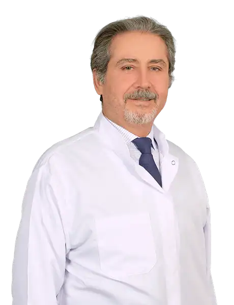
Prof. Mehmet Veli Karaaltın, MD

Aesthetic Plastic & Reconstructive Surgery
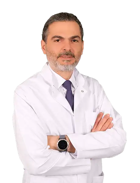
Assoc. Prof. Erdem Güven, MD

Aesthetic Plastic & Reconstructive Surgery
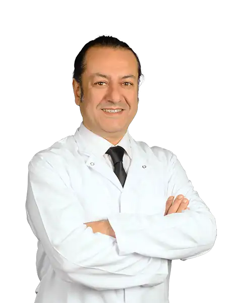
Assoc. Prof. Ahmet Küçükçelebi, MD
Aesthetic Plastic & Reconstructive Surgery
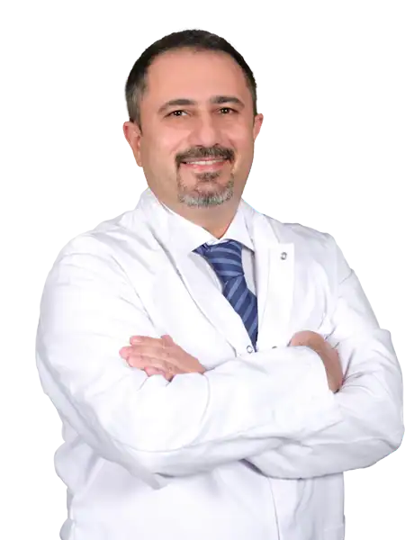
Assoc. Prof. Mehmet Altıparmak, MD

Aesthetic Plastic & Reconstructive Surgery