

Wilms Tumor Diagnosis and Staging: What Imaging, Kidney Tests and Pathology Tell the Team

Key Takeaways
- Wilms tumor is the most common kidney cancer in children and is usually found between the ages of three and four, most often as a painless abdominal swelling.
- Ultrasound is the first test, but the stage can only be assigned after CT or MRI of the abdomen, a chest CT, surgery and the pathologist's examination of the whole tumor.
- Tumor size does not determine stage; whether the tumor stayed within its capsule, was fully removed, and whether lymph nodes contain cells are what separate Stages I through III.
- Stage III means tumor remains in the abdomen after surgery, through positive lymph nodes, involved margins, spillage, peritoneal deposits, piecemeal removal or a pre-operative biopsy, without distant spread.
- Diffuse anaplasia on histology and loss of heterozygosity at chromosomes 1p and 16q move a child into a higher-risk group even when the stage is low.
- Surgery-first and chemotherapy-first pathways use the same stage numerals at different points in treatment, which explains many apparent contradictions between what different families are told.
Wilms tumor diagnosis and staging usually begins with an abdominal ultrasound, followed by CT or MRI of the abdomen and a chest CT to check the lungs, plus blood and urine tests of kidney function. The tumor is then staged I to V based on surgical and pathology findings, and classified as favorable or anaplastic histology. Together these results guide the treating team's plan.
It often starts in the bath. A parent soaps a toddler’s belly and feels a firmness on one side that was not there last week. The child is cheerful, eating well, chasing the dog. Nothing about the moment feels like an emergency, which is exactly why so many families remember it so vividly afterward.
Wilms tumor, also called nephroblastoma, is a kidney cancer that mostly affects young children, with most cases found between the ages of three and four. Because it tends to grow quietly, the first clue is frequently a painless swelling rather than a child who seems ill. From that first examination, wilms tumor diagnosis staging follows a fairly predictable path: pictures of the kidney, tests of how the kidneys and blood are working, and finally a pathologist’s report on the tumor itself.
This explainer walks through what each of those steps tells the team, what the stage numbers mean, and which questions are worth asking along the way.
Why a kidney lump in a young child gets examined so carefully
Children’s kidneys sit deep in the back of the abdomen, protected by the lower ribs and a cushion of muscle. A mass has to grow to a considerable size before anyone can feel it, so by the time a parent notices a swelling, the tumor may already be larger than the kidney it started in. That is one reason pediatricians treat an abdominal lump in a young child as something to image promptly rather than watch.
Wilms tumor is the most common kidney cancer in children, and it behaves differently from adult kidney cancers. It arises from cells left over from fetal kidney development that failed to mature, which is why it appears almost exclusively in early childhood and why the microscopic pattern often resembles embryonic kidney tissue. A pathologist, the doctor who examines tissue under a microscope, can usually recognize this appearance quickly.
The condition can show up in a few other ways besides a lump. Some children have blood in the urine, abdominal pain, a fever that does not settle, or a rise in blood pressure, because a kidney tumor can disturb the hormones the kidney uses to regulate pressure. Many children, though, have none of these and simply have a belly that looks or feels asymmetric.
Because the tumor is often confined to the kidney at diagnosis and is sensitive to treatment, the main job of the first weeks is not just to confirm what it is, but to map precisely where it is and whether it has traveled. That mapping is what the word staging describes, and it determines nearly everything that follows.
How does Wilms tumor diagnosis and staging actually work?
The pathway has three layers that build on one another. The first is imaging, which answers the question “where is the tumor and what is it touching?” The second is laboratory testing, which answers “how are the kidneys, blood and liver coping?” The third is pathology, which answers “what exactly is this tissue, and how aggressive does it look?” Only when all three are complete can the team assign a stage and a risk group.
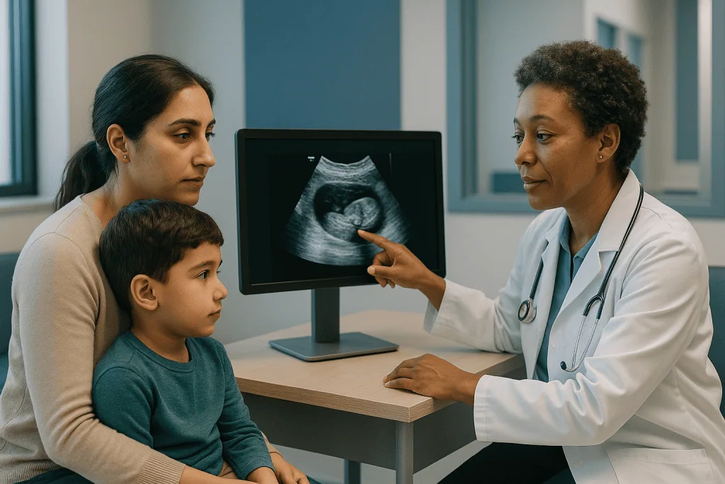
In practice the sequence is usually this. A doctor examines the child and arranges an ultrasound of the abdomen. If the ultrasound shows a solid mass in the kidney, the child is referred to a pediatric cancer center for CT or MRI of the abdomen and a CT scan of the chest. Blood and urine tests are drawn at the same visits. The team then decides whether to remove the kidney first and stage afterward, or to begin chemotherapy first and operate later, two approaches explained further down.
Staging itself is partly a surgical observation and partly a pathology finding. The surgeon records whether the tumor was removed whole, whether it had grown through the kidney’s outer capsule, whether lymph nodes (small bean-shaped filters in the body’s immune system) contain tumor cells, and whether any tumor spilled during the operation. The pathologist confirms these details under the microscope and adds the histology, the cell pattern, to the report.
What families should take from this is that wilms tumor diagnosis staging is a process rather than a single test. A scan alone cannot assign a stage, and neither can a blood test. The number appears only once the surgical and microscopic pieces are in place, which is why the team may describe a “provisional” or “suspected” stage for a while before the final one is settled.
What is the primary cause of Wilms tumors?
There is no single cause, and nothing a parent did or did not do during pregnancy or infancy is known to trigger it. The most honest answer is that Wilms tumor results from changes in the genes that control how kidney cells mature during fetal development. When those cells fail to finish developing, clusters of immature tissue can persist after birth and, in a small number of children, grow into a tumor.
Two gene regions are best understood. Changes in a gene called WT1, which directs kidney and genital development, are found in some tumors. A second region on chromosome 11, often labelled WT2, is linked to growth regulation. Most of these changes arise in the tumor cells only and are not inherited; they are sometimes called sporadic. A minority of children carry a change in every cell of the body, and these children may have an associated syndrome.
Several syndromes raise the risk. WAGR syndrome combines Wilms tumor risk with absence of the iris, genitourinary differences and developmental delay. Denys-Drash syndrome involves kidney disease and genital differences. Beckwith-Wiedemann syndrome is an overgrowth condition in which one side of the body or one limb may be larger than the other. Children with these diagnoses are usually offered regular ultrasound surveillance, discussed in the next section.
Family history matters, but less than many people assume. Only a small share of children with Wilms tumor have a relative who had it. When two or more relatives are affected, or when a child has one of the syndromes above, the team may suggest genetic counselling for the family, with any testing decision left to the parents and the clinicians caring for them.
Who is usually tested promptly, and who is asked to wait and watch
Any child with a new abdominal mass, unexplained blood in the urine, or a persistent one-sided swelling is examined and imaged without delay. Waiting to see whether a lump resolves is not the usual approach in this age group, because a solid kidney mass in a toddler has a short list of possible explanations and most of them need treatment.

A different group is asked to wait in a planned, structured way. Children known to have a predisposition syndrome such as Beckwith-Wiedemann, WAGR or Denys-Drash are offered surveillance ultrasounds at regular intervals through early childhood, because their lifetime risk is higher and the tumor is easier to manage when it is small. For these families “watching” means scheduled scans with a specialist, not simply keeping an eye on the belly at home. The exact interval and the age at which surveillance stops are decided by the child’s genetics and oncology team.
Siblings of a child with sporadic Wilms tumor are not routinely scanned, because their risk is close to that of any other child. If genetic testing shows an inherited change, that advice can shift, which is one reason the team may raise testing even when it seems unlikely to change the patient’s own care.
A third form of waiting happens within the diagnostic pathway itself. In centers that treat with chemotherapy before surgery, the definitive stage is deliberately postponed until the operation, and a biopsy may be skipped if the imaging picture is typical. Families sometimes experience this as uncertainty; in fact it is a recognized strategy with its own staging rules, and the team should be able to explain why it was chosen for this child.
What the first ultrasound shows, and what it cannot tell you
Ultrasound is almost always the first picture. It uses sound waves rather than radiation, needs no sedation, and can be done at the bedside while a parent holds the child. The radiologist is looking for three things: whether the mass is solid or fluid-filled, whether it arises from the kidney or merely pushes against it, and whether the large veins that drain the kidney and run up to the heart contain any tumor.
That third point is more important than it sounds. Wilms tumor has a known tendency to grow along the renal vein, the vessel carrying blood out of the kidney, and sometimes into the inferior vena cava, the body’s largest vein. Ultrasound with Doppler, a setting that shows blood flow in color, is good at spotting this. Finding tumor inside a vein changes the surgical plan, so it is checked early and again before any operation.
Ultrasound also looks at the other kidney. Because a small percentage of children have tumors in both kidneys, the radiologist examines both sides deliberately rather than concentrating only on the obvious mass. The liver is scanned at the same time.
What ultrasound cannot do is confirm the diagnosis or assign a stage. Several childhood tumors can look similar on a grey-scale image, including neuroblastoma, which arises from nerve tissue near the kidney rather than from the kidney itself. Ultrasound also measures size imprecisely and sees lymph nodes poorly. Think of it as the test that says “this is a solid kidney mass and here is roughly where it reaches,” after which more detailed imaging takes over.
CT, MRI and the chest scan: mapping the tumor and where it might travel
Cross-sectional imaging gives the surgeon the map. A CT scan uses X-rays from many angles to build slices of the body; an MRI uses magnets and radio waves and involves no radiation but takes longer and usually requires sedation in young children. Either can be used for the abdomen, and the choice depends on the center, the child’s age, and how urgently the pictures are needed. Both are typically done with contrast, a dye given through a small cannula that makes blood vessels and tumor tissue stand out.
The abdominal scan answers specific questions. How large is the tumor, and does it stay within the kidney’s capsule? Does it press on or invade neighboring organs such as the liver, spleen or bowel? Are the lymph nodes near the aorta enlarged? Is there tumor within the renal vein or vena cava? Is there any sign of a second tumor in the other kidney? Each answer feeds directly into the stage.
The chest needs its own scan because the lungs are the most common site to which Wilms tumor spreads. A chest CT can detect small nodules that a plain chest X-ray would miss. The radiologist reports their number and size, and the oncology team decides whether the nodules are large enough to count toward a Stage IV designation or whether they are too small to be certain about.
Bone scans and brain imaging are not routine for Wilms tumor with favorable histology, because spread to those sites is uncommon. They are considered for specific situations, including certain unfavorable cell types and children with symptoms that suggest involvement. Families can reasonably ask why a particular scan is or is not being ordered; there is usually a clear, evidence-based reason.
Kidney tests, blood counts and urine: what the laboratory adds
Laboratory tests do not diagnose Wilms tumor, but they tell the team what the child’s body can tolerate and whether the tumor has disturbed normal function. A complete blood count checks red cells, white cells and platelets. Anemia can occur if the tumor has bled internally, and the count also establishes a baseline before chemotherapy, which temporarily lowers blood counts.
Kidney function is measured with blood tests for creatinine and urea, waste products that healthy kidneys clear, along with electrolytes such as sodium and potassium. Because the standard operation removes an entire kidney, the team needs reassurance that the remaining one is working well. In children with tumors in both kidneys, these numbers become central to planning how much kidney tissue can be preserved.
Liver function tests and a clotting profile are also drawn. A small number of children with Wilms tumor develop an acquired form of von Willebrand disease, a clotting disorder, which matters before any surgery and is correctable once identified. Urinalysis looks for blood and protein; protein in the urine can hint at an underlying kidney condition such as the one seen in Denys-Drash syndrome.
One test deserves special mention because it is used to rule something out rather than in. Urine catecholamine metabolites, chemicals produced by neuroblastoma, are often measured when the imaging leaves any doubt about whether the mass is truly from the kidney. Normal results support the Wilms diagnosis.
Blood pressure is recorded at every visit. Roughly a quarter of children have raised pressure at diagnosis because of renin, a hormone the compressed kidney releases, and it usually settles after the tumor is removed. Reporting dizziness, headaches or vision changes to the team is worthwhile.
Biopsy, surgery first or chemotherapy first: the two main pathways
Here families encounter a genuine difference in practice around the world, and it is worth understanding because it changes when the stage is assigned. In North America, the standard approach has been to remove the affected kidney first, a procedure called nephrectomy, and to stage the tumor from the surgical and pathology findings. In much of Europe, children typically receive several weeks of chemotherapy before surgery to shrink the tumor, and staging happens after that pre-treated tumor is removed. Both approaches are well studied and both are considered acceptable.
Each pathway has trade-offs. Operating first gives the pathologist an untreated tumor, which makes the cell pattern easier to classify, and it avoids giving chemotherapy to a child whose mass turns out to be something else. Treating first makes a large tumor smaller and less likely to rupture during the operation, which can lower the chance of a higher stage from spillage. The team will explain which protocol they follow and why.
A needle biopsy, taking a sliver of tissue through the skin, is not routine in either system. In the surgery-first approach it is unnecessary, and in the chemotherapy-first approach the imaging is usually typical enough that treatment can begin without it. A biopsy is considered when the picture is unusual, the child is older than expected, or the team suspects a different tumor type. Notably, in the North American staging system a biopsy before removal upstages the tumor to Stage III, because the capsule has been breached, so it is never done casually.
Whichever path is chosen, the decision rests with the treating team after reviewing the scans, the child’s age and overall health, and the family’s understanding of the plan.
Wilms tumor stages explained: the diagnosis and staging summary table
Stage describes how far the tumor has spread at the time it is assessed. The system most families will hear about runs from I to V. Stages I through IV describe a single tumor in one kidney with increasing extent; Stage V is reserved for tumors in both kidneys, regardless of how far each has reached.
| Stage | What the surgeon and pathologist found | Main test that establishes it |
|---|---|---|
| I | Tumor confined to the kidney and removed completely; capsule intact, no tumor at the cut edges, no vessel involvement outside the kidney | Surgery and pathology of the whole specimen |
| II | Tumor grew beyond the kidney, into surrounding fat or nearby vessels, but was removed completely with clear margins | Surgery and pathology |
| III | Tumor remains in the abdomen after surgery: positive lymph nodes, tumor at the margins, spillage, peritoneal deposits, piecemeal removal, or pre-operative biopsy | Surgery, pathology and abdominal imaging |
| IV | Spread through the bloodstream to lungs, liver, bone or brain, or to lymph nodes outside the abdomen and pelvis | Chest CT and other imaging, sometimes confirmed by biopsy |
| V | Tumor in both kidneys at diagnosis; each side is also staged separately | Abdominal imaging, then surgery |
Two details often surprise families. First, size does not determine stage; a very large tumor that was removed intact with clear margins is still Stage I. Second, the stage is not the whole story. The team combines stage with histology, the microscopic cell type, and in some protocols with genetic markers in the tumor, to form a risk group that actually drives treatment. A child with Stage III favorable histology and a child with Stage I diffuse anaplasia sit in different risk groups despite the stage numbers suggesting otherwise.
In the chemotherapy-first system, the same Roman numerals are used but are applied to a tumor that has already shrunk, and the pathologist also grades how much of the tumor the chemotherapy killed. If the family has read staging explanations online that seem to contradict what their team said, this difference in timing is frequently the reason.
What are the characteristics of a Stage 3 Wilms tumor?
Stage III is the stage families most often ask about, partly because it sounds advanced and partly because its definition is broader than the others. In plain terms, Stage III means the tumor is still confined to the abdomen, with no spread to the lungs or other distant organs, but the surgeon could not be certain it was all removed. The reasons fall into a handful of categories.
The first is lymph node involvement. If tumor cells are found in the lymph nodes near the kidney or along the aorta, the stage becomes III even when the kidney itself was removed cleanly. This is why surgeons sample several lymph nodes during every Wilms operation; without that sampling, a Stage III tumor can be mistakenly recorded as Stage I or II. The second category is tumor at the surgical margin, meaning microscopic cells reach the edge of what was removed. The third is spillage, where the tumor ruptured or leaked before or during the operation. The fourth is tumor implants on the peritoneum, the thin lining of the abdominal cavity. The fifth is removal in pieces rather than as one specimen, and the sixth, in the North American system, is any biopsy performed before the kidney came out.
What Stage III does not mean is that the disease has traveled to distant sites. Lung nodules, liver deposits or bone involvement would place the tumor at Stage IV instead. Nor does it mean the tumor is of an aggressive cell type; the histology is assessed separately.
Treatment plans for Stage III typically include chemotherapy and radiation to the abdomen in addition to surgery, with the exact combination determined by the protocol and the histology. The team, not the stage number alone, decides what that looks like for a particular child.
Wilms tumor histology: favorable, anaplastic and what the molecular tests add
Once the kidney is removed, the pathologist examines slices of the whole tumor under a microscope. Most Wilms tumors show a mixture of three cell types that echo early kidney development, and when the cells look orderly the report says “favorable histology.” This describes the great majority of cases and is associated with a good response to standard treatment.
The pathologist is searching specifically for anaplasia, a term for cells with very large, irregular nuclei and abnormal patterns of division. Anaplasia is further divided into focal, meaning a single confined area, and diffuse, meaning scattered through the tumor or present at a margin or in a lymph node. Diffuse anaplasia is the finding that shifts a child into a higher-risk group, because these cells respond less well to chemotherapy. Reading anaplasia reliably takes experience, and specimens are often reviewed by a second pathologist at a central laboratory as part of clinical trial protocols.
The report also notes nephrogenic rests, patches of immature kidney tissue in the surrounding kidney. Their presence suggests the other kidney could develop a tumor later and may lead the team to recommend longer imaging follow-up.
Molecular testing is now part of pathology for many children. Loss of heterozygosity at chromosome regions 1p and 16q, meaning one copy of those regions is missing in the tumor cells, is linked in large cooperative-group studies to a higher chance of recurrence in favorable-histology tumors and can intensify treatment under some protocols. Gain of material on chromosome 1q is also being studied as a marker. These tests take time to return, sometimes longer than the main histology, which can delay the final risk assignment by days to weeks.
What is the rule of 10 for Wilms tumors?
The “rule of 10” is a teaching mnemonic that medical students use to remember rough proportions about Wilms tumor. In its common form it states that about 10 percent of tumors involve both kidneys, about 10 percent occur in a child with a recognized syndrome, about 10 percent show anaplasia, about 10 percent have already spread at diagnosis, and so on. Some versions add that around 10 percent are familial or that the tumor is found in roughly one in ten children with certain syndromes.
Its usefulness is as a memory aid, not as a set of precise figures. The real numbers vary across studies and populations, and several of them are closer to 5 percent than to 10. Bilateral disease, for example, is reported in a range of roughly 5 to 10 percent of cases depending on the series. Treating the rule as exact can lead a worried parent to overestimate how likely a given complication is for their own child.
What the mnemonic does capture correctly is a set of questions the team asks every time: Is the other kidney involved? Does the child have a syndrome that changes surveillance for the family? Is the histology anaplastic? Has the tumor spread? Those four questions, answered through the imaging and pathology described above, are the real backbone of risk assessment.
If a clinician mentions the rule of 10 in conversation, it is reasonable to ask which of those categories apply to the child in front of them, rather than to the textbook average. The individual scan, surgical and pathology findings carry far more weight than any population estimate, and the team will base decisions on those.
What people often get wrong about Wilms tumor diagnosis and staging
The first misconception is that a bigger tumor means a higher stage. It does not. Stage depends on whether the tumor stayed inside its capsule and whether it was fully removed, not on centimeters. Some of the largest tumors ever removed were Stage I, and some small ones were Stage III because a lymph node contained cells.
The second is that a blood test can detect or stage the tumor. There is no tumor marker in the blood for Wilms tumor. Blood tests measure how the kidneys and other organs are coping; imaging and pathology do the diagnosing.
The third is that a parent could have caused it or caught it earlier. Wilms tumor arises from developmental changes in kidney cells, not from diet, infection, injury or anything done in pregnancy. Its tendency to grow silently deep in the abdomen means that discovering it as a painless lump is the typical story, not a sign of delay.
The fourth is that “Stage IV” carries the same meaning it does in adult cancers. Children with favorable-histology Wilms tumor that has spread to the lungs are treated with intent to control the disease completely, and outcomes depend heavily on how the lung nodules respond to early treatment. Stage describes extent; it is not by itself a verdict.
The fifth is that a biopsy is always the first step in diagnosing a cancer. For Wilms tumor it is often deliberately avoided, and in one staging system it changes the stage. The sixth is that online staging descriptions all mean the same thing. The surgery-first and chemotherapy-first systems use the same numerals at different moments, so comparing a child’s stage to a cousin’s diagnosed in another country can be misleading. The team can reconcile the two.
What the following days and weeks usually look like
The diagnostic phase tends to move quickly once a solid kidney mass is seen, because the team wants treatment to begin without unnecessary delay. A typical week might include the ultrasound, a referral to a pediatric oncology center, the abdominal and chest scans, and the first blood and urine tests. Parents often describe this period as a blur of appointments; writing down each test and its purpose can help the information settle.
Preparation for imaging is mostly about comfort. Young children usually need sedation or a general anesthetic for MRI and sometimes for CT, so the child will fast for a period beforehand according to the hospital’s instructions. Contrast is given through a cannula, and numbing cream on the skin before insertion is commonly offered. Bringing a familiar toy or blanket into the scanner room, where allowed, makes a measurable difference to how a toddler copes.
If the center operates first, surgery usually follows within days of the scans. The child spends a few days in hospital, pain is managed by the anesthetic and surgical team, and most children are eating and moving around again before discharge. The pathology report arrives over the following week or so, with molecular results sometimes taking longer. The team then meets, often in a multidisciplinary tumor board, and presents the final stage and risk group to the family along with the treatment plan.
If the center treats first, chemotherapy begins soon after imaging, scans are repeated after several weeks to measure response, and surgery is scheduled afterward. Either way, the family should expect a formal conversation at the end of the staging process where the stage, histology and plan are explained in full and questions are welcomed.
Questions to ask your care team
Parents are rarely short of questions in the first weeks; the difficulty is remembering them in the room. A short written list, handed to the oncologist or nurse, works better than trying to hold everything in mind. The questions below are ones families most often wish they had asked earlier.
- Which imaging has been done so far, and is anything still outstanding before the stage can be confirmed?
- Does our center operate first or give chemotherapy first, and how does that affect when we will know the final stage?
- Did the scans show tumor in the renal vein or vena cava, and does that change the surgical plan?
- How is the other kidney, and is there anything in the pathology, such as nephrogenic rests, that affects long-term follow-up?
- What is the histology, and have molecular tests such as 1p and 16q been sent? When will those results return?
- What risk group does the combination of stage and histology place our child in, and which protocol or trial is being followed?
- Will radiation be part of the plan, and if so, to which area?
- Should our family be referred for genetic counselling, and does the diagnosis change anything for siblings?
- What symptoms should we watch for at home during the staging period, and whom do we call out of hours?
- Who is our main point of contact, and how will results be communicated to us?
None of these questions has a single correct answer, which is the point. The purpose is to understand how the evidence from imaging, laboratory tests and pathology has been assembled for this particular child, so that the plan the team proposes makes sense to the people who will live with it day to day.
When to call your doctor
During the diagnostic and staging period, and in the weeks that follow surgery or the start of chemotherapy, certain signs warrant a same-day call to the oncology team or a visit to the emergency department rather than waiting for the next appointment. A fever in a child receiving chemotherapy is treated as urgent, because chemotherapy lowers the white cell count that fights infection; the team will have given a specific temperature threshold and a number to call at any hour.
Sudden worsening abdominal pain, a belly that becomes rapidly tense or more swollen, or a child who becomes pale, sweaty and listless can signal bleeding into or around the tumor, which is uncommon but needs immediate assessment. Visible blood in the urine that is new or heavier than before, or passing very little urine over a day, should also be reported promptly, as should severe headache, vision changes, repeated vomiting or a seizure, any of which can accompany dangerously high blood pressure in a child whose kidney is releasing excess renin.
Breathing difficulty, a persistent cough or chest pain are reasons to seek care quickly given the lungs’ role as the most common site of spread. After surgery, redness, discharge or opening of the wound, or a fever, should be assessed by the surgical team.
Families should never feel they are bothering the team by calling about something that turns out to be minor. Pediatric oncology units expect and welcome these calls. The red flags above are not a self-diagnosis checklist; they are prompts to pick up the phone so that the people who know the child’s scans and results can decide what the next step should be.
Frequently asked questions
What are the characteristics of a Stage 3 Wilms tumor?
A Stage III Wilms tumor is confined to the abdomen but was not completely removed or has reached nearby lymph nodes. The category includes tumor cells in abdominal lymph nodes, tumor at the surgical margin, spillage before or during surgery, deposits on the abdominal lining, removal in pieces, or a biopsy taken before the kidney came out. There is no spread to the lungs or other distant organs.
What is the primary cause of Wilms tumors?
Wilms tumor arises from changes in genes that control kidney development, most often the WT1 gene and a region called WT2 on chromosome 11, which leave immature kidney cells able to grow after birth. Most changes occur only in the tumor and are not inherited. A minority of children have an associated syndrome such as WAGR, Denys-Drash or Beckwith-Wiedemann.
What is the Wilms tumor rule of 10?
The rule of 10 is a teaching mnemonic summarizing rough proportions: about one in ten tumors involve both kidneys, about one in ten occur with a syndrome, about one in ten show anaplasia, and about one in ten have spread at diagnosis. The true figures vary between studies and several are lower, so it should be treated as a memory aid rather than a precise statistic.
Can Wilms tumor be treated successfully?
Most children with Wilms tumor are treated successfully with a combination of surgery, chemotherapy and, for some stages, radiation, according to national cancer information services. The outlook depends on the stage, the histology, molecular markers in the tumor, and how the disease responds to early treatment. Only the treating team, with the child’s own results in hand, can give a meaningful individual outlook.
How are Wilms tumor stages explained to families?
Stages run from I to V. Stage I is confined to the kidney and fully removed; Stage II extends beyond the kidney but is fully removed; Stage III leaves tumor in the abdomen or involves abdominal lymph nodes; Stage IV has spread to the lungs, liver, bone or distant nodes; Stage V means both kidneys are involved. Stage is combined with histology to set a risk group.
Which imaging tests are used to diagnose Wilms tumor?
Ultrasound of the abdomen is usually first, because it shows whether a mass is solid and arises from the kidney and whether tumor has grown into the renal vein. CT or MRI of the abdomen then maps size, margins, lymph nodes and the other kidney. A CT scan of the chest checks the lungs, the most common site of spread. Bone and brain imaging are reserved for specific situations.
What does favorable histology mean in a Wilms tumor pathology report?
Favorable histology means the tumor cells, viewed under the microscope, show the orderly mixture of cell types typical of Wilms tumor without anaplasia. Anaplasia refers to cells with very large, abnormal nuclei and irregular division, and it is classed as focal or diffuse. Favorable histology describes the great majority of Wilms tumors and is associated with a good response to standard treatment.
Is a biopsy needed to diagnose Wilms tumor?
Usually not. In the surgery-first approach the whole kidney is removed and examined, making a biopsy unnecessary, and in the chemotherapy-first approach the imaging is typically clear enough to begin treatment without one. A needle biopsy is considered only when the picture is unusual. In the North American staging system, a biopsy before removal automatically places the tumor at Stage III.
Why is the chest scanned when the tumor is in the kidney?
The lungs are the most common place Wilms tumor spreads, because tumor cells can travel there through the bloodstream. A chest CT can find small nodules that a plain X-ray would miss. Whether those nodules are present, how many there are and how they respond to early treatment all influence the stage and the treatment plan the team recommends.
What blood and kidney tests are done for Wilms tumor?
The laboratory work includes a complete blood count, creatinine and urea to measure kidney function, electrolytes, liver function tests, a clotting profile and a urine test for blood and protein. Urine catecholamine metabolites may be checked to rule out neuroblastoma. These tests do not diagnose or stage the tumor; they show how the kidneys, blood and other organs are coping and set a baseline before treatment.
This article is for general information only and is not a substitute for professional medical advice. Please consult a qualified doctor about your individual situation.

More from the Blog

How Acute Myelogenous Leukemia Treatment Is Planned When Time Matters
Acute myelogenous leukemia treatment is usually planned within days of diagnosis because immature blood cells can crowd the bone marrow quickly. The care team…

Skin Surveillance After Basal Cell Carcinoma: How Often Checks Happen and What Is Examined
After treatment for basal cell carcinoma, most people are asked to return for a skin examination every 6 to 12 months for several years…

Living With Chronic Graft-Versus-Host Disease: Skin, Eye Dryness and Long-Term Follow-Up
Living with chronic GVHD means the donor's immune cells keep reacting against your own tissues, most often the skin, mouth and tear glands, for…
Recovering After Lung Cancer Surgery: Breathing Exercises, Walking and Chest Drains
After lung cancer surgery, recovery usually rests on three habits: slow deep breaths, often with an incentive spirometer, taken several times every waking hour;…

Flying Home After a Mastectomy: Drains, Compression and How Long International Patients Stay
There is no guideline that fixes a single safe interval for flying after a mastectomy. Surgical teams generally prefer that drains are out, which…

Supplements, Herbal Products and Chemotherapy: Why Your Oncologist Asks Before You Start
Oncologists ask about supplements during chemotherapy because some vitamins, herbs and extracts can change how chemotherapy drugs are absorbed, broken down or cleared, raise…