Quick answer
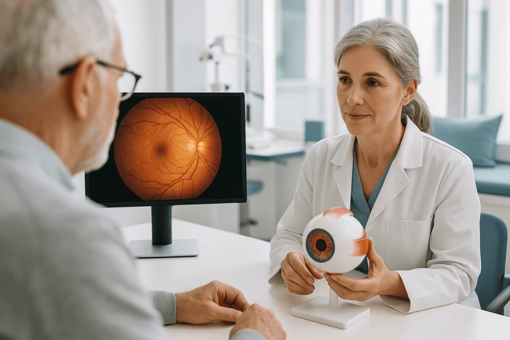
Macular degeneration is an age-related eye disease that damages the macula, the central part of the retina responsible for sharp, detailed vision. Treatment depends on whether the condition is dry or wet and may include monitoring, lifestyle support, medications injected into the eye, and laser-based approaches, with diagnosis and care guided by detailed retinal imaging and eye examinations at Acibadem…
What is macular degeneration?
Macular degeneration is an eye condition that damages the macula, the small central area of the retina at the back of the eye. The retina is the light-sensitive tissue that captures images and sends them to the brain, and the macula is the part responsible for sharp, detailed central vision: the vision you use for reading, recognizing faces, driving, and seeing fine detail. When people ask what is macular degeneration, the simplest answer is that it is a gradual breakdown of this central portion of the retina, which blurs or distorts the center of your visual field while usually leaving side (peripheral) vision intact.
The most common form is age-related macular degeneration, often abbreviated AMD, which mainly affects people over the age of 50 and becomes more common with each decade of life. It is one of the leading causes of vision loss in older adults worldwide. Macular degeneration is classified under the medical code ICD-10 H35.30 when the type is unspecified.
There are two main types:
- Dry (atrophic) macular degeneration: the more common form, accounting for the large majority of cases. It develops slowly as the macula thins and small yellow deposits called drusen (waste material under the retina) build up. Vision loss is usually gradual.
- Wet (neovascular or exudative) macular degeneration: less common but more aggressive. Abnormal, fragile blood vessels grow beneath the retina and can leak fluid or blood, damaging the macula. Vision loss can occur quickly, sometimes over days or weeks.
Dry macular degeneration can progress to the wet form, which is one reason ongoing monitoring matters. Importantly, macular degeneration does not typically cause complete blindness. Because peripheral vision is usually preserved, most people retain some useful sight, although advanced disease can make everyday tasks that depend on central vision very difficult.
Symptoms of macular degeneration
Macular degeneration symptoms often begin subtly, and in the early stages many people notice nothing at all. The condition is frequently first detected during a routine eye examination before any symptoms appear. When symptoms do develop, they affect central vision in one or both eyes.
Common macular degeneration symptoms include:
- Blurred or fuzzy central vision, especially when reading or looking at fine detail
- Distorted vision (metamorphopsia)straight lines, such as door frames or lines of text, appear wavy, bent, or crooked
- A dark, blurry, or blank spot in the center of your vision (a scotoma)
- Difficulty recognizing faces, even when peripheral vision seems normal
- Colors appearing less bright or vivid than before
- Needing brighter light for reading and close work
- Slow adjustment when moving from bright to dim environments
- Words disappearing or jumbling while reading
Symptoms differ by type and stage. In early dry macular degeneration, there may be no noticeable change at all, or only mild blurring and a need for more light. As the dry form advances, blurring becomes more pronounced and a gradual blind spot may develop in the center of vision. In the most advanced stage of dry disease, called geographic atrophy, patches of retinal cells waste away and central vision loss becomes more significant.
Wet macular degeneration tends to cause more sudden and dramatic symptoms. A rapid onset of visual distortion, straight lines suddenly appearing wavy, or a quickly enlarging dark spot in central vision can signal leaking blood vessels under the retina. Because prompt treatment of wet macular degeneration can help limit vision loss in many cases, any sudden change in central vision should be assessed urgently by an eye specialist.
If only one eye is affected, the healthy eye often compensates, so people may not notice symptoms until the disease is advanced or affects both eyes. This is another reason regular eye exams are important for people over 50.
Causes and risk factors
Macular degeneration causes are not fully understood, but the condition is thought to result from a combination of aging, genetics, and environmental factors. Over time, the cells of the macula and the supporting tissue beneath it can become less efficient at processing and clearing waste. Deposits of this waste material (drusen) accumulate, and the light-sensing cells of the macula gradually stop working properly. In the wet form, the eye responds by growing new, abnormal blood vessels under the retina, a process called choroidal neovascularization, and these vessels leak fluid and blood that damage the macula.
Known and suspected risk factors include:
- Age: the strongest risk factor. Risk rises significantly after age 50 and continues to increase with each decade.
- Family history and genetics: having a parent or sibling with macular degeneration raises your risk. Several genes have been linked to the condition.
- Smoking: current or past smoking substantially increases risk and can speed progression. It is the most important modifiable risk factor.
- Cardiovascular disease: conditions affecting the heart and blood vessels, including high blood pressure, may increase risk.
- Obesity: excess body weight has been associated with progression to more advanced disease.
- Diet: diets low in leafy green vegetables, fish, and antioxidant-rich foods may contribute to risk.
- Race: the condition is more common in people of European ancestry.
- Prolonged unprotected sun exposure: ultraviolet and high-energy light exposure may play a role, although the evidence is less definitive.
You cannot change your age or genes, but stopping smoking, managing blood pressure, maintaining a healthy weight, eating a balanced diet rich in vegetables and fish, and protecting your eyes from bright sunlight are sensible measures that may lower risk or slow progression. None of these steps guarantees prevention, but they support overall eye health.
Diagnosis
Macular degeneration diagnosis is made by an ophthalmologist (a medical doctor specializing in eye care) or, initially, by an optometrist during an eye examination. Because early disease often causes no symptoms, diagnosis frequently begins with a routine dilated eye exam. Several tests are commonly used, both to confirm the diagnosis and to distinguish the dry form from the wet form: a distinction that directly shapes treatment.
- Dilated eye examination: eye drops widen the pupil so the doctor can examine the retina and macula directly. The presence, size, and number of drusen, changes in retinal pigment, areas of thinning (atrophy), or signs of fluid and bleeding help establish the diagnosis and stage.
- Visual acuity testing: a standard eye chart measures how well you see at various distances and helps track changes over time.
- Amsler grid test: a simple grid of straight lines used to detect central vision distortion. If lines look wavy, broken, or missing, this can indicate macular disease. Many patients are given an Amsler grid to check their vision at home between visits.
- Optical coherence tomography (OCT): a painless, noninvasive scan that uses light waves to create detailed cross-sectional images of the retina. OCT can reveal retinal thinning, drusen, and fluid under or within the retina, and it is central to diagnosing wet macular degeneration and monitoring the response to treatment.
- Fluorescein angiography: a dye is injected into a vein in the arm, and a special camera photographs the dye as it travels through the retinal blood vessels. Leaking or abnormal vessels, the hallmark of wet macular degeneration, show up clearly. A related test using a different dye, indocyanine green angiography, is sometimes used for additional detail.
- OCT angiography: a newer, dye-free imaging method that can visualize abnormal blood vessel networks in some patients.
Doctors also stage the disease, early, intermediate, or advanced, based on the findings, because staging guides how often you need monitoring and whether treatment or nutritional supplementation may be appropriate. If wet macular degeneration is suspected, evaluation is usually arranged urgently, since earlier treatment is generally associated with better preservation of vision.
Treatment options
There is currently no cure for macular degeneration, but macular degeneration treatment can often slow progression and, in wet disease, help preserve or sometimes partially improve vision. The right approach depends on the type and stage of the disease. In hospital settings such as Acibadem, macular degeneration is managed within the ophthalmology department, typically by retina specialists. An overview of how the condition is evaluated and treated is also available on the dedicated macular degeneration treatment page.
Monitoring and lifestyle measures (dry macular degeneration)
For early dry macular degeneration, there is often no active treatment; instead, doctors recommend regular monitoring, sometimes called watchful waiting, along with lifestyle changes. This usually includes stopping smoking, eating a diet rich in leafy greens and fish, managing blood pressure, and checking your vision at home with an Amsler grid so that any progression is caught early.
Nutritional supplements
For people with intermediate dry macular degeneration or advanced disease in one eye, specific combinations of vitamins and minerals, based on large clinical studies known as the AREDS and AREDS2 trials, may reduce the risk of progression to advanced disease in many cases. These formulations typically include vitamins C and E, zinc, copper, lutein, and zeaxanthin. Supplements do not restore lost vision and are not appropriate for everyone, so they should be taken only on a doctor’s advice.
Anti-VEGF injections (wet macular degeneration)
The standard treatment for wet macular degeneration is injection of anti-VEGF medication into the eye. VEGF (vascular endothelial growth factor) is a protein that drives the growth of the abnormal, leaky blood vessels; anti-VEGF drugs block this protein. The injections are given into the white of the eye under numbing drops in an outpatient setting. They usually need to be repeated at regular intervals, often every one to three months at first, and treatment may continue long term. In many patients, anti-VEGF therapy stabilizes vision, and some patients experience a degree of improvement, although results vary and no outcome can be guaranteed.
Other procedures
Less commonly used options include photodynamic therapy, in which a light-activated drug is injected into the bloodstream and then activated by a laser aimed at the abnormal vessels, and thermal laser photocoagulation, which seals leaking vessels in selected cases. These procedures are used far less often since the introduction of anti-VEGF drugs but may still be considered in specific situations. Conventional surgery has a very limited role in macular degeneration itself; however, low-vision devices and, in carefully selected patients with advanced disease in both eyes, an implantable miniature telescope have been used to make better use of remaining vision.
Emerging treatments
Research is ongoing into treatments for geographic atrophy (the advanced dry form), longer-acting injections, gene therapy, and cell-based approaches. Some newer medications for geographic atrophy have become available in certain countries; whether they are suitable for an individual patient is a decision for a retina specialist.
Low-vision rehabilitation
When vision loss has occurred, low-vision rehabilitationincluding magnifiers, specialized lighting, electronic reading aids, and training in techniques that make the most of peripheral vision, can meaningfully improve daily functioning, even though it does not treat the disease itself.
Living with macular degeneration and outlook
The outlook with macular degeneration varies widely. Many people with early dry disease keep good functional vision for years, particularly if progression is slow. Others, especially those with untreated wet disease or advanced geographic atrophy, may lose significant central vision. Because peripheral vision is usually spared, complete blindness is uncommon, but advanced central vision loss can affect reading, driving, and independence.
Practical steps that often help include:
- Attending all scheduled eye appointments and injections, since consistent treatment of wet disease is closely tied to preserving vision
- Checking each eye separately with an Amsler grid as often as your doctor advises, and reporting new distortion promptly
- Using good lighting, high-contrast materials, and magnification for reading and close tasks
- Asking about formal low-vision rehabilitation services and occupational therapy
- Not smoking, eating a nutritious diet, and managing cardiovascular health
- Discussing driving safety honestly with your doctor, as legal vision requirements vary by country
Vision loss can affect mood and confidence, and depression is more common in people with significant sight impairment. Support groups, counseling, and family involvement can make a real difference. It is reasonable to be hopeful, treatment has improved considerably in recent years, while understanding that no therapy can promise a specific result.
Frequently asked questions
What is macular degeneration in simple terms?
Macular degeneration is the gradual wearing out of the macula, the small central part of the retina that provides sharp, detailed vision. As the macula deteriorates, central vision becomes blurred, distorted, or blocked by a dark spot, while side vision usually remains. It most often affects people over 50, in which case it is called age-related macular degeneration.
Can macular degeneration be cured or reversed?
There is currently no cure, and vision that has been lost usually cannot be fully restored. However, treatment can often slow the disease. For wet macular degeneration, anti-VEGF injections stabilize vision in many patients and sometimes lead to partial improvement. For dry disease, nutritional supplements and lifestyle changes may reduce the risk of progression in appropriate patients. Research into new treatments is active and ongoing.
Does macular degeneration lead to total blindness?
In most cases, no. Macular degeneration affects central vision but typically spares peripheral (side) vision, so most people retain some useful sight even with advanced disease. That said, severe central vision loss can make reading, driving, and recognizing faces very difficult, which is why monitoring and timely treatment matter.
What are the first symptoms of macular degeneration?
Early macular degeneration often causes no symptoms at all and may only be found during a routine eye exam. When symptoms begin, they commonly include mild blurring of central vision, needing brighter light to read, straight lines starting to look wavy, or a small blurry spot in the center of vision. Sudden distortion or a rapidly growing dark spot may indicate the wet form and needs urgent assessment.
How is macular degeneration diagnosed?
Diagnosis is made through an eye examination with dilating drops, which lets the doctor inspect the macula for drusen, pigment changes, thinning, or fluid. Supporting tests include visual acuity testing, the Amsler grid, optical coherence tomography (OCT) imaging, and sometimes fluorescein angiography, a dye test that reveals leaking blood vessels in the wet form of the disease.
How serious is macular degeneration?
Seriousness depends on the type and stage. Early dry macular degeneration may cause little or no vision change for years, while untreated wet macular degeneration can cause substantial central vision loss within weeks to months. Because the wet form is treatable when caught early, prompt evaluation of any new symptoms is important. Your ophthalmologist can explain the stage of your disease and what it means for you.
Are the eye injections for macular degeneration painful, and how long is recovery?
Most patients tolerate anti-VEGF injections well. The eye is numbed with drops beforehand, and the injection itself takes only a few seconds; many people describe pressure rather than pain. Mild irritation, redness, or floaters for a day or so afterward are common. Serious complications such as infection are rare but possible, so worsening pain, increasing redness, or decreasing vision after an injection should be reported to your doctor immediately.
When to see a doctor
Anyone over 50 should have regular comprehensive eye examinations, even without symptoms, because early macular degeneration is often silent. If you have already been diagnosed, keep to the monitoring and treatment schedule your eye specialist recommends.
Seek prompt medical attention, ideally within days, not weeks, if you notice any of the following red-flag warning signs:
- Sudden distortion of vision: straight lines such as door frames, tiles, or text suddenly appear wavy, bent, or broken
- A new or enlarging dark, gray, or blank spot in the center of your vision
- A rapid decline in central vision in one or both eyes over days or weeks
- Sudden difficulty reading or recognizing faces that was not present before
- New changes on your home Amsler grid check, if you have been given one
- After an eye injection: increasing eye pain, worsening redness, light sensitivity, or falling vision, which can signal infection and is an emergency
Sudden central vision changes can indicate wet macular degeneration or another serious retinal problem, and earlier treatment generally offers the best chance of preserving sight. If you experience any of these symptoms, contact an eye care professional urgently rather than waiting for your next routine appointment.
Medically reviewed by the Acıbadem International Medical Board September 2, 2026
See our medical review board →
Update history
- PublishedJune 14, 2026
- Medical review approvedSeptember 2, 2026
- Last content updateSeptember 2, 2026