3D Mammography vs Standard Mammogram: What Is the Difference?
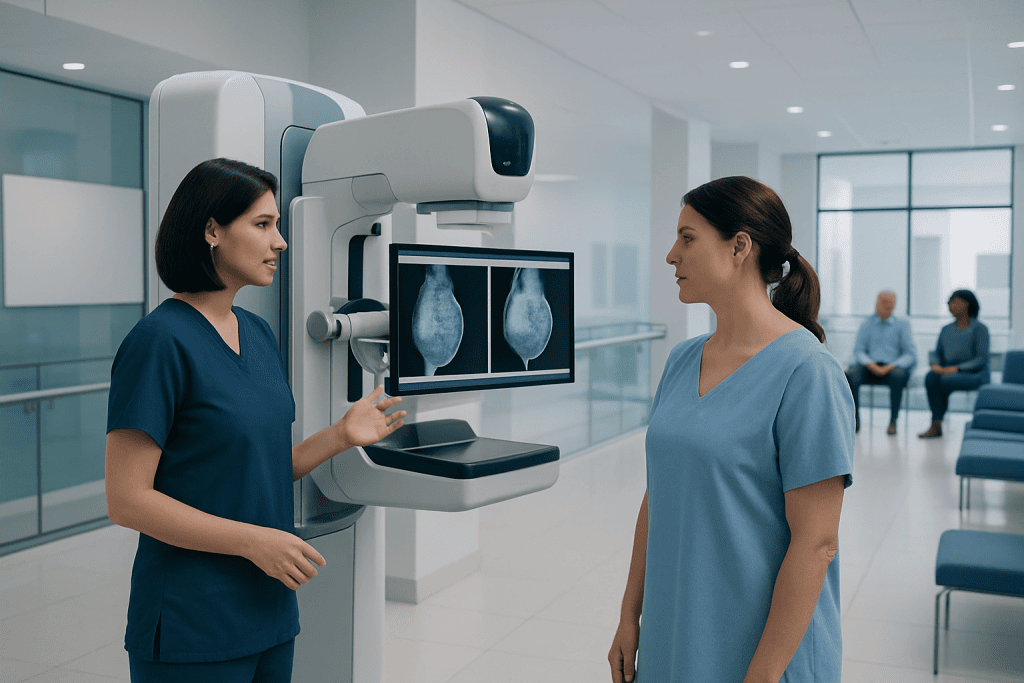
Both 2D and 3D mammograms are effective tools for breast cancer screening. 3D mammography takes multiple low-dose images to create a layered view of the breast.
Key Takeaways
- Both 2D and 3D mammograms are effective tools for breast cancer screening.
- 3D mammography takes multiple low-dose images to create a layered view of the breast.
- 3D screening may reduce call-backs for extra imaging and improve cancer detection in some patients.
- The best test depends on breast density, personal risk factors, age, and local availability.
- A mammogram may still feel briefly uncomfortable, but the compression and exam time are usually short.
3D mammography and standard mammography are both important breast imaging tests used to screen for breast cancer. The main difference is that 3D mammography creates multiple thin images of the breast, which can make some abnormalities easier to see, especially in dense breast tissue.
Overview: how these two mammograms differ
When comparing 3D mammography vs standard mammogram, the key difference is how the breast images are captured and viewed. A standard mammogram, often called 2D mammography, takes X-ray pictures of the breast from a few fixed angles. A 3D mammogram, also known as digital breast tomosynthesis, takes many low-dose images from different angles and reconstructs them into thin layers.
This layered view can help radiologists look through overlapping breast tissue more clearly. In a standard 2D image, normal tissue may overlap and either hide a small abnormality or create an area that looks suspicious when it is not. By reducing this overlap, 3D mammography can make interpretation easier in some cases.
Both methods are used to look for early signs of breast cancer before symptoms appear. Neither test is perfect, and no single screening method is best for everyone. The right choice depends on personal risk, breast density, age, prior imaging findings, and the recommendations of a qualified doctor or breast imaging specialist.
How a standard mammogram works
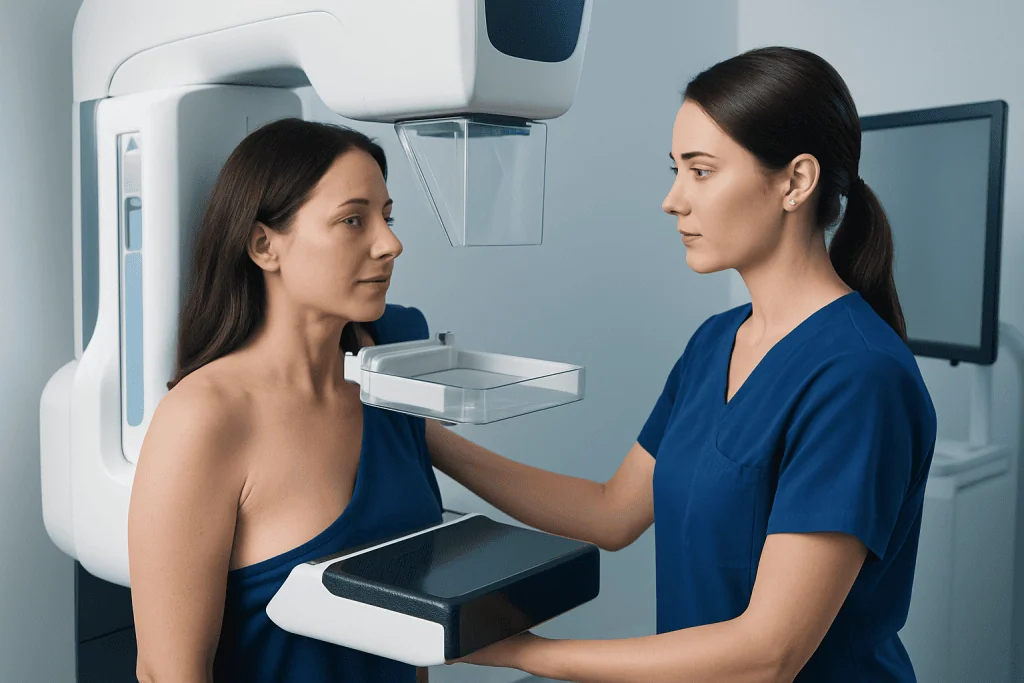
A standard mammogram uses low-dose X-rays to create two-dimensional images of the breast. During the test, the breast is placed on a flat plate and gently compressed for a few seconds while images are taken. Compression is important because it helps spread the tissue, improves image quality, and keeps the radiation dose as low as possible.
Most screening mammograms include two views of each breast. These images allow radiologists to check for masses, calcifications, asymmetry, or other changes that could need closer evaluation. Standard mammography has been used for many years and remains a widely available and well-established tool for breast cancer screening.
For many people, a standard mammogram provides enough information for routine screening. It is especially useful when compared with earlier mammograms, because even small changes over time can help guide next steps. If anything appears unclear, additional views or another imaging test may be recommended.
How 3D mammography works
3D mammography is performed in a way that feels very similar to a standard mammogram. The breast is still compressed, but the X-ray arm moves in a small arc over the breast while taking multiple images. A computer then combines these images to create a series of thin slices that the radiologist can review one layer at a time.
This technology can be especially helpful when breasts contain overlapping tissue, which is common and normal. With a layered image set, the radiologist may be able to separate normal structures from areas that deserve more attention. This can improve confidence in reading the exam and reduce the chance that normal tissue will mimic a problem.
Many centers now use 3D mammography for routine screening, while others offer it selectively. Patients who are called back after screening may also need extra breast imaging such as mammography, targeted ultrasound, or in some situations breast MRI to clarify a finding.
Potential benefits and limitations of each option
The main advantage of 3D mammography is improved visualization of breast tissue. Studies have shown that it can increase cancer detection in some groups and lower the number of call-backs for additional imaging. This may be particularly helpful for women with dense breasts, where overlapping tissue can make a 2D image harder to interpret.
Another benefit is that 3D imaging may help identify invasive cancers more clearly. At the same time, it is important to remember that 3D mammography does not prevent cancer and does not find every cancer. Some abnormalities may still be too small, hidden, or biologically difficult to detect on mammography alone.
Standard mammography remains an effective and valuable screening test. It is widely available, familiar to most patients, and often sufficient for routine screening. In some healthcare settings, 3D mammography may cost more or may not be available everywhere. For that reason, screening plans should balance technology, access, and individual risk.
Neither approach replaces careful clinical follow-up if there is a lump, nipple discharge, skin change, or another breast symptom. In that situation, diagnostic imaging is usually needed rather than routine screening alone, and the doctor may also evaluate for conditions such as breast cancer.
Breast density, risk factors, and who may benefit most
Breast density describes how much fibroglandular tissue and fatty tissue are seen on a mammogram. Dense breasts are common, and they are not abnormal. However, dense tissue can make cancers more difficult to see on a standard mammogram because both dense tissue and many cancers appear white on the image.
For women with dense breasts, 3D mammography may offer a clearer view by reducing the masking effect of tissue overlap. Some patients with a higher-than-average risk of breast cancer may also benefit from a more individualized screening plan that combines mammography with other tests. These plans may include ultrasound or MRI depending on the person’s history.
Risk factors that can affect screening decisions include age, family history, certain inherited gene changes, previous chest radiation, personal history of breast disease, and prior abnormal imaging. If the risk is elevated, a doctor may recommend a dedicated evaluation through breast check-up services or referral to a breast specialist.
Even so, average-risk women may also benefit from 3D mammography if it is available. The choice is not always about high risk versus low risk. It is often about what imaging method offers the clearest, most appropriate screening for the individual patient.
What to expect during the exam and understanding radiation
For the patient, a 3D mammogram and a standard mammogram feel very similar. In both tests, the breast is compressed briefly to obtain clear images. The discomfort is usually temporary and lasts only a few seconds for each image. Scheduling the test when the breasts are less tender, such as after a menstrual period for premenopausal women, may help.
Before the exam, patients are often advised not to use deodorant, powder, lotion, or cream on the underarms or breasts because these products can affect the images. It is also helpful to bring previous mammograms if the imaging is being done at a new center, since comparison with older studies can improve accuracy.
Radiation exposure from mammography is low. 3D mammography uses low-dose X-rays as well, and current systems are designed to keep the total dose within accepted safety standards. For most patients, the benefits of appropriate breast cancer screening outweigh the small risks associated with radiation exposure.
How doctors choose between 2D and 3D screening
Doctors do not usually choose between 2D and 3D mammography based on one factor alone. They consider age, breast density, prior mammogram results, symptoms, family history, and whether the exam is for routine screening or diagnostic evaluation. Availability and insurance or payment considerations may also influence the decision.
If a patient has had repeated call-backs after standard mammograms because of overlapping tissue, 3D imaging may be particularly useful. If previous imaging has always been clear and breast density is low, standard mammography may continue to be a reasonable choice. The most important point is regular screening with a method that is accessible and appropriate.
When a mammogram shows an area that needs clarification, radiologists may recommend additional tests. These can include spot compression views, ultrasound, or breast ultrasound to look more closely at a mass or focal finding. In select situations, tissue sampling may be needed to make a definite diagnosis.
When to speak with a doctor
Patients should speak with a doctor if they are unsure when to start screening, how often to have a mammogram, or whether 3D mammography is right for them. A personalized discussion is especially useful for women with dense breasts, a family history of breast cancer, previous abnormal breast imaging, or a known elevated risk.
Medical advice should also be sought promptly if there are breast symptoms such as a new lump, skin dimpling, nipple inversion, persistent breast pain in one area, bloody nipple discharge, or swelling under the arm. These symptoms do not always mean cancer, but they deserve proper medical assessment.
For international patients who need evaluation, Acibadem International’s multidisciplinary specialists and JCI-accredited hospitals provide diagnosis and treatment for breast conditions using modern imaging and coordinated care. A doctor can explain whether screening, further imaging, or follow-up is most appropriate for the individual situation.
Frequently asked questions
Is 3D mammography better than a standard mammogram?
3D mammography can offer advantages, especially in women with dense breast tissue or in those who have been called back after screening in the past. It may improve detection of some cancers and reduce false alarms, but it is not automatically the best choice for every patient. A doctor can help decide which option fits a person’s needs.
Does a 3D mammogram hurt more than a regular mammogram?
For most people, the experience is very similar. Both tests require brief breast compression, which can feel uncomfortable but usually lasts only a few seconds at a time. The level of discomfort varies from person to person rather than from the imaging type alone.
Is radiation higher with 3D mammography?
3D mammography uses low-dose X-rays and is designed to stay within accepted safety limits. The exact dose can vary depending on the equipment and technique used. In general, the radiation amount remains low, and the benefits of appropriate screening usually outweigh the small risk.
Who should consider 3D mammography?
Women with dense breasts, prior call-backs, or certain risk factors may particularly benefit from 3D imaging. However, it may also be a reasonable screening option for many average-risk women if it is available. The best approach depends on personal history and medical advice.
Can a 3D mammogram miss breast cancer?
Yes. Like all screening tests, 3D mammography can miss some cancers. That is why regular screening, comparison with prior images, and prompt medical evaluation of any breast symptoms remain important.
If my mammogram is abnormal, does it mean I have cancer?
Not necessarily. Many abnormal screening results turn out to be benign changes, overlapping tissue, cysts, or other noncancerous findings. An abnormal result usually means that more imaging or testing is needed to understand the finding clearly.
References
- World Health Organization
- American Cancer Society
- U.S. Food and Drug Administration
- Radiological Society of North America
- American College of Radiology
This article is for general information only and is not a substitute for professional medical advice. Please consult a qualified doctor about your individual situation.
Oncology care in Turkey — second opinion and treatment plan
JCI-accredited · board-certified surgeons · reply within 24h
Add us as a Preferred Source to see more of our trusted health content across Google Search, AI Overviews and Discover.